Investigations into the role of ErbB4 in mouse
development.
Hester Tidcombe
Supervisor: Martin Gassmann
Submitted towards the degree of Doctor of Philosophy,
University College London.
-ProQuest Number: U643888
All rights reserved
INFORMATION TO ALL USERS
The quality of this reproduction is dependent upon the quality of the copy submitted.
In the unlikely event that the author did not send a complete manuscript and there are missing pages, these will be noted. Also, if material had to be removed,
a note will indicate the deletion.
uest.
ProQuest U643888
Published by ProQuest LLC(2016). Copyright of the Dissertation is held by the Author.
All rights reserved.
This work is protected against unauthorized copying under Title 17, United States Code. Microform Edition © ProQuest LLC.
ProQuest LLC
789 East Eisenhower Parkway P.O. Box 1346
Acknowledgements
As countless people have said about many diverse undertakings, and more will no doubt say in the future:
There are many people without whom this project would have been impossible. There are others without whom it would have been one heck of a lot easier...
Many thanks to all the following, who all fall in the former category:
John Atkinson, Amanda Barlow, Joe Brock, Jim Burt, Marion Caulfield, Elena Grigorieva, Ian Harragan, Frank Johnson, Stuart Law, Lesley McNeill, Kristine Cronhelm, Rod King, Tim Mohun, Dipa Natarajan, Randa Pember, Justin Peter, Melvyn Sherwood, Derek Stemple, Masa Tada, Paula Towers, Vicky Tsoni and Peter Woodhams.
Particular thanks must go to Robb Krumlauf, my second supervisor, for his feedback. Also to Anita Mynett and Monica Dixon for their suggestions, help, and invaluable moral support, and to Monica for her meticulous proofreading.
To Jon Golding, current acting supervisor, for endless patience and advice on both experiments and this thesis.
And thanks to my primary supervisor, Martin Gassmann, for all his guidance and support over the last four years.
AC KNO WL E DGE ME NT S ...2
A B S T R A C T ... 6
1. I N T R O D U C T I O N ... 7
1.1 Re c e p t o r t y r o s in e k i n a s e s... 7
1.2 Er bB r e c e p t o r s a n d n e u r e g u l in s...8
1.2.1 ErbB receptors...8
1.2.2 Overview o f EGFR and the ErbB receptors...8
1.3 Ne u r e g u l in s...13
1.3.1 Neuregulin-1 splice variants...13
1.3.2 Neuregplin structure...14
1.3.3 Other neuregulin-like proteins...16
1.4 Er bB a n d n e u r e g u l in m u t a n t s... 19
1.5 Th e Hi n d b r a i n... 19
1.6 Th e He a r t a n d Er bB r e c e p t o r s a n d l i g a n d s... 27
1.6.1 Heart trabeculation...27
1.6.2 ErbB4 and the heart...28
1.6.3 ErbB3 and the heart...30
1.7 Ph e n o t y p e s o f Er bB 2 , Er bB 3 a n d N R G k n o c k o u t s... 30
1.7.1 ErbB4 knockout neural phenotype...34
1.8 Po s s ib l e r o l e o f Er bB 4 in t h e h in d b r a in... 35
1.9 Re s c u e of t h e Er bB 4 -/- m o u s e... 37
Fig u r e 1.1 St r u c t u r e of Er bB r e c e p t o r s... 39
Fig u r e 1.2 St r u c t u r e of n e u r e g u l in s...4 0 Fig u r e 1.3 Lig a n d b in d in g t o Er bB r e c e p t o r s... 41
Fig u r e 1.4 De v e l o p m e n t o f t h e m o u s e h in d b r a in... 42
Fig u r e 1.5 Hy p o t h e t ic a l Er bB 4 a c t iv it y m e c h a n is m... 43
2. M A T E R I A L S A N D M E T H O D S ...44
2.1 An i m a l s... 4 4 2.1.1 Acquisition o f embryos...44
2 .2 Ge n o t y p in g...45
2.2.1 Tail biopsies...45
2.2.2 DNA isolation from tissue...45
2 .3 P C R OF GENOMIC D N A ... 4 6 2.3.1 ErbB4 alleles...46
2 .4 Pl a s m id s u b c l o n in g a n d t r a n s f o r m a t io n... 4 9 2.4.1 HER4 plasm id...49
2.4.2 Isolation o f cH4M2 plasm id...51
2.4.3 Concentration and purity o f D N A...51
2.4.4 Sequencing o f D N A...51
2.4.5 Other plasm ids...53
2.5 C r e a t i o n o f r e s c u e c o n s t r u c t ... 54
2 .6 Cr e a t io n o f t r a n s g e n ic m ic e... 58
2 .7 Id e n t if ic a t io n o f t r a n s g e n ic m ic e b y P C R ... 59
2 .8 An a l y s is o f w h e r e c o n s t r u c t is e x p r e s s e d: R T -P C R ...60
2.8.1 RNA isolation from E l 0.5 embryos...60
2.8.2 RNA isolation from older embryos, newborn pups, and adult organs...61
2.8.3 Trizol protocol fo r RNA isolation:...62
2.8.4 Reverse transcription o f RN A...64
2.8.5 PCR o fcD N A...66
2.9 A n a l y s i s o f r e s c u e p h e n o t y p e ... 67
2.9.1 Whole mount antibody staining...67
Clearing o f whole mount antibody-stained embryos...68
2.9.2 Whole mount in situ hybridisation...69
-2.9.2.1 Making the Sox 10 probe...69
2.9.2.2 In situ hybridisation protocol...70
2.9.2.3 Solutions:...72
2.9.3 Histology...73
2.9.3.1 Phospho-StatS staining...74
2.9.3.2 Counterstaining...75
Fig u r e 2.1 Ge n o t y p in g Er bB 4 m i c e... 77
Fig u r e 2 .2 Pl a s m id Ma p s... 78
Fig u r e 2.3 Cl o n in g s c h e m e... 79
Fig u r e 2 .4 Ac t u a l cH 4 M 2 s e q u e n c e in c l u d e s e x t r a Xb aI s it e... 80
Fig u r e 2 .5 P C R p r im e r s u s e d f o r g e n o t y p in g r e s c u e d a n i m a l s...81
3. RESULTS:... 82
3.1 ERBB4 -/- MICE RESCUED USING ΠM H C -H E R 4-PA CONSTRUCT... 82
3 .2 Re s c u e d Er bB 4 -/- m o t h e r s fa il t o r a is e p u p s...83
3 .3 Fe w e r r e s c u e d m ic e b o r n t h a n e x p e c t e d...84
3 .4 Th e H E R 4”^ ^ c o n s t r u c t is h e a r t sp e c if ic... 85
3.5 He a r t h is t o l o g y is n o r m a l in t h e r e s c u e d m i c e...85
3.5.1 Trabeculation...85
3.5.2 Heart valves...86
3 .6 Mis g u id e d n e u r a l c r e s t in r e s c u e d e m b r y o s... 86
3 .7 Mis r o u t e d a x o n s in r e s c u e d e m b r y o s... 87
3.7.1 Ectopic nerve at later ages...87
3.8 Pa n c r e a s h is t o l o g y... 88
3.8.1 ErbB receptors are expressed in the developing pancreas...88
3.8.2 Wild type and rescue sections o f pancreas appear identical...88
3 .9 Br e a s t h is t o l o g y...89
3.9.1 Mammary gland development...89
3.9.2 Control o f mammary gland development...90
Fig u r e 3.1 P C R pr im e r s u s e d fo r g e n o t y p in g...92
Fig u r e 3 .2 Co m p a r is o n o f P C R pr im e r p a ir s... 93
Fig u r e 3.3 Mo s t p u p s b o r n t o Er bB 4 -/- HER4™'^^ m o t h e r s d i e... 94
Fig u r e 3 .4a Er bB 4 -/- m o t h e r s h a v e s m a l l e r l it t e r s... 95
Fig u r e 3 .4b Co m p a r it iv e w e ig h t g a in o f p u p s w ith Er bB 4 + /- a n d Er bB 4 -/- m o t h e r s...96
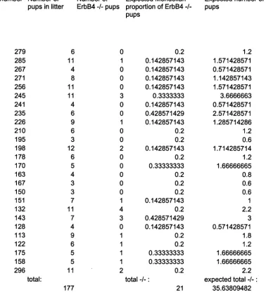
Fig u r e 3.5 Fe w e r Er bB 4 -/- m ic e b o r n t h a n e x p e c t e d... 97
ErbB4 - / - HER4^^‘”^ pups are not born in the expected proportions from ErbB4 + / - mothers with and without the HER4 allele...97
Fig u r e 3 .5a Ac t u a l a n d e x p e c t e d n u m b e r s of Er bB 4 -/- p u p s f r o m Er bB 4 + / - m o t h e r s 99 Fig u r e 3 .5b Ca l c u l a t in g e x p e c t e d n u m b e r of Er bB 4 -/- p u p s: ... 100
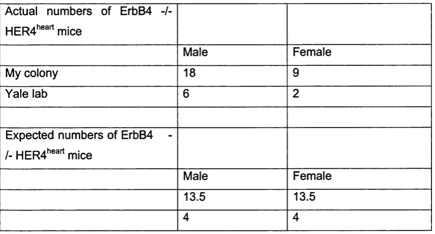
Fig u r e 3 .6 Fe w e r Er bB 4 -/- HER4"^"'^ m ic e a r e f e m a l e t h a n e x p e c t e d...101
Fig u r e 3 .7 R T -P C R ... 102
RT-PCR demonstrates that the oMHC-HER4-pA construct produces RNA in the heart from E10.5 onwards but in no other tissue examined....102
Fig u r e 3.8 Er bB 4 -/- H E R 4“ '^^ h e a r t h is t o l o g y is n o r m a l a t E 1 0 .5 ...103
FIGURE 3 .9 HEART VALVES A T E 17.5 APPEAR NORMAL IN RESCUED EMBRYOS... 104
Fig u r e 3 .1 0 Va l v e s in n e w b o r n Er bB 4 -/- HER4"^'^^ h e a r t s a p p e a r n o r m a l...105
Fig u r e 3.11 Re s c u e d m u t a n t s s h o w a b e r r a n t m ig r a t io n o f c r a n ia l n e u r a l c r e s t... 106
Fig u r e 3 .1 2 Ne u r o f il a m e n t s t a in in g o f E l 1.5 e m b r y o s... 107
ErbB4 - / - rescue embryos show links between trigeminal and facial ganglia...107
Fig u r e 3.1 3 Ne u r o f il a m e n t s t a in in g o f E 1 2.5 e m b r y o s... 108
Links between the trigeminal and facial ganglia persist at E l 2.5...108
Fig u r e 3 .1 4 His t o l o g y o f t r ig e m in a l a n d f a c ia l g a n g l ia a t E 1 4 .5 ... 109
Fig u r e 3 .1 5 His t o l o g y o f f a c ia l a n d t r ig e m in a l g a n g l ia a t E l 7 . 5 ...I l l Fig u r e 3 .1 6 Pa n c r e a s h is t o l o g y o f Er bB 4 r e s c u e s a p p e a r s n o r m a l...112
Fig u r e 3 .1 7 Co m p a r is o n o f w il d t y p e a n d Er bB 4 r e s c u e b r e a s t... 113
Fig u r e 3 .1 8 St a t5 is n o t p h o s p h o r y l a t e d in Er bB 4 -/- l a c t a t in g b r e a s t... 114
4. DISCUSSION... 115
4.1 Re d u c e d n u m b e r s o f Er bB 4 -/- HER4™'^^m i c e...115
4.1.1 Reduced numbers o f female ErbB4 -/- m ice...116
Ro l e s o f Er bB 4 in v a r io u s o r g a n s... 116
4 .2 Er bB 4 a n d t h e h in d b r a in...117
4.2.1 Neural crest phenotype...117
4.2.2 Axon Phenotype...117
4.2.3 Possible clinical relevance o f ectopic nerve...119
4 .3 Er bB 4 a n d to o t h d e v e l o p m e n t... 121
4 .4 Er bB 4 a n d h e a r t d e v e l o p m e n t... 122
4 .5 Er bB 4 a n d t h e p a n c r e a s...123
4 .6 Er bB 4 a n d m a m m a r y g l a n d s... 124
4.6.1 ErbB receptors and ligands in the mammary gland...124
4.6.2 Dominant-negative ErbB4...125
4.6.2.1 Comparison with dominant negative EGFR or ErbB2...126
4.6.2.2 Comparison o f the ErbB4 rescue mouse to the dominant negative ErbB4 mouse...127
4.6.3 Previous investigations into Stat5 activation in the breast...127
4.6.3.1 Prolactin had been thought to regulate StatS...129
4.6.4 ErbB4 conditional knockout...130
4.7 CONCLUSIONS... 132
4 .8 Fu t u r e DIRECTIONS... 133
5. A STUDY OF MESENCHYME COMPONENTS OUTSIDE RHOMBOMERE 3 ...135
5.1 CRYOCULTURE ASSAY ...135
5 .2 C S P G EXPRESSION STUDY... 137
5.3 Re s u l t s... 138
5.3.1 CSPG distribution...138
5.3.2 C-6-SPG distribution...139
5.3.4 Versican distribution...140
5.3.5 Heparan sulphate proteoglycans...141
5 .4 S P G Co n c l u s io n s... 141
5.4.1 Sulphated proteoglycans and ErbB4...141
5.5 Ma t e r ia l s a n d Me t h o d s...143
5.5.1 Immunohistochemistry...143
5.5.2 Cryosections...143
5.7.3 Polywax-embedded sections...144
5.7.4 Antibody staining o f fixed, blocked sections:...145
Fig u r e 5.1 Hy p o t h e t ic a l Er bB 4 a c t iv it y m e c h a n is m... 147
Fig u r e 5 .2 Ch o n d r o it in su l p h a t e p r o t e o g l y c a n s t r u c t u r e... 148
Fig u r e 5.3 C S P G d is t r ib u t io n in t h e h in d b r a in...149
Fig u r e 5 .4 Spe c ific it y o f C -6-S P G a n d C SP G a n t ib o d ie s...150
Fig u r e 5.5 H S P G s t a i n i n g...151
REFERENCES... 152
Abstract
ErbB4 is the fourth and, to date, final member o f the ErbB family o f receptor tyrosine
kinases, which includes the EGF receptor. ErbB receptors are expressed in many tissues
during development, and thought to have roles in proliferation, survival, and
differentiation. Additionally, their expression is deregulated in many tumour types.
M ice have previously been created which lack the ErbB4 receptor. These show defects
in cranial neural crest cell migration and axon pathfinding, but die by embryonic day
(E) 10.5 due to failure o f trabeculation to occur in the heart. To circumvent this mid-
embryonic lethality and to allow the study o f later genotypes, I have rescued the cardiac
defect in ErbB4 -/- mice using a construct expressing human ErbB4 (HER4) under a
cardiac-specific promoter. Expression o f this construct is shown to be specific to the
heart. ErbB4 -/- HER4*’®^ mice survive to adulthood and appear superficially normal.
However, fewer ErbB4 -/- rescued pups are born than predicted by Mendelian genetics.
Additionally, ErbB4 -/- HER4^®^ mothers fail to rear most o f their pups, probably due
to a lactation defect. I demonstrate that ErbB4 -/- HER4^®^ breast fails to differentiate
correctly during pregnancy, and also that ErbB4 regulates phosphorylation o f Stat5, a
central molecule in milk production.
The ErbB4 -/- neural crest and cranial nerve phenotype is shown to be replicated by
ErbB4 -/- HER4^®^ embryos, and an aberrant nerve linking the trigeminal and facial
ganglia persists at E l 7.5.
I also show results o f a separate study into the roles o f sulphated proteoglycans in wild
type and ErbB4 -/- embryonic hindbrain regions.
-1. Introduction
1.1 Receptor tyrosine kinases
One of the largest families o f cell surface receptors is the receptor tyrosine kinases
(RTKs). Receptor tyrosine kinases have intrinsic protein tyrosine kinase activity,
catalysing the transfer o f phosphate from ATP to hydroxyl groups on tyrosines of the
target protein, and this catalytic ability is activated when ligand binds the receptor. RTKs
have a ligand-binding extracellular domain, which is generally glycosylated, and this
ligand-binding domain is linked to the intracellular domain via a single transmembrane
helix. The intracellular domain contains a conserved protein tyrosine kinase domain.
Receptor tyrosine kinases (except the insulin receptor family) exist in the cell membrane
as monomers, and are induced to dimerise when ligand binds them. Some ligands are
bivalent, binding two receptor molecules simultaneously (e.g. growth hormone,
erythropoietin), some exist as homodimers (e.g. VEGF, PDGF), while others bind only
one receptor and induce dimérisation of the receptor with a coreceptor molecule, which
may be the same as or different from the activated receptor. Dimérisation in turn induces
autophosphorylation o f the receptor cytoplasmic domain at specific sites. Tyrosine
autophosphorylation is essential for these sites to be available for recruitment and
activation o f various signalling proteins, and thus RTKs also act as platforms for
recruitment o f specific sets of signalling proteins (Pawson and Schlessinger, 1993).
This thesis investigates one particular receptor tyrosine kinase, ErbB4, which belongs to
the ErbB subfamily.
-1.2 ErbB receptors and neuregulins
1.2.1 E rbB receptors
The ErbBs are a family of four receptor tyrosine kinases (RTKs), which includes the EGF
receptor (the first ErbB receptor to be discovered, also known as ErbBl), and three
closely related proteins. As RTKs, the ErbB receptors have a glycosylated ligand-binding
extracellular domain linked to an intracellular domain via a transmembrane helix, and an
intracellular domain which contains the protein tyrosine kinase region, plus various
regulatory sequences (reviewed by Schlessinger, 2000). ErbB receptors’ characteristic
structure includes two diagnostic cysteine-rich boxes in the extracellular domain. See figure 1.1 on page 39. As with most RTKs, the ErbB receptors (also known as neuregulin
receptors) are catalytically inactive until they bind a ligand, upon which the cytoplasmic
tyrosine kinase catalyses the cross-phosphorylation o f tyrosines, thus activating
downstream signal transduction cascades.
The term "ErbB receptor" derives from proteins found in the avian erythroblastosis virus.
This retrovirus carries two oncogenes, known as v-erbA and v-erbB. and erbB shows
great homology to the epidermal growth factor (EGF) receptor. So far in vertebrates
ErbBs 1-4 have been discovered. However, the EGF receptor (ErbBl) is somewhat
different from its three ErbB homologues found so far, in that it has not been found to
become activated in response to neuregulin in vivo.
1.2.2 Overview of EG FR and the ErbB receptors
The EGF receptor is expressed in many different tissues, particularly in basal cells of
epithelia and in proliferating tissues such as hair sheaths and sebaceous glands, and in
the brain, EGFR is expressed from E9.5 and is required for development of the
telencephalon, where it appears to be required in neural migration pathways. Postnatally
EGFR protein is seen in Purkinje and Golgi cells, and pontine and medullary nuclei, and
other areas o f the brain. EGFR expression has also been analysed in many tumour types
(Gullick 1991, and others). O f particular clinical interest is the finding that EGFR protein
levels vary between different breast tumours; there appears to be an inverse relationship
between EGFR levels and levels of oestrogen receptor (Sainsbury et a l, 1987), and expression of EGFR correlates with a poor response to endocrine therapy.
ErbB2, previously identified as the proto-oncogene c-neu, is similar in structure to EGFR,
although unfortunately for sensible nomenclature, c-neu/ErbB2/HER2 (Human EGF
Receptor 2) does not bind EGF, although it does function as a co-receptor. Also, ErbB2
cannot bind neuregulin directly, due to its ligand binding domain having very low affinity
for neuregulin (see section 1.2.3, ErbB dimérisation). ErbB2 protein is found in adult cells
derived from all three germ layers, including heart myocardium, peripheral neural tissues,
cerebellar granule cells, pancreatic cells, and squamous epithelia and hair follicles, among
others, but is not preferentially expressed in proliferating cells. In embryonic
development, ErbB2 appears to be ubiquitously expressed at low levels, with higher
levels in the heart and nervous system. It is involved in heart differentiation, and required
for migration of neural crest cells, for survival of Schwann cells and for nerve
myelination, and later in breast development (all described in more detail in later
sections). Overe?q)ression of ErbB2 is seen in various cancers (Slamon et al., 1989,
j
reviewed by Hynes and Stem, 1994); particularly in 20-30% of breast and ovarian
! cancers, gut and pancreatic cancers, and this overexpression correlates with a poor
prognosis (Holmes et at., 1992; Carraway et a l, 1995; Hynes and Stem, 1994).
-ErbB3 protein is seen in most tissues except the immune system. High levels are seen in
adrenal cortex, salivary gland ducts, and areas of the brain. Its expression is different to
that o f EGFR and ErbB2 in that ErbB3 protein is expressed in pancreatic islets, spinal
ganglion cells, adrenal cortex and testis, none o f which normally express EGFR or ErbB2.
In development, ErbB3 is known to be required for heart valve formation and
and also for migration o f neural crest cells and for survival of Schwann cell precursors (see
section 1.7), and to be expressed in the developing breast (see chapter 4). ErbB3 is also
expressed in tumours, notably in all gastric tumours in one study (Sanidas et al., 1993), 90% o f pancreatic tumours (Lemoine et al., 1992), and in a proportion o f ovarian, cervical, prostate and breast tumours (reviewed by Mason and Gullick, 1995). ErbB3's
catalytic domain appears to have no tyrosine kinase activity, so ErbB3 must dimerise with
another receptor in order to transduce a signal upon binding a ligand.
ErbB4 has both a functional ligand binding domain and tyrosine kinase domain. ErbB4
mRNA is found in adult brain (especially forebrain, pituitary and cerebellum), heart,
kidney, testis and breast at high levels, and at lower levels in thymus, lung, salivary gland
and pancreas. ErbB4 is not detected in liver, colon, bone marrow or ovary. In
development, it is required in heart myocardium, and also in the hindbrain, where it
influences neural crest and axon guidance (see sections 1.6.2 and 1.7.1 for more detail).
Later it is expressed in epithelial tissues, skeletal and cardiac muscle, and in PNS ^ d CNS
neurons (Srinivasan et a l, 199&). ErbB4 is the least understood ErbB receptor, in terms of its in vivo ligands and co-receptors, ^ d its involvement in cancers. Like the ntha" ErbB reçeptors, ErbB4 has beerr inqilicated irr various disease profiles and s h o w n to have altered expression in numerous tumour ceU lines, induding breast and pancreas (Mason and Gullick,
1995).
-The generation o f ErbB4 knockout mice has demonstrated ErbB4 to be required during
development, in the heart and also in the hindbrain, where it is involved in neural crest
and axon guidance (Gassmann et a i, 1995). This thesis describes experiments to elucidate the functions of ErbB4 in development, following on from information gained from the
ErbB4 knockout mouse, which dies in mid-embryogenesis due to heart defects
(Gassmann et a i, 1995). Thus it concentrates on the heart and hindbrain. My experiments demonstrate that ErbB4 is also required in the breast, which is discussed in chapter 4.
1.2.3 ErbB dimérisation and signalling
Being RTKs, ErbB receptors form dimers, but studies in vitro and in vivo show that ErbB dimérisation is not straightforward (reviewed by Carraway and Sweeney, 2001). It is
often thought that ErbB receptors are triggered to dimerise when they bind a ligand,
(Alroy and Yarden, 1997), but there are other possibilities: ErbB receptors may actually
form inactive dimers which become activated upon binding ligand, or active and inactive
dimers could exist in equilibrium, with ligand binding stabilising the active state. It could
even be that the receptors exist as homodimers which form active tetramers in response to
ligand (reviewed by Schlessinger, 2000).
However, in vitro studies, where cells not normally expressing ErbB receptors are made to express one or more receptor, have shown which combinations of receptors can
dimerise to produce functional signalling molecules (Riese et a i, 1995; Tzahar et a i, 1996). The EGF receptor forms homodimers in vivo, but can form active heterodimers in vitro with other ErbB receptors (Riese et a i, 1995). No in vivo EGFR heterodimers have
been found to date, ani, (,ack ^ ptUcr- ErLG
6X15^*.
-ErbB2 can form heterodimers with both ErbB3 and ErbB4, and both heterodimers are
capable o f cytoplasmic tyrosine phosphorylation in response to neuregulin. ErbB2
homodimers, however, cannot bind neuregulin directly, and are therefore incapable of
phosphorylating internal tyrosines in response. ErbB3 forms active heterodimers with
ErbB2 or ErbB4, while ErbB4 can also form active homodimers. ErbB3 homodimers can
bind neuregulin, but do not undergo tyrosine phosphorylation, as their tyrosine kinase
domains are virtually catalytically inactive (Riese et a l, 1995). ErbB2 and ErbB3 can thus compensate for the other’s defects by forming heterodimers, see figure L I, page 39.
However, just because a configuration is capable of being activated in vitro does not mean that it will lead to downstream signalling in vivo. The only known in vivo ErbB receptor pairings so far (apart from EGFR homodimers) are ErbB2 with either ErbB4
(found in the heart) (Gassmann et al., 1995; Lee et al., 1995) or with ErbB3 (sympathetic nervous system and glial cells) (Britsch et al., 1998; Erickson et al., 1996). In the mammary gland, for example, the EGF receptor and all three ErbB receptors are
expressed, but it is unclear which bind ligand and which function as coreceptors
(Olayioye et al., 2000). In parts o f the hindbrain, ErbB4 is expressed but EGFR, ErbB2 and ErbB3 are not. This suggests, but does not prove, that active ErbB4 homodimers
exist. An alternative possibility is the existence o f another ErbB receptor, which was not
picked up by the low stringency screening of human and rat cDNA libraries using viral v-
ErbB probe (ErbB2), nor by degenerate oligos corresponding to conserved regions of the EGFR and ErbB2 receptors - the method used to discover ErbB3 and ErbB4. 4
tip
6
/664
- migklr new/ fce^fiofs. fqckscreen
(las 6ec»v
h
Downstream signalling cascades activated by the ErbB receptors include the ras -> raf ->
MAP kinase pathway. Activation of ErbB3 and at least one isoform o f ErbB4 can lead to
activation of PI-3-kinase, but there are most likely other unknown downstream targets
(Elenius et al., 1999). Recently it has been shown that BrbB4 activation can lead to phosphorylation and entry into the nucleus o f the signal transduction factor Stat5 in the
mammary gland, presumably via Jak2. Upregulation of ErbB receptors is also observed in
numerous cancers, so the downstream targets are o f clinical interest.
1.3 Neuregulins
The neuregulins are a set o f polypeptide growth factors with diverse structures and
functions which are now known to all be encoded by a single neuregulin gene, NRG or
NRG-1, with the diversity created by initiation from different promoters and alternative splicing of NRG-1 transcripts (Garratt et al., 2000). Three further neuregulin gaies (NRG2-4 ) have been found, but these gene products tend to be referred to by number and have not
generally been included under ‘neuregulin’.
1.3.1 Neuregulin-1 splice Tariants
NRG-1 isoform? been shown to bind ErbB receptors in vitro, with varying affinities. The in vivo roles that have been described include:
- Glial Growth Factor (GGF), a Schwann cell mitogen which also suppresses neuronal
differentiation while promoting or permitting glial differentiation (Marchionni e t a l , 1993; Shah era/., 1994);
- Acetylcholine Receptor Inducing Activity (ARIA), a trophic factor isolated from
extracts of chick brain or spinal cord, which induces acetylcholine receptor translation in
skeletal muscle, and also accelerates formation of oligodendrocytes (Falls et al., 1993; Vartanian er a/., 1994);
- neu differentiation factor (NDF), which stimulates tyrosine phosphorylation of the neu
proto-oncogene product, inducing differentiation o f mammary tumour cells to
producing, growth-arrested cells, and is also known to be a survival factor for astrocytes
(Pinkas-Kramarski et a l, 1994; Wen et a l, 1992).
- Heregulin, also identified as specifically inducing phosphorylation o f ErbB2, appears to
be a similar splice variant to NDF (Holmes et a/., 1992).
- sensory and motor neuron-derived factor (SMDF), which is the predominant neuregulin
isoform in sensory and motor neurons, whereas other neuregulin variants are not
expressed in the peripheral nervous system. It also activates ErbB2 in cell lines. (Ho et a l, 1995)
The continued expression o f NRGs and ErbBs in the adult suggests roles in tissue
maintenance and repair, and possibly degenerative diseases might be alleviated by
augmenting NRG-ErbB signalling (Marchionni, 1995).
1.3.2 Neuregulin structure
All NRG-1 isoforms include a single EGF-related motif, with the same distance between
the third and fourth cysteine residues as in EGF and other molecules which bind the EGF
receptor. A subset o f NRG-1 isoforms (called type I) have an Ig domain, an a - or p-
variant EGF-like domain, variable extracellular juxtamembrane sequences, numbered 1-5,
a hydrophobic domain of uncertain function but likely to be an internal signal sequence
for protein secretion, and variable C-terminal sequences referred to as a, b or *
(representing a stop codon). These include NDF/heregulin and ARIA. Thus an individual
protein may be referred to, for example, as NDF-P or NRGp, indicating the type o f EGF-
like domain, or more precisely to indicate the exact juxtamembrane splicing variant, as
NRG I P2, for example (Liu et a l, 1998; Mason and Gullick, 1995).
14-Type II isoforms include GGF, and contain a signal peptide and a kringle-like sequence,
which are absent in type I neuregulins, plus an Ig and p-EGF-like domain. Type III
isoforms, originally identified as SMDF, share only the P-variant EGF-like domain with
other isoforms, and exhibit a hydrophobic domain within a cysteine-rich sequence in the
N-terminal domain, known as an SMDF region.
The transmembrane region is highly conserved across all NRG-1 isoforms, although
alternative splicing can produce soluble isoforms (reviewed by Lemke, 1996). The length
o f the cytoplasmic regions of NRGs has led to speculation that these domains could
participate in cell autonomous signalling, i.e. act as receptors as well as ligands (Shum et a l, 1994). See figure 1.2, page 40.
All NRG-1 proteins can interact directly with ErbB3 and ErbB4, and can stimulate ErbB2
via heterodimerisation of these receptors. Type I isoforms show higher affinity for ErbB4
than ErbB3 (Tzahar et al., 1994), and binding affinity varies among type I isoforms containing a or p-type EGF-like domains (Marikovsky et al., 1995). The hierarchy of ErbB dimérisation choices and the influence of EGF-like or neuregulin ligand are
complex. In general, however, ErbB heterodimers that include ErbB2 have their
formation favoured in response to neuregulin (Alroy and Yarden, 1997; Tzahar et al., 1996). NRG-1 isoforms do not bind EGFR or any other known receptors.
In situ analysis using isoform-specific probes has been used to analyse NRG-1 expression during mouse development (Meyer et al., 1997). To summarise: type I isoforms are predominant during early embryogenesis, with types II and III first detectable at
midgestation. Distinct isoform expression patterns are observed in development of the
-peripheral and central nervous system, indicating distinct functions. Type I NRG is
needed for development o f crest-derived sensory neurons in cranial ganglia and for heart
trabeculation, whereas type III is necessary for Schwann cell lineage development. Type
II mRNA is seen in the subventricular zone of neuroepithelium at e l2 and type III in the
outer layer o f the telencephalon, while at that age type I is restricted to a few cells in the
ventricular zone. NRG-1 type I mRNA has been identified in mouse hindbrain in
rhombomeres 2 ,4 , and 6 (Meyer and Birchmeier, 1995; Meyer et al., 1997)
1.3.3 Other neuregulin-like proteins
In addition to NRG-1, three further neuregulin-like genes have recently been cloned:
NRG-2, -3, and -4. These are separate genes to NRG-1 but encode products with similar
EGF-like domains to neuregulins and contain similarly-located alternative splicing sites
to NRG-1.
NRG-2 (Chang et al., 1997) encodes a ligand of ErbBS and ErbB4 in vitro, but does not activate the same profile of ErbB receptors as NRG-1 in vivo (Carraway et al., 1997). NRG-1 and NRG-2 expression is complementary in many areas; e.g. in the heart NRG-1
is expressed strongly in ventricular endocardium and weakly in atrial endocardium,
whereas for NRG-2 highest mRNA concentration is in the atria, with lower expression in
ventricular endothelium. Unlike NRG-1, NRG-2 is not found in the embryonic hindbrain,
although there is neural NRG-2 expression in adult olfactory bulb, dentate gyrus, and
most strongly in the cerebellum. There are two alternative EGF-like domains that
neuregulin-2 molecules can have, and these are termed neuregulin-2a and neuregulin-2p
respectively. Neuregulin-2 proteins are most similar to heregulin-pi o f the neuregulins:
neuregulin-2p has 45% amino acid homology with heregulin-pi and 40% with GGFII.
Certain regions are even more similar: there is 91% homology between the
-transmembrane domains of neuregulin-2 p and heregulin-pi, and parts o f the cytoplasmic
tails are also highly conserved, suggesting possible important functions.
NRG-3 binds the extracellular domain of ErbB4 in vitro, and also when ErbB4 is expressed in a stable cell line (Zhang et al., 1997), inducing ErbB4 tyrosine kinase phosphorylation. NRG-3 does not bind any other known receptors. The NRG-3 amino
acid sequence is 62% homologous to heregulin-pi, with a similar C-terminal hydrophobic
region and a similar N-terminal internal signalling sequence, and also contains an EGF-
like domain and glycosylation sites, but it has no extracellular Ig-like or kringle domains.
Its EGF-like domain is distinct enough from those of NRG-1 and NRG-2 to show that it is
not merely a novel NRG-1 or NRG-2 isoform produced by alternative splicing. NRG-3
mRNA is expressed in adult brain, and at E l6 in brain, spinal cord, trigeminal, vestibulo
cochlear and spinal ganglia, as well as in differentiating areas of the telencephalon. NRG-
3 mRNA exists in the nervous system at E l 3 in mice, but earlier time points have not
been examined using in situs, although NRG-3 mRNA has been detected at E l l (but not E4) using Northern blots. It has been suggested that NRG-3 could encode a ligand for
ErbB4 in the hindbrain (Zhang et a l, 1997).
A fourth neuregulin (NRG-4) can induce growth of cultured cells made to ectopically
express ErbB4, and can displace NRG-1 bound to ErbB4. It has an EGF-like domain
showing 32% amino acid homology to NRG-ip, including six cysteines characteristic to
all NRG gene products, but apart from the transmembrane region has no other motifs
similar to any seen in other NRG proteins. NRG-4 mRNA is detected in adult pancreas
and weakly in muscle, but no other tissues, indicating a different physiological role to that
-of the other neuregulins (Harari et al., 1999), which are predominantly involved in the heart and nervous system.
There are also other ligands, non-NRG members o f the EGF-related family, which
activate ErbB3 and ErbB4 in vitro. These are heparin-binding EGF (HB-EGF) (Higashiyama et a l, 1991), amphiregulin (Shoyab et a l, 1989) and betacellulin (Beerli and Hynes, 1996; Riese et a l, 1996). All these EGF-like ligands are products o f separate but related genes. These three ligands are known to bind the EGFR in vivo but it is unclear whether they also bind ErbB4 in vivo, for example in the mammary gland. See figure 1.3 fo r summary o f ErbB receptors and their ligands. Recently, our lab
demonstrated that HB-EGF is not expressed in mouse embryonic hindbrain, and thus is
not ErbB4's ligand there (Tsoni et al, manuscript in preparation).
18-1.4 ErbB and neuregulin mutants
Transgenic mouse technology has been used to study the roles o f ErbBs and
neuregulins during development. Mice with null mutations o f ErbB2, ErbB3, ErbB4
and NRG-1 have been generated (Lee et al., 1995; Erickson et al., 1997; Riethmacher et al., 1997; Gassmann et al., 1995; Meyer and Birchmeier, 1995; M eyer et a l, 1997).
In the NRG knockout, as well as in ErbB2 -/- and ErbB4 -/- mice, and in mice containing
a deletion o f the Ig or intracellular domain of NRG-1, homozygotes express no detectable
protein, and die between ElO and E l l (Kramer et al., 1996; Meyer and Birchmeier, 1995). The heterozygotes o f all three mutants are fertile and superficially normal. Thus all
these genes are proven essential, and are not entirely functionally or developmentally
redundant throughout the embryo, although they may be in certain areas.
The systems most affected by loss o f an ErbB receptor or neüregulin are the embryonic
nervous system and the heart. Information from these knockouts has led to further
experiments explaining more o f the roles of the individual receptors and ligands in these
systems. The next sections describe in more detail tissues where ErbBs and their ligands
are expressed concurrently (i.e. hindbrain and heart), and thus ligand-receptor interaction
is thought to have a role in development and in some cases, also in the adult.
1.5 The Hindbrain
The hindbrain is an established model for studying mechanisms o f cell signalling and
migration during vertebrate development, notably in mouse, chick, and zebrafish. The
hindbrain develops to form, in the adult, the cerebellum, pons, and medulla.
19-The nervous system derives from a specialised part of the embryonic ectoderm; in
neurulation the ectodermal layer of the gastrula becomes partitioned into the neural tube,
epidermis, and neural crest. Cell shape changes and movements delineate the neural plate
and later produce the neural tube, with overlying ectoderm and neural crest cells, which
delaminate and migrate away from the dorsal lip of the tube around the time o f neural
tube closure. The trunk region o f the neural tube is destined to become spinal cord, and
remains a comparatively simple tube, while the cephalic region, which will form the
brain, forms a series of bulges which subdivide it both physically and molecularly and
enable different parts of it to specialise, as detailed below.
The first cephalic bulges, known as primary compartments, are (anterior to posterior) the
prosencephalon, mesencephalon, and rhombencephalon, destined to form forebrain,
midbrain, and hindbrain respectively. These are seen at about E7.5 in mouse. The
rhombencephalon (from its rhombus shape) subsequently divides into the metencephalon
and myelencephalon, and then (E8.5 - E l l in mouse, stages 9-12 in chick), is transiently
segmented into structural repeating units, known as rhombomeres (summarised by
Gilbert, 1994).
The rhombomeres divide the hindbrain into compartments, where cells from different
rhombomeres are prevented from mixing, and exhibit different patterns o f gene
expression, lineage restriction, and neuronal differentiation. Thus cells are segregated and
their fate determined to enable the fixture formation o f more specialised structures. This
makes the hindbrain a comparatively convenient system for study of CNS patterning, as it
is accessible, easy to visualise the different rhombomeres, and contains a relatively low
number o f cell types. Additionally, lessons learnt from one segment may be applicable to
another, and perhaps have relevance to other segmented systems. Segmentation is
frequently used in animal development to produce homologous but distinct structures
from repeated tissue blocks along the AP axis; the tissue becomes segmented into
repeating lineage-restricted blocks, which then differentiate somewhat differently
according to their position along the body axis. Hindbrain segmentation, although
restricted to vertebrates, bears superficial resemblance to metamerism seen in completely
different animals such as the Drosophila larva, and also to somite formation in vertebrates. Although the segmentation strategy is believed to have evolved
independently many times (Newman, 1993), these examples share conceptual features,
such as the idea o f a developmental compartment.
A developmental compartment is considered to be a block o f cells which do not mingle
with adjacent cell populations, and where the block corresponds with a distinct profile of
gene expression and action (Lawrence and Struhl, 1996). Thus the cells have a collective
identity and can follow a co-ordinated differentiation programme. Compartments may
also help limit cell proliferation. However, the morphological boundary, which separates
many developmental compartments, including the rhombomeres, is not a requirement, as
other mechanisms are available which can prevent intermingling between adjacent cell
populations, such as differential cell adhesion or repulsion in different rhombomeres.
Rhombomere boundaries are known to exhibit differences to the rest o f the rhombomere;
cells are altered in shape (Heyman et a l, 1993) and have reduced rates of cell division (Guthrie et a i, 1991), and show larger extracellular spaces and altered extracellular matrix (Heyman et al., 1995). However, experimental evidence shows that it is not these physical properties so much as inherent cell-specific differences which cause boundary
-formation: rhombomere-specific gene expression is observed before morphological boundaries can be seen (e.g. H o x -^W ilk in so n et al., 1989b)); boundaries regenerate after being surgically removed (Guthrie and Lumsden, 1991); grafting and transplant
experiments show that cells from adjacent rhombomeres (e.g. r4 and r5) will sort out if
mixed in vitro (Guthrie et a l, 1993; Wizenmann and Lumsden, 1997), while cells o f two even-numbered or two odd-numbered rhombomeres will mix, though to a lesser extent
than cells o f the same rhombomere (Guthrie and Lumsden, 1991; Wizenmann and
Lumsden, 1997). Small clumps o f cells can maintain their rhombomere identity when
transplanted into other rhombomeres, provided the cells remain in contact, but if
dispersed they will acquire the gene expression identity o f the new rhombomere (Trainor
and Krumlauf, 2000). This indicates that cell repulsive interactions or differences in
adhesion is what keeps cells in their rhombomeres, and that alternate rhombomeres (e.g.
r2 and r4, or r3 and r5) have similar, but not identical, properties.
The rhombomeres (rl to r8) give rise to migrating neural crest cells and later to cranial
motor nuclei (Lumsden et a l, 1991). {See figure 1.4, page 42.) Neural crest cells are produced all along the neural tube, but within the hindbrain exit the neuroepithelium only
from specific rhombomeres to form segregated streams. Thus, neural crest cells enter the
mesenchyme only from rl+ 2 , r4, and r6+7, with proportions of these streams migrating to
populate the first, second, and third branchial arches respectively, while another proportion
ceases ventral migration lateral to the hindbrain, forming the cranial ganglion. Hence the
neural-crest-derived components o f the trigeminal and facial ganglia derive from r2 and r4
respectively, and also in normal mouse or chick there is no neural crest seen in
mesenchyme outside rhombomeres 3 or 5. Crest cells in the forming cranial ganglia are
joined by presumptive neuroblasts migrating medially from epidermal placodes. See figure
1.6.
Meanwhile, branchiomotor neuron organisation shows similar spacing. Groups of
branchiomotor neurons with axons innervating a single branchial arch (IJl, or 3) occupy
22-pairs o f segments. Thus motor axons from r2+3 innervate branchial arch 1 and axons
extending from r4+5 and r6+7 innervate the second and third arches respectively.
Similarly, sensory axons extending from the trigeminal and facial ganglia enter the
neuroepithelium only at specific entry points in r2 and r4 respectively, and neural
crest contributions to the trigeminal (V) ganglion come from r2 and to the geniculate
(VII) ganglion from r4. The petrosal ganglion (^X ) has neural crest contributions
from r6 and r7, and the jugular (gX) from r7. The vestibulo-acoustic ganglion (gVIII)
is exclusively placode-derived.
See figure 1.6, showing cranial ganglia formation, and 1.7 which contrasts the different neural components o f cranial and dorsal root ganglia.
In the hindbrain, both neural crest and the ectodermal placodes contribute to the
cranial ganglia, with placode-derived neurons being located distal to crest-derived
neurons (D’Amico-Martel and Noden, 1983).
The relative contribution o f neural crest and ectodermal placodes to the various
cranial ganglia has been mapped using quail tissue transplants (D’Amico-Martel and
Noden, 1980, 1983). The trigeminal ganghon, associated with cranial nerve V, is the
most rostral sensory ganglion, and contains both small proximal crest-derived neurons
and larger distal placode-derived neurons in one large ganglion. Neurons in the
ganglia associated with cranial nerves VII, IX and X, however, form two separate
ganglia, of crest and placodal origin respectively. These include the root (proximal)
and geniculate (distal) ganglia of nerve VII (facial nerve), the superior (proximal) and
and nodose (distal) of nerve X (vagus nerve). The proximal (vestibular) and distal
(acoustic) ganglia of nerve VIII (acoustic nerve) are both composed of otic-placode-derived
neurons (D’Amico-Martel, 1982). Supporting cells in the cranial ganglia, including
Schwann cells, are also neural crest-derived. In the trunk, in contrast, spinal ganglia are
solely derived from neural crest.
Hindbrain neural crest cells will populate sensory ganglia, but also go on to form a wide
variety o f other muscular, skeletal, neurogenic and vascular structures, including the
bones and other structures o f the inner ear, the jaw and palate (Gilbert, 1994; Koentges
and Lumsden, 1996).
Neural crest cells contain some cell-autonomous information with them about their
origins (Prince and Lumsden, 1994; Saldivar et aL, 1996). However, their paths are also influenced by the environment. The destination and fate o f crest cells depends partly on
the age o f surrounding mesenchyme, as shown by transplanting crest cells o f various
origins and ages into older or younger embryos. Thus the mesenchyme can influence
crest cell migration, although the effect also depends on crest cell density - so-called
'community effects'. (Raible and Eisen, 1996). It has been unclear to what extent crest is
pre-pattemed and how much role the mesenchyme has to play (see (Golding et aL, 2000), and chapter 5). It is plausible that the mesenchyme through which neural crest cells pass
can affect the cells in different ways, causing them to adopt different fates.
23-1.5.2 Gene expression in the hindbrain
This thesis looks at aspects of cell segregation and patterning in the hindbrain that are
regulated by molecular segmentation o f the hindbrain. This section describes some of the
genes that illustrate the molecular segmentation of the hindbrain.
Various members o f different families o f signalling molecules are known to be
expressed in and to have functional roles in the hindbrain, such as Eph receptors and
their ligands the ephrins, and Hox genes (Becker et a l, 1994; Hunt et a l, 1991b; reviewed by McGinnis and Krumlauf, 1992) . These and other genes often have the
anterior and/or posterior boundaries o f their expression coinciding with rhombomere
boundaries.
The Hox genes are found in all vertebrates, in four clusters (Hoxa, Hoxb, Hoxc and
Hoxd) which have arisen by duplication and evolution o f a single cluster o f homeotic
genes found in Drosophila^ which are organised in order corresponding to their expression along the AP axis o f the embryo. Hox genes play a central role in specifying
tissue identity along the AP axis of all multicellular animals from C elegans to vertebrates (reviewed by Krumlauf, 1994). As in Drosophila, vertebrate Hox genes are organised along the chromosome within their cluster in the same order as they are
expressed, with genes at the 5' end o f the complex being most posteriorly expressed, and
genes further 3' having more anterior limits of expression.
In the hindbrain, the anterior limits of Hox gene expression correspond with various
rhombomere boundaries (Wilkinson, 1989; Hunt et a l, 1991a), so Hoxb2 reaches as far anterior as the r2/3 boundary, while Hoxb3 and Hoxb4 expression only reach the r4/5 and
-the r6/7 boundaries respectively (Wilkinson, 1989). However, rhombomere-specific
variations in amounts of Hox gene expression are superimposed on top o f this pattern, so
Hox gene 5’-3’ expression cannot be the only patterning mechanism that controls Hox
gene expression in the rhombomeres. In vertebrates, as in Drosophila, disruption of Hox gene expression leads to homeotic transformation o f body parts in their anterior regions of
expression, disrupting AP axis patterning (reviewed by Krumlauf, 1994). Hox genes are
known to be controlled by retinoids, which bind retinoic acid/ retinoid X receptors (RARs
/ RXRs), which in turn bind c/5-acting retinoic acid response elements (RAREs) in the
regulatory regions o f the various Hox genes (Gould et al., 1998; Studer et al., 1998).
Other genes showing rhombomere-specific expression are known to be vital for setting up
or maintaining segmentation o f the hindbrain. The transcription factor Krox20 is
expressed in r3 and r5 only (Wilkinson et al., 1989a), and when mutated, although r3 and r5 are initially formed correctly, they are rapidly lost. The transcription factor kreisler, on
the other hand, expressed in r5 and r6, is required to specify r5 identity; in kreisler
mutants rl-3 appear normal but at the r4-7 level rhombomere identity breaks down, so the
neural tube is smooth and morphologically unsegmented.
Eph receptor tyrosine kinases and their ligands, the ephrins, are known to be involved in
establishing regions o f differential adhesion and/or repulsion, and were in fact initially
studied in the hindbrain where they exhibit rhombomere-specific patterns (Nieto et a l, 1992). They are also involved in axon pathfinding, neural crest migration and retino-tectal
mapping, among others. In the hindbrain, Eph receptors EphA4, EphB2 and EphB3 show
complementary expression patterns to their ligands, being expressed in rhombomeres 3
and 5, while the ephrins ephrinBl, B2 and B3 are all transmembrane ligands expressed in
-rhombomeres 1, 2, 4 and 6 (Flenniken et al., 1996; Gale et a l, 1996). Ephrins are only usually active when membrane-bound, and ephrin-B proteins appear in fact to be capable
of bidirectional signalling when they bind receptors (Henkemeyer et al., 1996; Mellitzer et al., 1999),
When a dominant negative EphA4 receptor is overexpressed in r3 and r5, disrupting
receptor function, hindbrain patterning is disrupted (Xu et al., 1995): ectopic r3 and 5 cells are observed in r2, 4 and 6. Also, neural crest migration is disrupted; crest which
normally migrates to the third branchial arch migrates to the second and fourth arches as
well (Smith et al., 1997). Overexpression of ectopic ephrinB2 has a similar effect. In vitro analysis ("fishball assays") has shown that Ephs and ephrins directly enable cell sorting,
restricting intermixing of odd and even-numbered rhombomeres.
Migration o f the neural crest is influenced not only by the rhombomeric origin of crest
cells, but also by their age, and presumably by the mesenchyme through which they pass.
The mesenchyme will also affect extension o f axons growing through it towards entry
sites in the neuroepithelium.
Previous studies have suggested that the mesenchyme outside the hindbrain has a role in
influencing^cre^t cell and axon pathfinding, i.e. that these cells respond to their
environment rather than their destinations being entirely pre-pattemed. In particular, the
mesenchyme next to r3 and r5 contains an activity capable o f excluding neural crest cells
(Farlie et al., 1999, Keynes and Cook review 1995), implying that it is not only properties of the crest cells that leads them to emanate only from rhombomeres 2, 4, and 6 into the
mesenchyme.
In the trunk, the mesenchyme is clearly segmented; the somites are divided into rostral and
caudal portions with different properties, leading to segmentation of neural crest and motor
axons due to repellent properties of the caudal somite. However, there is no sudh obvious
mesenchyme segmentation in the head.
Tam and Meier (1982), on the basis of scanning electron microscope (SEM) studies, claimed
the existence of mesenchyme segments in the head, which th ^ termed somitcaneres. However
somitomeres’ existence has not been supported by any other methods such as gene e^qpression
studies. Transplantation of mesoderm from near the neuroepithelium from one cranial axial
level to another suggests that the cells migrate to the nearest branchial arch, implying that there
is no difference in the environment to attract the mesoderm cells to one arch rather than another
(Hacker and Guthrie, 1998). Given that the neural tube is segmented in the hindbrain, unlike in
the trunk, and that this segmentation enables neural crest cells to be created with different
prqjerties along the A-P axis (e.g. differential Hox gene expression), the consensus of opinion
holds that that head m^enchyme was unsegmented and the only A-P differential was intrinsic
to the neural tube.
Grafts of neuroepithelium posterior to the otic vesicle result in reprogramming of Hox gene
expression, reflecting cells' new location along the AP axis. This plasticity is mediated by
somitic mesoderm which lies caudal to the otic vesicle (post-otic) (Itasaki et al., 1996; Gould et a i, 1998). In pre-otic grafts in chick, however, rhombomeres autonomously maintain the Hox expression patterns of their original AP position (Prince and Lumsden, 1994). These
differences correlate with differences in mesoderm in pre- and post-otic regions; pre-otic does
not express somitic markers, nor show epithelial-type condensation, staying more diffuse, and
is thougfrt to have only a passive role in head patterning, responding to signals from neural
crest (Traincff and Krumlauf, 2000).
The fixed neural crest prepatteming model comes from experiments in chick involving large
Lumsden, 1994). Recently, however, Krumlauf and Trainer (2000) have used small groups of
labelled mouse cells in cultured embryos for transplantation experiments, which suggest that
head mesoderm does after all have scxne part to play in neural patterning. Heterotopic grafts
(r3, 4 or 5 into r2) showed that tlK main grafts maintained Hox repeater gene expression, but
cells dispersed fi’om the graft lost reporter expression, indicating plasticity of Hox expression,
and that community effects are important for expression. Neural crest cells produced from
rhombomere grafts migrate towards the branchial arches consistent with the host environment,
indicating influence of the environment on migrating crest.
When grafts of seccsid arch crest cells and mesodam were transplanted into branchial arch 1,
unlike grafts of crest alone, the reporter gene was reactivated. Thus mœoderm is capable of
maintaining neural crest cell Hox gene expression in an ectq)ic environment, although it
cannot induce ectopic reporter expression by itself. This signal maintenance implies a role of
cranial mesoderm in maintaining the AP register between different tissue primordia (Trainor
and Krumlauf 2001).
As well as prepatteming and mesodermal maintenance, there is evidence of a third influence on
crest cells: signals fix>m the rhombomeres can influence the environment. In ErbB4 mutant
embryos, loss of ErbB4 signal in r3 and r5 leads to ectopic anterior migration of r4-derived
crest cells. ErbB4 mutants in a Hoxb2/L^:Z reporter background show downregulatioe of Hox
genes in the ectopic locatiæ (Golding et a l, 2000). See section 1.8 for more detail. This shows there is interplay between instructive signals in the neuroectoderm and permissive signals in
the mesenchyme, perhaps needed to regulate neural crest cell identity and migration pathways.
Therefœe we have analysed potential signalling molecules in the hindbrain.
Chapter 5 contains a stu<fy of molecules which are present in mesoichyme adjacmt to the
1.6 The Heart and ErbB receptors and ligands
At embryonic day 9.5, the mouse heart ventricles consist o f three layers: the outer
pericardium, the thick myocardium, consisting mainly o f developing myocytes, and the
inner endocardium, where mesenchyme and myocytes form the endocardial cushions. The
atria have much thinner walls, due to a very thin myocardium and almost complete lack
o f endocardial cushions.
1.6.1 Heart trabeculation
In NRG -/-, ErbB2 -/- and ErbB4 -/- mice, embryonic death is due to lack of
differentiation o f heart muscle. Neuregulin-1 is expressed at high levels in the endothelial
lining of the heart ventricles (Meyer and Birchmeier, 1994), in direct contact with
developing heart muscle, or myocardium, where developing cardiac myocytes express the
receptors ErbB2 and ErbB4. E9 hearts in these embryos appear normal, but by E l0.5 the
embryos are dead. In the intervening period, wild type heart muscle contains some
myocytes which develop into sponge-like regions o f myocardium, a process known as
trabeculation (Sedmera and Thomas, 1996). Trabeculae are totally absent in all these
knockout mice.
The ligand-expressing cells and receptor-expressing cells are directly apposed. The NRG-
1 gene product is acting non-cell-autonomously; myocardial development is halted but the
endocardial cells which usually express NRG appear normal. In contrast, ErbB2 and
ErbB4 receptors act cell-autonomously. Evidently all three signalling components -
NRG, ErbB2 and ErbB4 - are necessary for trabeculation; the fact that loss of either
-ErbB2 or ErbB4 is enough to cause death implies that in myocardium between E9 and
E l0.5 ErbB2/ErbB4 heterodimers are required, and that the NRG-1 isoforms present are
incapable o f activating ErbB4 homodimers. Thus neither receptor is functionally
redundant, even when there are receptors present in the same cell which can act in vitro as homodimers. NRG-2 is expressed in endocardium of the atria, directly apposed to ErbB2
and ErbB4 in the atrial myocardium. It is tempting to assume that it acts in a similar
induction process for differentiation o f the atrial walls in development.
1.6.2 ErbB4 and the heart
In the adult heart, ErbB4 RNA is known to be present in muscle o f both atria and
ventricles. ErbB4 is specific to myocytes during development and in adult; Northern blots
do not detect ErbB4 in myocyte-depleted cardiac cell populations. ErbB2 is also seen in
ventricular myocytes during development and in adult, but at lower levels compared to
ErbB4. ErbB2 and ErbB3 also exist in myocyte-depleted adult cultures, unlike ErbB4
(Zhao et a/., 1998). ErbB4 has been shown to contain a caveolar binding motif and be localised to caveolae in cardiac myocytes: caveolae are flask-shaped invaginations of the
plasma membrane, which contain high levels o f cholesterol and similar molecules which
mediate endocytosis and movement across the membrane in a clathrin-independent
manner. Various signalling cascades utilise caveolar localisation, which is presumed to
facilitate activation o f signal transduction by ligands, and to enable cross-talk between
different signalling pathways (Zhao et al., 1999). Upon NRG-1 binding, ErbB4 is rapidly translocated out o f caveolae, which presumably enables the receptor to become
desensitised in the presence of ligand, although interestingly ErbB2 stays put.
-Upon activation, ErbB4 becomes cleaved by protein kinase C, yielding a soluble 120kD
fragment from the extracellular domain, and an 80 kD membrane-anchored fragment
representing the entire cytoplasmic and transmembrane domains. This presumably
regulates the distribution o f extracellular ErbB4 at the cell surface, s '^nce. unlike EGFR and many other RTKs, ErbB4 is not internalised by endocytosis (Vecchi et a l, 1996).
A second mechanism o f ErbB4 cleavage has been established in which a metalloprotease
also cleaves the extracellular domain, and levels of the membrane-tethered product are
then controlled by proteasomes. The tethered 80 kD fragment, which retains tyrosine
kinase activity, is polyubiquitinated, unlike intact ErbB4, which is presumably why
proteasome activity does not act on the ErbB4 receptor before metalloprotease cleavage
(Vecchi and Carpenter, 1997), Thus ErbB4 receptor metabolism demonstrates a novel
coupling o f two separate protease activities, acting sequentially, to enable ligand-
independent ErbB4 degradation and yet preventing cytoplasmic buildup of the 80 kD
fragment. The fragment may have ligand-independent kinase activity, which would make
its destruction particularly important, but why the 80 kD fragment is constitutively
produced by ErbB4-expressing cells is unclear. It may act as a membrane-bound docking
molecule for signalling molecules, such as SH2 domain proteins, PI-3-kinase or Grb-2,
which appear to require docking at the plasma membrane before tyrosine
phosphorylation. PLC-y and She both precipitate with the 80 kD fragment (Vecchi et al., 1996; Vecchi and Carpenter, 1997).
1.6.3 ErbB3 and the heart
ErbBB RNA is found in endocardial cushion and bulbus cordis mesenchyme during
development (Meyer and Birchmeier, 1995). The bulbus cordis is part o f the heart tube
which derives from the top o f the ventricle and will form the aorta and pulmonary artery.
In wild type mouse hearts at E9.5, ventricular endocardial cushions are apposed, with a
thick core o f mesenchyme and myocytes. In ErbB3 -/- embryos, however, mesenchyme is
lacking in the cushions, and the cushions are distinctly thinner. The cushions give rise to
the atrio-ventricular (AV) valves, so it is not surprising that by E l3.5 the AV valves in
ErbB3 -/- embryos are hypoplastic, little larger than at ElO. Videos o f valves at E l 3.5
showed blood reflux through the valves, which is presumably the cause o f death at E l3.5
(Erickson er fl/., 1997),
1.7 Phenotypes of ErbB2, ErbB3 and NRG knockouts
Although the NRG -/-, ErbB2 -/-, and ErbB4 -/- mice have identical phenotypes in the
heart, and all three exhibit neural phenotypes, the neural phenotypes are all different.
ErbB3 -/- embryos also exhibit a neural phenotype.
NRG-1 is normally expressed in midembryonic neuroepithelium. In the hindbrain, NRG
is restricted to neuroepithelium of, and neural crest cells from, rhombomeres 1+2, 4, and
6. These crest streams, plus placodal derivatives, normally form cranial ganglia V,
V IW ill and IX (trigeminal, facial/acoustic and glossopharyngeal, respectively). These
are structures o f the peripheral nervous system (PNS) and form immediately adjacent to
the hindbrain.
In the NRG mutant, however, most trigeminal ganglion (gV) cells, and a proportion o f
those in the other ganglia mentioned, are missing (Meyer and Birchmeier, 1995). The
remaining parts of the ganglia are more distant to the hindbrain than usual, and it is
assumed that most o f the normally NRG-positive neural crest cells that usually contribute
to these ganglia have died, so that