___________________________________________________________________________________________ 1(4): 459-468, 2011
SCIENCEDOMAIN international
www.sciencedomain.org
Vitamin D Receptor BsmI Gene Polymorphisms
and Gestational Diabetes Mellitus:
A Saudi Study
M. A. Tawfeek
1, F. A. Habib
2and Entessar Emam Mouhamed Saultan
3*1
Clinical Pathology Department, Obstetric College of Medicine, Taibah University, Al – Madina Al - Monawara, Saudi Arabia.
2
Obstetric and Gynecology Department,College of Medicine, Taibah University, Al – Madina Al - Monawara, Saudi Arabia.
3
Internal Medicine Department, College of Medicine, Taibah University, Al – Madina Al - Monawara, Saudi Arabia.
Received 5th June 2011 Accepted 14th June 2011 Online Ready 28th September 2011
ABSTRACT
Background: Both vitamin D deficiency and Gestational diabetes (GDM) are common among Saudis. The vitamin D receptor (VDR) gene is a candidate gene for susceptibility to several diseases. Studies on association between VDR polymorphisms and risk of GDM in Saudi populations are yet inconclusive.
Objective: to evaluate the association between Vitamin D receptor gene polymorphisms and genetic susceptibility to gestational diabetes (GDM) in pregnant Saudis.
Subjects & Methods: thirty pregnant Saudi women with diabetes (17 GDM and 13 type 2 diabetes (T2DM) with past history of GDM) were compared to 14 pregnant women with normal glucose tolerance. Patients and controls were recruited at their third trimester from Taibah University medical unit from January to July 2010. Genomic DNA was extracted and the genotyping related to vitamin D receptor BsmI gene single – nucleotide polymorphisms was carried out by polymerase chain reaction and restriction fragment length polymorphism (PCR-RFLP) analysis.
Results: The gene frequency, allele frequency and carriage rate of the VDR polymorphism BsmI did not differ between patients and controls with no significant association with any clinical parameters. The 25 hydroxyl Vitamin D level but not the
gene frequency was a significant predictor of history of abortion among diabetics (OR=-0.29, 95% CI -0.081-0.0, p=0.047).
Conclusions: Vitamin D receptor BsmI gene polymorphisms is not associated with gestational diabetes among Saudis. Further studies of other Vitamin D receptor gene polymorphism in combination are required.
Keywords: Gestational diabetes; VDR polymorphism; 25 hydroxyl; vitamin D.
1. INTRODUCTION
The study of the genetics of gestational diabetes mellitus (GDM) provides excellent opportunities to link molecular insights with epidemiological data. A minority of women have evidence for evolving type 1 diabetes with the highest frequency in populations with the highest rates of type 1 diabetes but does not appear to be >20% in any population . Causes of beta cell dysfunction vary among women without evidence for beta cell autoimmunity. Some have specific genetic problems affecting beta cells, such as genetic variants that also cause maturity-onset diabetes of the young (MODY). Most others have clinical obesity and physiological insulin resistance and poor beta cell compensation characteristics that suggest evolving type 2 diabetes (T2D) supporting the idea that it is unlikely that GDM has a unique genetic predisposition but rather is a manifestation of multiple susceptibility variants for T2D (Anonymous, 2004).
Despite the present evidence of simple, reliable means to identify people who are at risk at GDM, still identification of genetic variants linked to GDM will contribute to our understanding of the physiopathology of GDM and to the development of more aggressive preventive strategies in a subset of women at higher risk (Anonymous, 2004).
Genes involved in the metabolic pathway of insulin have been regarded as good candidates for both type 1 and type 2 diabetes pathogenesis. One of these genes is vitamin D receptor (VDR) gene (Zhang et al., 2008). In addition, it has long been known that vitamin D deficiency is prevalent among pregnant women and has been associated with elevated risk for pregnancy complications and a number of serious short- and long-term health problems in off springs (Dent, 1975; Bodnar et al., 2007). Identification of genetic variants underlying GDM will not have a major impact on clinical care for patients with GDM but in case of association with VDR polymorphism if proven, vitamin D supplementation will represent a promising preventive strategy for both GDM and T2D later in life. This will be of utmost benefit among population with prevalent GDM, T2D and vitamin D deficiency like Saudi women. Therefore, the aim of this study was to study the association of VDR polymorphisms with GDM in Saudi pregnant women.
2. SUBJECTS & METHODS
diabetics had family history of Type 2 diabetes. Screening and management of diabetes during pregnancy were done according to the American Diabetes Association guidelines (Anonymous, 2004). A questionnaire was used to record clinical history including history of maternal and neonatal complications. Fourteen age-matched normal healthy pregnant controls with no past history of GDM or past history of bad obstetric outcomes or family history of DM were enrolled for the same unit. All patients were receiving multivitamins at the time of the enrolment. They were screened with standard oral glucose tolerance test at their third trimester. Women known to have type 1 DM, multiple pregnancies and women with medical disease (hypothyroidism, renal, hepatic, hypertension ....etc) were excluded. All participants gave written consent to participate in the current study. The mean age of the diabetic group was 35.73±5.33 years, 19 (63%) of them were on insulin treatment and the remaining 11 patients (36.7%) were on diet therapy alone. The mean systolic blood pressure was 110 ±10.8 mmHg, and diastolic blood pressure was 73.46 ±8.00 mmHg. The most common bad obstetric history among diabetics was abortion (24 (80%): 15 patients (50%) had 1 abortion, remainder 9 patients (30%) had more than one abortion), followed by big baby (5(16.7%)). Other obstetric history included pregnancy induced hypertension (1(3.3%)), stillbirth (1 (3.3%)) and accidental hemorrhage (1 (3.3%)). Six patients (20%) were primigravida, 19(63.3%) delivered normally, 10% (33.3 %) delivered by CS and 5 patients (16.6%) delivered normally but had CS for the last deliveries because of obstetric complications (table 1).
At entry to the study nine ml venous blood was collected under complete aseptic technique after overnight fasting without venostasis. Three ml. blood were equally delivered into three sterile EDTA coated tubes. One for the PCR technique, the other is for complete blood count using electronic cell counter CD 1700 (USA). The third tube for separation of plasma which were kept at -20 degree and then tested for 25–hydroxyl vitamin D (25 OHD) by ELISA Kit (Immunodiagnostic System Inc IDS Inc USA) (Atkins et al., 2007). The minimal detetion level was 0.5 ng/ml, the normal range: 9–37.6 ng/ml. Subjects were classified as vitamin D– deficient (VDD), insufficient, and sufficient on the basis of serum 25-OHD concentrations of less than 20 ng/ml, 20 to 32 ng/ ml, and more than 32 ng/ml, respectively (Grant, and Holick 2005), but >150 ng/ml was considered toxic (Atkins et al., 2007). A serum 25-OHD level below 20 ng/mL (50 nmol/L) will be considered as mild VDD. When the serum 25-OHD level is below 10 ng/mL (25 nmol/L), a moderate VDD was considered. Severe VDD was defined when 25-OHD is below 5 ng/mL (12.5 nmol/L) (Julie and Althea, 2008).
Six ml. were delivered into plain tube and then clear non–hemolysed sera were separated by centrifugation for analysis of fasting blood glucose (Trinder, 1969), liver function tests (Reitman, and Frankel 1975), renal function tests (Newman and Price 1999) and serum calcium (Goswami et al., 2004). Another venous blood sample of one ml. was withdrawn into a plain tube after one hour, two hours and three hours after 100 grams oral glucose load and serum glucose is estimated in the three samples (Trinder 1969, Anynoumous, 2004).
Diagnosis of GDM with a 100-g oral glucose load:
mg/dl mmol/l
Fasting 95 5.3
1-h 180 10.0
2-h 155 8.6
Two or more of the venous plasma concentrations must be met or exceeded for a positive diagnosis. The test should be done in the morning after an overnight fast of between 8 and 14 h and after at least 3 days of unrestricted diet (≥150 g carbohydrate per day) and unlimited physical activity. The subject should remain seated and should not smoke throughout the test (Trinder 1969; Anonymous, 2004).
2.1 Molecular Analysis
Genomic DNA was extracted from EDTA blood sample using Wizard® Genomic DNA Purification Kit Promega USA (.Miller et al., 1988). For all forty four subjects, VDR gene SNP was analyzed by using Polymerase Chain Reaction–Restriction Fragment Length Polymorphism (PCR–RFLP) analysis.
.
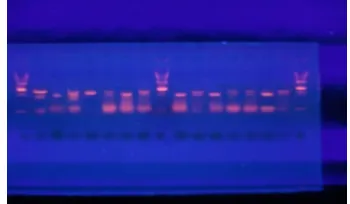
Fig. 1A. Controls and a 100 bp DNA ladder
Fig. 1B. Diabetic patients and the DNA ladder
Figure (1): Polymerase chain reaction–restriction fragment length polymorphism (PCR– RFLP) analysis of VDR single nucleotide polymorphism (SNPs) BsmI. Agarose gel (1.2 %) electrophoresis after digestion of PCR products with BsmI restriction enzyme at 37 degree. Two genotypes were detected, homozygous BB subjects showing an undigested 822-bp product; heterozygous Bb subjects showing 822-, 650-, and 172-bp fragments; homozygous bb subjects showing 650– and 172– bp fragments.
Mva12691) and the digested products was electrophoresed on ethidium-stained 1.2 % agarose gel in 0.5% Tris-borate/EDTA buffer for 1h and was visualized by using a ultraviolet transilluminator. For the statistical analysis, capital letters denote the absence and lowercase letters denote the presence of the site of the restriction enzymes BsmI (B/b), Genotype will be determined from the length of fragments i.e. Homozygote (BB) subjects at 822- base pair product, Heterozygote (Bb) subjects at 822-, 650- and 172- base pair product and Homozygote (bb) subjects at 650- and 172- base pair products (Figure 1-A and B).
The SNP leading to A---G substitution in interon 8 of the VDR gene leads to the generation of a BsmI restriction site. Homozygous subjects with allels containing nucleotide A at this position showed an intact 822 – bp band and were designated as having BB BsmI genotype. Homozygous subjects with alleles containing G at this position showed 2 bands of 650 –bp and 172 – bp and were designated as bb. Subjects with heterozygote status at this position showed all 3 bands: 822 – bp, 650 – bp and 172 – bp and were designated as Bb (Vandevyver et al., 1997).
2.2 Statistical Analysis
Allele frequency was calculated as the number of occurrences of the test allele in the population divided by the total number of alleles. Carriage rate was calculated as the number of individuals carrying at least one copy of test allele divided by the total number of individuals. Allele frequencies, genotype frequencies and carriage rates of the alleles in all the groups were compared by using Chi square test (Fisher's exact test). Other data comparison was calculated by one way ANOVA test. Linear regression was calculated using bad obstetric history as the dependant variable and both vitamin D level and gene frequency as predictors. All P values were two sided, and differences were considered statistically significant for P< 0.05.
3. RESULTS
The clinical and biochemical characteristics of the patients are summarized in Table 1. The mean fasting blood glucose was 108.33 ± 13.92 mg/dl, post prandial was 146.16 ± 35.76 mg/dl, and A1c was 5.92±0.18. The control group mean age was 33.28±4.06 years, their mean FBG was 72.21±4.79 mg/dl, mean SBP was 98.214 ± 6.96 mmHg, and mean DBP was 63.92±5.25 mmHg (data not shown).
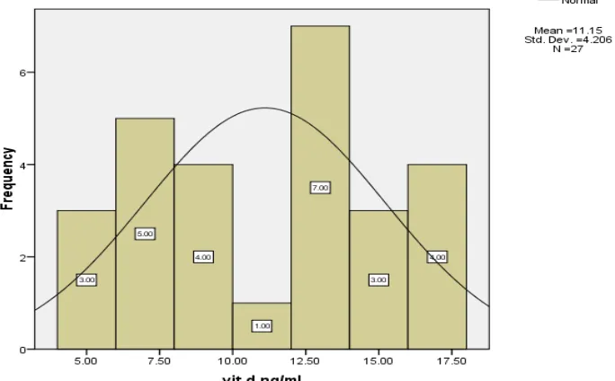
All participant pregnant women had no clinical or biochemical evidence of vitamin D deficiency. The mean Vitamin D level was low in both the diabetes and the control groups (11.15± 4.2 and 11.26± 4.20 ng/ml respectively). In the diabetes group, mild vitamin D deficiency was detected in 18 patients (60%), moderate vitamin D deficiency in 10 patients (33.3%) and severe vitamin D deficiency in 2 patients (6.7%). Figure 2 shows the frequency of vitamin D level among diabetics (Fig. 2-A) and non-diabetics (Fig. 2-B).
significant negative predictor to obstetric history of abortion among diabetics (OR=-0.29, p=0.047, 95% CI -0.08-0.00).
Table 1. Clinical and laboratory characteristics of pregnant women with diabetes
Variables Patients
n=30
Age: years 35.73±5.33
Medical nutrition treatment only 11 (36.7%)
Insulin treatment 19 (63%)
Cesarean section delivery Abortion
Big baby
Pregnancy induced hypertension Stillbirth Abruption placenta 10 (33.3%) 24 (80%) 5 (16.7%) 1 (3.3%)) 1 (3.3%) 1 (3.3%) Fasting blood glucose mg/dl 108.33 ± 13.92 Postprandial blood glucose mg/dl 146.16 ± 35.76
Glycated hemoglobin A1c % 5.92±0.18
Table 2. Comparison of the genotype frequency, allele frequency and carriage rate of BsmI between diabetic and control groups
Parameters Diabetic group N=30. Control group N=14. Genotype frequency: BB Bb bb 3 (10%) 18 (60%) 9 (30%) 1 (7.1%) 8 (57.1%) 5 (35.7%) Allele frequency: B B 24 (40%) 36 (60%) 10 (36%) 18 (64%) Carriage rate: B B 21 (70%) 27 (90%) 9 (64.3%) 13 (92.9%) P > 0.05 not significant
Table 3. Comparison of different parameters between different BsmI genotypes
Parameters BB Bb Bb
Age: years 35.00±4.24 36.08±4.58 36.83±6.911
SBP mmHg 115.00±21.21 108.33±7.52 110.00±12.24
DBP mmHg 77.50±10.60 73.33±8.16 72.00±8.36
FBG mg/dl 105.00±2.82 103.20±15.78 114.80±13.82
PP mg/dl 123.50±3.53 144.80±41.22 156.60±37.67
A1C 6.13 20 5.89 18 5.9 0.13
Vitamin D level ng/ml 10.43±9.10 11.79±3.61 10.43±4.43
Fig. 2A. Frequency distribution of serum vitamin D among diabetic pregnant women
Fig. 2B. Frequency distribution of vitamin D level among control group (non-diabetic pregnant women)
4. DISCUSSION
pregnant Saudis. We reported no evidence of allelic or genotypic association of the BsmI of the VDR gene with GDM in our studied population.
Not surprisingly, the results obtained by different investigators who studied BsmI polymorphism vary even among type 1 DM. BsmI polymorphism has been linked to susceptibility to present the disease in Southern Indians (McDermott et al., 1997), Taiwanese (Chang et al., 1997), Croatians (Skrabic et al., 2003) and Japanese (Motohashi et al., 2003). In Finnish (Turpeinen et al., 2003) and Chilean (Angel et al., 2004) populations the link could not be established. In type 2 DM a link between BsmI and the onset of the disease has been found in Hungarians (Speer et al., 2001) and Germans (Ortlepp et al., 2001) but not in French (Ye et al., 2001), Bangladeshi (Hitman et al., 1998) or Polish (Malecki et al., 2003) populations. Similar results have been found regarding the other most common polymorphisms (Ye et al., 2001; Malecki et al., 2003).
The apparent discrepancies between different studies could be explained by the effect of ethnic differences related to the distribution of VDR polymorphisms in these populations, as well as to interactions with other genetic or environmental factors involved in the pathogenesis of type 2 diabetes mellitus. Furthermore, because these polymorphisms, have no known functional effects, the VDR itself may not be the disease affecting locus, but rather a marker locus in linkage disequilibrium with the redisease locus, and the discrepant findings may reflect variable strengths of linkage disequilibrium in different populations.
In this study, the mean level of vitamin D was consistent with a diagnosis of vitamin D deficiency (<20 ng/mL) in both diabetic and non diabetic groups (12.62±4.693 and 11.26±4.20 ng/ml, respectively). Vitamin D deficiency is common in pregnant women (5-50%) despite the widespread use of prenatal vitamins, because these are inadequate to maintain normal vitamin D levels (>or=32 ng/mL). Middle Eastern are considered risk of vitamin D deficiency (Mulligan et al., 2010). The finding of vitamin D deficiency among both diabetic and non diabetic pregnant women could mean that both groups might have similar genetic and environmental backgrounds that predispose to vitamin D deficiency. In support of the assumption of similar genetic background is the absence of any difference in the studied genotype frequency, allele frequency or carriage rate between both groups as shown in Table 2.
Adverse health outcomes such as preeclampsia, low birth weight, and neonatal hypocalcaemia have been linked to low vitamin D levels during pregnancy (Mulligan et al., 2010). In our study, the vitamin D level but not the gene frequency was significant negative predictor to abortion, the most common bad obstetric history among diabetics (Table 4). The major limitation of the current study is low sample size and assessment of a single polymorphism. An Indian study suggested that VDR gene polymorphism in combination of genotypes is associated with the risk of Type 2 DM (Kumar et al., 2009).
5. CONCLUSION
• Vitamin D receptor BsmI gene polymorphisms is not associated with gestational diabetes among Saudis.
of these polymorphisms on GDM; also, the studies should be carried out on a large cohort of population.
REFERENCES
Anonymous. (2004). Gestational diabetes mellitus. American Diabetes Association, (Position Statement). Diabetes Care., 27 (Suppl. 1), S88–S90, 6-7.
Angel, B., Santos, J.L., Carrasco, E., Albala, C., Perez-Bravo, F. (2004). Vitamin D receptor polymorphism and susceptibility to type 1 diabetes in Chilean subjects: a case–parent study. Eur. J. Epidemiol., 19(12), 1085–7.
Atkins, G. J., Anderson, P. H., Findlay, D. M., Welldon, K. J., Vincent, C., Zannettino, C. W., O’Loughlin, P. D., Morris, A. (2007). Metabolism of vitamin D3 in human osteoblasts: Evidence for autocrine and paracrine activities of 1a,25-dihydroxyvitamin D3. Bone, 40, 1517-1528.
Bodnar, L. M., Simhan, H. N., Powers, R. W., Frank, M. P., Cooperstein, E., Roberts, J. M. (2007). High prevalence of vitamin D insufficiency in black and white pregnant women residing in the northern United States and their neonates. J. Nutr., 137, 447–452. Chang, T. J., Lei, H. H., Yeh, J. I., Chiu, K. C., Lee, K. C., Chen, M. C., Tai, T. Y., Chuang, L.
M. (2000). Vitamin D receptor gene polymorphisms influence susceptibility to type 1 diabetes mellitus in the Taiwanese population. Clin. Endocrinol., 52(5), 575–80. Dent, C. E. (1975). Plasma 25-hydroxyvitamin-D-levels during pregnancy in Caucasians and
in vegetarian and non-vegetarian Asians, Lancet, 2, 1057–1060.
Goswami, R., Brown, E. M., Kochupillai, N.(2004). Prevalance of calcium sensing receptor antibodies in patients with idiopathic sporadic hypoparathyrodism. Eur. J. Endocrinol., 150, 9-18.
Grant, W. P., Holick, M. F. (2005). Benefits and requirements of vitamin D for optimal health: A review. Altern. Med. Rev., 10, 94-111.
Hitman, G. A., Mannan, N., McDermott, M. F., Aqnna, E., Oqunkolade, B. W., Hales, C. N., Bouchard, B. J.(1998) .Vitamin D receptor gene polymorphisms influence insulin secretion in Bangladeshi Asians. Diabetes, 47(4), 688–90.
Julie, R., Althea, G. M. (2008).The genetics of gestational diabetes mellitus: evidence for relationship with type 2 diabetes mellitus. Genet Med., 10(4), 240-50, 10(4).
Kumar, H. R., Konwar, R., Aggarwal, C. G., Gautam, C., Saxena, M., Nayak, V. L., Banerjee, M. (2009). Vitamin D receptor (FokI, BsmI and TaqI) gene polymorphisms and type 2 diabetes mellitus: A North Indian study. J. global infectious Dis., 63(5), 187-194. Malecki, M. T., Frey, J., Moczulski, D., Klupa, T., Kozek, E., Sieradzk, I. J. (2003). Vitamin D
receptor gene polymorphisms and association with type 2 diabetes mellitus in a Polish population. Exp. Clin. Endocrinol. Diabetes, 111(8), 505–9.
McDermott, M. F., Ramachandran, A., Ogunkolade, B. W. (1997). Allelic variation in the vitamin D receptor influences susceptibility to IDDM in Indian Asians. Diabetologia., 40(8), 971–5.
Miller, S. A., Dykes, D. D., Polesky, H. F. (1988). A simple salting out procedure for extracting DNA from human nucleated cells. Nucl. Acids Res., 16, 1215.
Motohashi, Y., Yamada, S., Yanagawa, T. (2003). Vitamin D receptor gene polymorphism affects onset pattern of type 1 diabetes. J Clin. Endocrinol. Metab., 88(7), 3137–40. Mulligan, M. L., Felton, S. K., Riek, A. E., Bernal-Mizrachi, C. (2010). Implications of vitamin
Newman, D.J., Price, C. B. (1999). Renal function and nitrogen metabolites. In: Brutis, C. A., Ashwood, E. R., eds, Tietz. Textbook of Clinical Chemistry, 3rd ed., Philadelphia: WB Saudnders Company, 1204 – 65.
Ortlepp, J. R., Lauscher, J., Hoffmann, R., Hanrath, P., Joost, H. G. (2001). The vitamin D receptor gene variant is associated with the prevalence of Type 2 diabetes mellitus and coronary artery disease. Diabet Med., 18(10), 842–5.
Reitman, S., Frankel, S. (1975). Colorimetric methods for determination of serum transaminase activity. Am. J. Clin. Path., 10, 262.
Skrabic, V., Zemunik, T., Situm, M., Terzic, J. (2003). Vitamin D receptor polymorphism and susceptibility to type 1 diabetes in the Dalmatian population. Diabetes Res. Clin. Pract., 59(1), 31–5.
Speer, G., Cseh, K., Winkler, G., Vargha, P., Braun, I., Takacs, E., Lakatos, P. (2001). Vitamin D and estrogen receptor gene polymorphisms in type 2 diabetes mellitus and in android type obesity,'' Eur. J. Endocrinol., 144(4), 385–9.
Trinder, A. (1969). Determination of glucose in blood using glucose oxidase with an alternative oxygen acceptor. Ann. Clin. Biochem., 6:24.
Turpeinen, H. R., Hermann, S. VaaraLaine, A. P., Simell, O., Knip, M., Veijola, R., Ilonen, J. (2003). Vitamin D receptor polymorphisms: no association with type 1 diabetes in the Finnish population. Eur. J. Endocrinol., 149(6), 591–6.
Valdivielso, J., Fernandez, E. (2006) .Vitamin D receptor polymorphisms and diseases. Clinica. Chimica. Acta., 371, 1–12.
Vandevyver, C., Wylin, T., Cassiman, J. J., Raus, J., Geusens, P. (1997). Influence of the vitamin D receptor gene alleles on bone mineral density in postmenopausal and osteoporotic women. J. Bone Miner. Res., 12, 241–7.
Ye, W. Z., Reis, A. F., Dubois-Laforgue, D., Bellanne-Chantelot, C., Timsit, J., Velho, G. (2001). Vitamin D receptor gene polymorphisms are associated with obesity in type 2 diabetic subjects with early age of onset. Eur. J. Endocrinol., 145(2), 181–6.
Zhang, C., Qiu, C., Hu, F.B., David, R. M., van, Dam, R. M., Bralley, A., Williams, M. A. (2008). Maternal Plasma 25-Hydroxyvitamin D Concentrations and the Risk for Gestational Diabetes Mellitus, PLoS ONE., 3(11), e3753.