| FLYBOOK DEVELOPMENT AND GROWTH
Development and Function of the
Drosophila
Tracheal System
Shigeo Hayashi*,†,1and Takefumi Kondo‡,§
*Laboratory for Morphogenetic Signaling, RIKEN Center for Developmental Biology, Kobe, Hyogo 650-0047, Japan,yDepartment of Biology, Kobe University Graduate School of Science, Nada-ku, Hyogo 657-8501, Japan,zGraduate School of Biostudies, Kyoto University andxThe Keihanshin Consortium for Fostering the Next Generation of Global Leaders in Research (K-CONNEX), Kyoto, Sakyo-ku 606-8501, Japan ORCID IDs: 0000-0001-7785-1290 (S.H.); 0000-0001-8127-1141 (T.K.)
ABSTRACTThe tracheal system of insects is a network of epithelial tubules that functions as a respiratory organ to supply oxygen to various target organs. Target-derived signaling inputs regulate stereotyped modes of cell specification, branching morphogenesis, and collective cell migration in the embryonic stage. In the postembryonic stages, the same set of signaling pathways controls highly plastic regulation of size increase and pattern elaboration during larval stages, and cell proliferation and reprograming during metamorphosis. Tracheal tube morphogenesis is also regulated by physicochemical interaction of the cell and apical extracellular matrix to regulate optimal geometry suitable for airflow. The trachea system senses both the external oxygen level and the metabolic activity of internal organs, and helps organismal adaptation to changes in environmental oxygen level. Cellular and molecular mechanisms underlying the high plasticity of tracheal development and physiology uncovered through research onDrosophilaare discussed.
KEYWORDStrachea; development; tubulogenesis; branching; physiology; FlyBook
TABLE OF CONTENTS
Abstract 367
Introduction 367
Placode Specification and Branching 368
Primary Branching and Guidance 369
Terminal Branching and Internal Lumen Formation 371
Tip Cell Specification and Branch Fusion 371
Tube Geometry: Control of Length and Diameter 372
Tube Maturation 373
Spiracles 373
Larval Trachea and Metamorphosis 374
Future Challenges 374
Copyright © 2018 by the Genetics Society of America doi:https://doi.org/10.1534/genetics.117.300167
Manuscript received December 20, 2017; accepted for publication February 12, 2018.
I
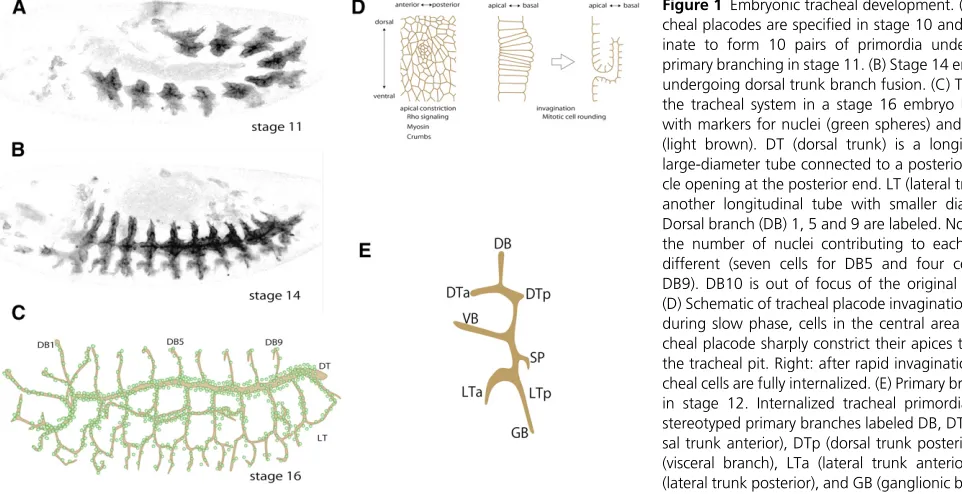
NSECT locomotion is fueled by the efficient exchange of gases through a dedicated respiratory organ, the trachea, to power muscles forflight and walking, the gut for taking in nutrition, and the central nervous system for neural process-ing. The evolutionary innovation of the tracheal system helped terrestrial arthropods and annelids expand their hab-itat and morphological diversity. The tracheal system, which consists of a network of branched tubules extending through-out the body cavity, allows airflow through openings in the body wall, called spiracles, and delivers it to highly ramified tracheal termini, called tracheoles. The exchange of gasses through the large surface area of the tracheole lumen causes waste gas (carbon dioxide) to diffuse from the terminus to the spiracles and fresh air (oxygen) to diffuse in the opposite direction (Wigglesworth 1972).Tracheal system development has been studied extensively in the fruitflyDrosophila(Whitten 1980; Manning and Krasnow 1993; Samakovlis et al.1996a; Affolter and Caussinus 2008; Schottenfeldet al.2010; Loganathanet al.2016). During em-bryogenesis, the tracheal primordia appear as 10 pairs of tracheal placodes that invaginate into the body cavity while maintaining epithelial integrity, and undergo stereotyped branching and fu-sion processes to form a network of tubular epithelium (Figure 1). The largest tubule is the dorsal trunk, which serves as the major airway connecting the open posterior spiracle and the anterior spiracle, which opens in the second larval instar. The dorsal trunk is a multicellular tubule, with multiple cells contributing to the luminal surface except at the fusion point, which is formed by a pair of torus-shaped fusion cells surrounding the lumen. Tubules branching off the dorsal trunk are thinner and consist mostly of tracheal cells with autocellular junctions (unicellular tubules).
The last embryonic mitosis (cycle 16) occurs during the invagination of the tracheal placode cells into the body. In the larval stage, most tracheal cells grow by endocycling. In the pupal stage, polyploid larval tracheal cells undergo apoptotic cell death, and the remaining diploid tracheal cells proliferate and remodel the tracheal system into the adult pattern. In this review, we summarizefindings from the past three decades of research on the genetic and developmental features of the Dro-sophilatracheal system.
Placode Specification and Branching
Tracheal development begins during stage 10 of embryogen-esis, about 5 hr after egg laying, with the specification of 10 pairs of tracheal placodes located in the lateral part of the second thoracic (T2) to eighth abdominal (A8) segments (Campos-Ortega and Hartenstein 1997) (Figure 1A). The
first metamere in T2 consists of80 cells, and the remaining metameres have 50 cells. During the G2 interphase after mitosis 15 (Hartenstein and Campos-Ortega 1985), tracheal placodes appear as aflattening of the apical surface, and cells in the center of each placode begin apical constriction to form the tracheal pit (Isaac and Andrew 1996; Wilket al.1996;
Nishimuraet al.2007; Kondo and Hayashi 2013) (Figure 1C). This slow invagination phase, which lasts30 min, is followed by a rapid invagination phase that internalizes all of the tra-cheal placode cells within60 min (Kondo and Hayashi 2013) (Figure 1C). Tracheal cells undergo cycle 16 mitosis during invagination and are then arrested in the G1 phase. Like most larval somatic cells, the majority of tracheal cells resume the cell cycle during the larval period, growing by endocycling and becoming polyploid (see section Larval Trachea and Metamorphosis).
Early research focused on identifying the determinants of tracheal cell fate and tracheal tissue-specific characteristics. The transcription factor Trachealess (Trh), which is expressed in all tracheal cells from the onset of tracheal placode spec-ification through adulthood (Isaac and Andrew 1996; Wilk
et al.1996), regulates a multitude of other tracheal genes, most notablybreathless(btl), which encodes the FGF receptor Breathless (Btl) (Ohshiro and Saigo 1997; Chung et al.
2011). The tracheal system does not develop iftrhfunction is absent, suggesting thattrhis a central regulator of tracheal cell identity. Trh expression is stimulated in the anterior com-partment by JAK/STAT signaling acting directly on the trh
enhancer element (Brown et al.2001; Sotilloset al.2010), but is repressed in the posterior compartment and in dorsal ectoderm bywingless(wg) anddpp, respectively (Isaac and Andrew 1996; Wilket al.1996). Althoughtrhmutants retain some early characteristics of tracheal cell identity, these char-acteristics are mostly lost in the Stat92E mutant (Li et al.
2003), suggesting that tracheal cell identity requires addi-tional transcripaddi-tional factors such asapontic(Liuet al.2003) and the POU-domain protein encoded by ventral veinless
(Andersonet al.1995; Llimargas and Casanova 1999; Boube
et al.2000; Zelzer and Shilo 2000).
The process of tracheal placode invagination involves sev-eral mechanisms. The constriction of apical cell surfaces in the middorsal placode shifts the apical surface of the epithelium basally to form the tracheal pit (Figure 1C). This process ad-vances throughEGF receptor(EGFR) signaling controlled by the EGF ligand activator moleculeRhomboid(Llimargas and Casanova 1999) and the intracellular signal transducersERK
andMEK, and is regulated by Rho GTPase signaling (Brodu and Casanova 2006). In a parallel process, an accumulation of the subapical proteinCrumbsalso contributes to apical con-striction (Letiziaet al.2011). EGFR signaling coordinates the formation of circular supracellular myosin cables around the tracheal pit, and these cables apply centripetal compression (Nishimuraet al.2007; Kondo and Hayashi 2013). A similar circular arrangement of cells and myosin cables is found in salivary gland primordia undergoing invagination, although the circles in the latter case remain in the same cell population (Röper 2012). How the circular patterns of contractile myosin cables emerge in the ectodermalfield under the control of anterior–posterior (Wg and Hh) and dorsal–ventral (Dpp and EGFR) coordinated systems remains to be explained.
stage, all of the placode cells are arrested in the G2 interphase of cycle 15. In most cases, thefirst cells to reenter cycle 16 are in or around the tracheal pit. Mitotic cell rounding triggers buckling instability in the densely packed placode epithelia; the cells in the tracheal pit are rapidly internalized, followed by the surrounding tracheal cells (just entering cycle 16), forming the completely internalized, sac-like tracheal primor-dium (Kondo and Hayashi 2013) (Figure 1D). Although both mitotic cell rounding and EGFR-dependent Myosin force are important in coordinating the site, timing, and cell shape changes of invagination, embryos with a single mutation blocking EGFR signaling or cycle 16 mitosis still formed in-vaginated tracheal primordia. However, the invagination of the tracheal placode fails severely in triple-mutant embryos lacking cycle 16 mitosis, FGF signaling (which is required for cell migration and plays a backup role in invagination), and EGFR signaling (Kondo and Hayashi 2013), demonstrating that these cellular processes provide high redundancy and robust support for tracheal placode invagination.
Primary Branching and Guidance
Genetic mosaic analysis indicated that tracheal primordial cells are not committed to any position or cell type at the time of placode specification (Samakovliset al.1996a). Therefore, the cell and branch type specification must be determined under the influence of the surrounding environment. Once internalized, the tracheal primordia start expressing the FGF receptor tyrosine kinase Btl (Glazer and Shilo 1991; Klämbt
et al.1992; Shishidoet al.1993), which is activated by one of the three FGF ligands in Drosophila, Branchless (Bnl)
(Sutherlandet al.1996), and the process of stereotyped pri-mary branching begins. Each branch migrates toward a spe-cific target tissue; for example, the dorsal branch toward the dorsal epidermis, the visceral branch toward the intestine, and the ganglionic branch toward the ventral nerve cord. Branch migration is guided by Bnl, which is expressed dynam-ically in a number of nontracheal tissues (Sutherland et al.
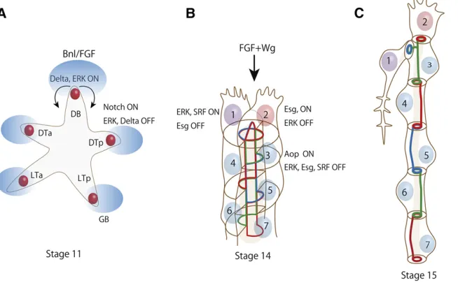
1996; Ohshiro et al.2002; Du et al.2017) (Figure 2A). At stage 15 and later, Bnl–Btl signaling stimulates the differenti-ation and outgrowth of a tracheal cell subtype called terminal cells, which send cytoplasmic branches out toward target tis-sues to support respiration (Figure 2, B and C).
The receptor tyrosine kinase Btl forms a complex with the FGFR adaptor protein Dof(Michelson et al.1998; Vincent
et al. 1998; Imamet al. 1999) and, when activated by Bnl, turns on the RAS pathway intracellular signaling cassette to activate ERK MAP kinase (Reichman-Friedet al.1994; Gabay
et al. 1997). The importance of the RAS–ERK pathway in tracheal branch migration was shown by the partial rescue of defective tracheal migration in btl mutants by constitu-tively active forms of Ras, Raf, and ERK (Reichman-Fried and Shilo 1995; Dossenbachet al.2001).
Bnl/FGF is important for guiding branch migration, and two models have been proposed to explain its influence. The chemical guidance model suggests that some upstream-facing cells sense and respond locally to a chemical gradient of guidance cues, as observed in the migration of solitary cells in culture (Artemenkoet al.2014). Tracheal tip cells exposed to Bnl/FGF extend actively movingfilopodia and lamellipo-dia in a Btl-dependent manner (Ribeiroet al.2002), and this activity is further stimulated by ectopic Bnl/FGF. However,
the direction of thefilopodia does not correlate well with the location of the Bnl/FGF (Okenve-Ramos and Llimargas 2014), arguing against the idea that thefilopodia sense and extend along the Bnl/FGF gradient. Alternatively, the collec-tive guidance model (Rørth 2007) posits that Btl signaling is activated only in one or two cells at the branch tip; in this model, only the tip cells respond to migratory cues, while the rest of the tracheal cells follow the tip. In late-stage tracheal branches, btltranscription is limited to the tip by positive feedback (Ohshiro et al. 2002). Genetic mosaic assays showed thatbtlactivity is required in tip cells but not in stalk cells (Ghabrial and Krasnow 2006), and that expressing a constitutively active form of Btl in a single cell is sufficient to specify leader cell properties and induce branch migration (Lebreton and Casanova 2014). Thesefindings suggest that limiting FGF signaling activity to a subset of tracheal cells is sufficient to differentiate two classes of cell types: tip cells, which sense Bnl/FGF and migrate toward the source, and tube-forming stalk cells that follow the tip cells. Thesefi nd-ings favor the collective guidance model for FGF-guided primary branch migration. Although there is no decisive ev-idence for the chemical guidance model in primary branching, this mechanism is likely to be active in terminal branching, since localized ectopic Bnl expression attracts excessively elongated terminal branches in the embryo (Miao and Hayashi 2015) and larva (Jareckiet al.1999).
The primary branches have unique identities that specify tube shape, cell composition, and target organ specificity. These branch identities are specified at least in part by the transcription factorsKnirps/Knirps-like(Kni/Knrl) andSpält
(Sal), known as the gap-class segmentation genes, in the
blastoderm (Kuhnlein and Schuh 1996; Chen et al. 1998; Franch-Marro and Casanova 2002). During tracheal placode specification, these genes are expressed in a complementary pattern: Sal is expressed in the dorsal region, which gives rise to the dorsal trunk and dorsal branch, and Kni/Knrl is expressed in the ventral region, which gives rise to the lateral trunk and the ganglionic and visceral branches. In the dorsal region, Sal is expressed in the dorsal trunk but is repressed in the dorsal branch by Dpp-induced Kni/Knrl (Chen et al.
1998). The dorsal trunk is unique in that the lumen surface (except for the fusion point) is covered by cells with hetero-cellular junctions, producing a multihetero-cellular tubule with a relatively large diameter. Other branches are small-diameter unicellular tubules, formed of single cells with autocellular junctions (Ribeiroet al.2004). Autocellular junctions in the dorsal branch are formed by cell intercalation, which is pro-moted by enhanced E-cadherin turnover (Shindoet al.2008) and is inhibited in the dorsal trunk by enhanced endosomal recycling (Shayeet al.2008) stimulated by Sal (Ribeiroet al.
2004). Self-adhesion interfaces do not form in most animal tissues, and are seen only in rare cases of tubular tissues such as blood vessels (Yuet al. 2015) and infish gill pillar cells (Katoet al.2007). The extent to which cell-autonomous my-osin contractility and external pulling forces contribute to the formation and stabilization of tracheal autocellular junc-tions is a matter of some debate (Caussinuset al.2008; Ochoa-Espinosaet al.2017). Interestingly, the tracheal system in
Triboliumlarvae consists exclusively of unicellular tubules with a spiracle opening in each metamere. Sal is not ex-pressed in theTriboliumdorsal trunk, which probably reflects an adaptation to the relatively dry environment found inflour
Figure 2Cell specification and cell rearrangement during branching morphogenesis. (A) Schematic im-age of stim-age 11 tracheal primordia undergoing pri-mary branching. Bnl/FGF (blue ovals) is expressed by groups of cells near invaginated tracheal primordia and activates Btl/FGFR and ERK activity in tracheal cells. One cell near the source of Bnl/FGF elevates Delta expression and ERK activity to the highest level and becomes a tip cell (red circle), and stimu-lates Notch signaling in the rest of the cells to re-duce Delta expression and ERK activity to become stalk cells. Tip cells become motile and track Bnl/FGF to elongate tubules. VB (visceral branch) is not shown in this picture for clarity. (B) Schematic image of a stage 14 DB (dorsal branch) at the beginning of secondary branching. Under the influence of Wg, tip cells turn on Esg and other genes to acquire FC (fusion cell) fate (cell number 2). Esg turns off ERK signaling and permits activation of ERK signal-ing in the neighborsignal-ing number 1 cell, which ac-quires TC (terminal cell) fate by activating ERK and
as opposed to the more humid environments inhabited by
Drosophilalarvae (de Miguelet al.2016).
Successful branch migration requires guidance from other signals in addition to Bnl/FGF. Hedgehog, which is expressed in stripes in the posterior compartment, inhibits the migration of the dorsal and terminal branches, effectively confining their position to the anterior compartment of each hemi-segment (Katoet al.2004). This segregates the branches from their anterior and posterior neighbors and assures a precise meta-mere-by-metamere arrangement. As the tip of the dorsal branch reaches the margin of the dorsal epidermis, Dpp em-anating from the leading edge of the epidermis inhibits the dorsal elongation of the terminal branch and promotes the typical“U-turn”shape. The combined inhibitory effects of Hh and Dpp restrict the extent of branch migration in each half-metamere, which avoids overlap between branches from dif-ferent segments.Slit-Robosignaling, which repels ganglionic branch migration in the CNS, has varying effects on dorsal branch migration (Englundet al.2002).
The visceral branches migrate toward the midgut and spread over the surface of the visceral mesoderm. This mi-gration requires two integrin subunits,aPS1expressed in the visceral branch and aPS2 expressed in the visceral meso-derm, respectively (Boubeet al.2001). These integrin-a sub-units both associate with the commonbPS subunit, and are thought to assemble basal lamina for cell migration by cap-turing specific laminin molecules (Martinet al.1999; Urbano
et al.2009). In addition, visceral branch migration requires extracellular matrix remodeling by AdamTS-A metallopro-tease (Ismatet al.2013).
Terminal Branching and Internal Lumen Formation
During secondary branching, tracheal branch termini become further elaborated through the appearance of the terminal branch, which is a single-cell extension that contains intra-cellular lumen and undergoes repeated elongation and branching, accompanied by lumen growth, to spread over the target organ (the epidermis, CNS, gut, or another internal organ). Cytoplasmic extensions from terminal cells depend on SRF (serum response factor) (Affolteret al.1994; Guillemin
et al. 1996), indicating that Bnl/FGF induces terminal cell differentiation. Bnl/FGF overactivation induces SRF expres-sion and excessive terminal cell differentiation (Lee et al.
1996; Sutherlandet al.1996; Miao and Hayashi 2015). Dur-ing the larval period, terminal cells undergo extensive growth and branching, covering increasingly larger areas, to keep up with the growth of the larva. Terminal cell growth is con-trolled by local oxygen demand, which stimulates Bnl/FGF expression in target tissues (Jareckiet al.1999) to promote terminal cell outgrowth, and by trachea-autonomous stimu-lation of FGF receptors (Centaninet al.2008). Both mecha-nisms are controlled by Sima, a Hypoxia-Inducible Factor-a homolog (Centaninet al.2008).
Lumen formation in terminal cells is unique in that it forms at a distance from the cell–cell adhesion interface
(Sigurbjörnsdóttir et al. 2014), and occurs by the inward growth of the apical plasma membrane at the interface of the terminal branch and stalk cells (Gervais and Casanova 2010). Lumen growth occurs in the direction of terminal branch outgrowth, most likely by following microtubule tracts (Gervais and Casanova 2010). Mutants with supernu-merary centrosomes form multiple terminal branches in sin-gle-terminal cells, suggesting that microtubule organization is the key initial step in terminal branching (Ricolo et al.
2016). In mutants with defective actinfilament organization, lumen growth fails to follow the direction of cell growth, resulting in misrouted lumen in the terminal branches as seen inikkemutants (Oshimaet al.2006), or in poor lumen for-mation as seen inbtszorSlikmutants (JayaNandananet al.
2014; Ukkenet al.2014). Larvae require a continuous supply of apical membranes as they grow, and the elongation of terminal branch lumen is supported by dynein-dependent transport (Schottenfeld-Roames and Ghabrial 2012) and re-stricted by endocytosis (Schottenfeld-Roameset al.2014).
Tip Cell Specification and Branch Fusion
Tracheal tip cells are ERK-active single cells, located at the tip of each primary branch, that lead tubule formation and branch migration, much like the tip cells in vascular sprouting in vertebrates (Herbert and Stainier 2011). The combined ac-tion of Bnl/FGF and Wingless increases the expression of the Notch ligand Delta in multiple cells (Chihara and Hayashi 2000; Llimargas 2000); from these, a single Delta-high cell is selected as the tip cell for the branch through lateral in-hibition (Figure 2A). The remaining Delta-low, Notch-active cells assume the stalk cell fate (Ikeya and Hayashi 1999; Llimargas 1999). The tip cell remains at the leading position throughout branch migration and fusion, unlike vertebrate endothelial tip cells, which are frequently exchanged with stalk cells (Jakobssonet al.2010). Activated Notch suppresses ERK signaling and restricts Bnl/FGF activity to the Delta-high tip cell (Ikeya and Hayashi 1999); this self-restrictive Notch mechanism limits the response of nontip cells to Bnl/FGF and shapes the tubular geometry.Anterior open, an ETS-domain transcriptional repressor, suppresses the tip cell fate in stalk cells by antagonizing ERK and Wingless signaling (Caviglia and Luschnig 2013).
Tip cells adhere to target tissues and follow sources of dynamically changing Bnl expression (Sutherlandet al.1996; Duet al.2017). Under the influence of Wingless signaling, the tip cells then begin expressing genes that specify a fusion cell fate,escargot(esg) (Samakovliset al.1996b; Tanaka-Matakatsuet al.1996; Chihara and Hayashi 2000; Llimargas 2000), followed bydysfusion(dysf) (Jiang and Crews 2003). At the same time, ERK expression diminishes in the tip cells while increasing in neighboring terminal cells, which begin to express SRF, a transcription factor encoded byblistered
from thefirst stage of FGF signaling in primary branch mi-gration to the second stage of FGF signaling in terminal cell differentiation is controlled by esg, which suppresses ERK activity in prospective fusion cells (Miao and Hayashi 2016). This allows a second tip cell to respond to Bnl/FGF and acti-vates the secondary branching program.
Branch fusion (anastomosis) is mediated by contact be-tween fusion cells andde novolumen formation (Caviglia and Luschnig 2014). Dorsal branch fusion at the dorsal midline has been studied extensively, because this is the most acces-sible region for high-resolution imaging (Samakovlis et al.
1996b; Tanaka-Matakatsu et al.1996; Gervaiset al.2012; Katoet al.2016). Fusion cells attached to the basal surface of the leading edge of the epidermis migrate toward the dorsal midline after closure of the dorsal epidermis. Fusion cells from contralateral sides contact each other as they approach the dorsal midline by extending numerous filopodia, and they establish adhesion by accumulating E-cadherin at the contact interface. Escargot stimulates thede novosynthesis of E-cadherin, which is essential for adhesion between fusion cells (Tanaka-Matakatsuet al.1996). Although thefilopodia from fusion cells come into contact with neighboring epider-mal and mesoderepider-mal cells, and sometimes with tracheal cells from the same side, stable E-cadherin adhesion is established only between fusion cells from contralateral sides. The mech-anism of this selectivity is currently unknown. Once a stable attachment is established, the paired fusion cells create new lumen at the contact interface by directing Golgi-mediated secretion to the contact site (Kato et al.2016). Fusion cell plasma membranes are fused internally to create their char-acteristic torus-like shape through a process requiring the endosomal protein ARF-like 3(Jiang et al. 2007; Kakihara
et al.2008), which acts through the secretory lysosomal
pro-tein Staccato/Unc-13-4(Cavigliaet al.2016). Interestingly,
this conversion of cell surface topology always occurs simul-taneously in paired fusion cells. Paired fusion cells at each fusion point produceCG13196, a specialized ZP (zona pelu-cida) domain-containing protein (Jaźwińska and Affolter 2004), and form a uniquely patterned cuticle that lacks taeni-dial folds (Matuseket al.2006). During larval molt, tracheal cuticles are broken at the fusion point and are extruded through a transient reopening of the tracheal pit. Although ARF-like 3 and Staccato/Unc-13-4 are required for all tra-cheal fusion events,esganddysfare dispensable for fusion of the dorsal trunk (Tanaka-Matakatsu et al.1996; Jiang and Crews 2006), suggesting that the regulation of fusion cells is different in this branch. It should be noted that the conversion of fusion cells into a torus is not the sole method of joining tracheal branches in insects; in the Manduca
moth, tracheal fusion points are formed by multiple prolif-erative cells (Nardi 1990).
Tube Geometry: Control of Length and Diameter
Researchers have conducted detailed morphometric analyses of the tracheal tubes (Beitel and Krasnow 2000). For tracheal
tubes to serve as airways, the length of each branch mustfit the size of the body. The number of cells in each tracheal branch is not strictly determined, and the number of cells varies even more when tracheal cell subsets are removed by apoptosis (Baeret al.2010). This variability in cell number is compensated for by aflexibility in individual tracheal cell shape. In dorsal branches, for example, the number of cells in each branch is not predetermined (Samakovliset al.1996a), and in branches with fewer cells, each cell covers a greater length of lumen (Figure 1C). Experiments that increased or decreased the number of tracheal cells demonstrated this
flexibility even more markedly (Caussinuset al.2008). While branch connections and tube fusion are nearing completion in stage 15, the second phase of tube maturation begins, and each branch further elongates and takes on a sinusoidal shape. Cell shape analysis revealed that the axial elongation of dorsal trunk cells contributes to tube elongation (Förster and Luschnig 2012; Nelsonet al.2012). Dorsal trunk elon-gation is limited by the core planar cell polarity pathway (Chung et al.2009; Nelson et al. 2012; Warrington et al.
2013) andYorkie, a target of Hippo pathway-dependent re-pression (Robbins et al. 2014). Axial polarity in the larval trachea is specified by a transient transversal cell junction accumulation ofatypical PKCthat targetsRhoAsignaling to longitudinal cell junctions, and this planar polarization aligns cortical actin cytoskeletons into parallel arrays to support axial elongation (Hosonoet al.2015).
The dorsal trunk’s largest tube diameter is at its posterior end, where it connects to the posterior spiracle that opens to take up air when the larva hatches. Although the dorsal trunk has a multicellular composition, the trunk diameter gradually decreases toward the anterior due to the differential effects of Hox genes in each metamere (Matsuda et al.2015). Cross-sectional views show that dorsal tube sections with a similar diameter can be formed by one tofive cells, demonstrating the greatflexibility of cell size and shape. Various unicellular branches also maintain a near-regular luminal diameter unique to the branch type. These observations indicate that tube geometry is controlled, at least partly, at the tissue level. Mutations that affect tracheal tube shape reveal three classes of genes that regulate tracheal tube geometry: those that primarily affect tube diameter (class I), tube length (class II), or both (class III) (Wu and Beitel 2004; Schottenfeldet al.
2010; Dong and Hayashi 2015). Class I mutations identify genes that affect the size and uniformity of tube diameter (Beitel and Krasnow 2000), including genes that affect components of chitinous fibers in the apical extracellular matrix (aECM) (Araújo et al.2005; Devine et al. 2005; Tonninget al.2005, 2006; Moussianet al.2006; Wanget al.
2006) or the intracellular processing and secretion of the aECM (Tsarouhaset al.2007; Griederet al.2008; Jayaram
et al.2008; Försteret al.2010; Norumet al.2010; Tiklová
Massive luminal accumulations of chitin and other secreted macromolecules are thought to supply the force for tube ex-pansion. In response to this secretory activity, F-actin accu-mulates on the apical plasma membrane facing the lumen in the form of multiple parallel cables, oriented in a circumfer-ential direction, which act as a platform for myosin-dependent constrictive force (Matusek et al. 2006; Kondo et al. 2007; Öztürk-Çolak et al. 2016a,b). This expansion-triggered me-chanical feedback and the viscoelastic property of the aECM (Försteret al.2010; Donget al.2014a) prevent excess tube expansion throughout the length of the tubules. The absence of chitin causes a loss of stability, and uncontrolled cycles of constriction and relaxation in the cortical membrane, resulting in irregular tube diameter (Hannezoet al.2015; Öztürk-Çolak
et al.2016b). How this mechanical feedback loop is equalized along the length of a tracheal tubule to create a uniform lumi-nal diameter is not fully understood. Biophysical alumi-nalysis of the aECM is necessary to identify mechanisms related to the tissue-level coordination of cell contractility.
Class II mutations cause excessive dorsal trunk elongation. These mutations can be grouped into three subclasses. The
first subclass causes excessive apical membrane biosynthesis. Shrub/Vps32 and the epithelial polarity proteins Yurt and Scrib regulate the localization and activity of the apical po-larity protein Crumbs (Lapriseet al.2006, 2010; Donget al.
2014a). Activated Crumbs promotes apical membrane bio-genesis (Tepass et al.1990). The deregulation of Crumbs inshrub,yurt, andscribmutants promotes excessive apical membrane synthesis and excessive tubule length. The second subclass affects aECM elasticity. Serpentine and Verm, which are chitin-binding proteins with domains homologous to bac-terial and fungal chitin deacetylase, are required to nega-tively control tube length (Luschniget al.2006; Wanget al.
2006). Deacetylation converts chitin into the more water-absorptive form chitosan. Therefore, Serp and Verm might increase the aECM elasticity. Another aECM component, Dumpy, is a large elastic protein that helps to restrict dorsal trunk length (Donget al.2014a) and taenidial fold pattern-ing (Wilkin et al.2000), and maintains the dorsal trunk’s mechanical strength under pulling force (Jaźwińska et al.
2003). The third subclass affects the apical membrane–aECM bond. ER-resident O-GlcNAc transferase (EOGT) modifies the extracellular domains of secreted and membrane pro-teins, including the EGF repeats of Dumpy. EOGT is required for maintaining connections between apical membranes and the aECM (Sakaidaniet al.2011). The chitin-binding protein Obst-A stabilizes chitinfiber organization, maintains cuticle– epidermis adhesion, and restricts tracheal tube length (Petkau
et al.2012). Taken together, the products of these three sub-classes of genes that regulate tube length constitute two cou-pled elastic modules, stretchable aECM bound by an expanding apical plasma membrane. A physical model of these coupled elastic modules reproduced a sinusoidal-curving, overelon-gated tube in mutant embryos (Donget al.2014a).
Class III mutants include septate junction proteins that are required for both tube length and diameter control (Behret al.
2003; Genova and Fehon 2003; Paul et al.2003; Wuet al.
2004; Nelsonet al.2010), and are thought to control tube diameter by regulating the secretion of the aECM through mechanisms that are independent of the epithelial barrier function of the septate junctions (Wu and Beitel 2004).
The epidermis of the body wall develops independently of the trachea and grows larger than the size of the egg shell, so that the mature epidermis is tightly folded under the vitelline membrane. After the larva hatches, the epidermis and the trachea stretch out to matching lengths, suggesting that the two axial tissues undergo systemic-level coordination. Serp is initially transcribed mainly in the trachea and later in the epidermis, where it affects the aECM properties and cuticle rigidity (Luschniget al.2006; Wanget al.2006). Serp is also produced in the mesodermal-tissue fat body, where excessive Serp accumulation was observed in a mutant with defective endocytosis and endosomal recycling (Dong et al.2014b). Fat body-derived Serp travels through the body cavity, reaches the tracheal lumen by transcytosis, and is retained by chitin binding (Donget al.2014b). This systemic regulation of the aECM may help to coordinate the elongation of the trachea and epidermis to matching lengths.
Tube Maturation
Once tracheal tubules reach theirfinal pattern and size, cuticle layers begin forming on the apical surface with a circumfer-ential ridge, called a taenidial fold, with a pattern correspond-ing to the cortical F-actin cable arrays formed at an earlier stage (Matuseket al.2006; Öztürk-Çolaket al.2016a,b). Inside the lumen, the aECM is degraded and absorbed by tracheal cells to clear a large fraction of the luminal macromolecules (Behr et al.2007; Tsarouhas et al.2007). Finally, a gas is generated independently of the outside atmosphere on part of the cuticle surface, and the gas quickly spreads over the entire tracheal lumen to completely fill the tube with gas. Although the mechanism of gas generation is not well un-derstood, it requires the apical membrane protein Uif (Zhang and Ward 2009). The identification of fatty acyl-CoA reduc-tase as a critical requirement for gas generation suggests that the formation of a hydrophobic wax layer on the cuticle surface may contribute to bubble generation from gas satu-rated in the luminal liquid (Jasperset al.2014), previously suggested as cavitation-bubble formation on a hydrophobic surface (Wigglesworth 1953).
Spiracles
Spiracles, the epidermal structures that connect the internal tracheal network to the outside environment, serve as gate-keepers to control gasflow and water loss, and protect the luminal environment. Although spiracles can form in every metamere from which tracheal primordium is derived, only the posterior spiracle is functional in first-instarDrosophila
(Castelli Gair Hombríaet al.2009). However, their primordia are juxtaposed and develop simultaneously, producing struc-turally interconnected mature tissue (Hu and Castelli-Gair 1999). The posterior spiracle, which is positioned in A8, con-sists of two parts: an internal tubule (called the spiracular chamber orfilzkörper) that connects the trachea to the out-side and the external stigmatophore. There are four sensory hairs at the exit of the spiracular chamber. Reflecting the mature structure, the posterior spiracle primordia develop in two parts: the central spiracular chamber part, marked by expression of the transcription-factor genes trh (Isaac and Andrew 1996),cut(Blochlingeret al.1990), andempty spiracle (Dalton et al. 1989), and the outer stigmatophore part, which expressessal (Hu and Castelli-Gair 1999). The central part also expresses the Unpaired ligand, which acti-vates the JAK/STAT pathway (Brownet al.2001) to promote invagination by the basolateral expansion and inward move-ment of prospective spiracular chamber cells (Lovegroveet al.
2006; Simões et al.2006; Tsikalaet al.2014). The cell be-haviors and mechanisms involved in posterior spiracle invag-ination appear to be distinct from those used in tracheal placode invagination. Furthermore, posterior spiracles utilize a distinct mechanism of air-filling that requires a scavenger receptor class B molecule (Wingenet al.2017).
Larval Trachea and Metamorphosis
After larval hatching, most tracheal cells stop proliferating and begin endocycling, increasing ploidy and cell size, and the tracheal tubules elongate and increase in diameter to match the growth of the larval body. During molting, tracheal cells renew their apical surface by planar polarization and by reorganizing cortical F-actin (Hosonoet al.2015), and they secrete cuticles with taenidial folds at a spacing of0.8mm at the beginning of the second and third instar (Glasheenet al.
2010). Taenidial fold spacing increases up to twofold during each instar as the tracheal tube elongates, and this cuticle plasticity depends on apically secreted matrix metallopro-tease 1 (Glasheenet al.2010).
Tracheal histoblasts (tracheoblasts), which are derived from cut-positive cells, populate the spiracular branch that connects the tracheal tubule to the epidermal region at the site of the initial tracheal pit formed at the beginning of tra-cheal development (Pitsouli and Perrimon 2010, 2013). These unique cells, which proliferate extensively in the third instar, replace larval tracheal cells and form adult-specific tracheal branches during metamorphosis. The spiracular branch does not have an open lumen and does not function as an airway except during the molt, when the branch opens and sheds the tracheal cuticles from the previous instar. Therefore, tracheal histoblasts are considered to be undiffer-entiated, dedicated adult precursor cells.
An additional source of adult precursor cells is found among the functional tracheal cells. Dorsal branch stalk cells in the second tofifth tracheal metameres (Tr2–Tr5) (Weaver and Krasnow 2008) and in the dorsal trunk in Tr2 (Guhaet al.
2008; Sato et al. 2008) enter the cell cycle at late L3 and proliferate to become adult tracheal cells. These cells enter quiescence after embryonic cycle 16 mitosis. While other tra-cheal cells start endocycling, these precursor cells retain their mitotic capacity. The nuclear proteins Htx and Exd, along with their binding partner Ubx, are implicated in maintaining cell cycle arrest and multipotency in these cells (Sato et al. 2008) by inhibiting the endocycle-promoting genefzr(Djabrayanet al.2014).
Air sacs are adult-specific tracheal tissues encased in the notum that supply air to flight muscles to meet their high oxygen demands duringflight. The trachea becomes associ-ated with the wing disc immediately after the wing disc invaginates in stage 15 of embryogenesis (Inoue and Hayashi 2007). In third-instar larvae, the air sac primordia are found as a part of the Tr2 tracheal metamere attached to the basal side of the presumptive notum of the wing imaginal disc, where the disc cells expressbnl. Tracheoblasts in the pri-mordia express btl, and the activation of FGF signaling promotes the migration of tracheoblasts toward the poste-rior (Satoet al.2002) through an invasive process involv-ing the extensive remodelinvolv-ing of basement membranes (Guhaet al.2009). FGF signaling also stimulates secretion of the ligand molecule Vein (Cruzet al.2015), which ac-tivates tracheoblast proliferation and survival (Cabernard and Affolter 2005).
Future Challenges
The genetic pathways that specify the basic grand plan of the embryonic tracheal system have been described extensively. One prominent discovery is that a surprisingly simple set of signaling pathways, most notably FGF signaling, are used repeatedly at multiple stages of tracheal development to model and remodel tracheal pattern and functions, keeping pace with the ever-changing respiratory needs of the larva, pupa, and adult. These discoveries set the stage for investi-gating even more complex unexploredfields.
Bnl/FGF, which attracts invasive tracheal growth into the T-tubule through openings at the plasma membrane (Peterson and Krasnow 2015). Thus, the reactivation of FGF signaling competence in tracheal cells, along with various Bnl/FGF expression patterns, form the basis for tracheal remodeling and tissue-specific tracheal morphologies. The transcrip-tional and post-transcriptranscrip-tional regulation of Bnl remains to be elucidated.
Studies of adult midgut development reveal indications of reciprocal cross talk between the trachea and target tissues. Terminal branch growth associated with part of the larval intestine is regulated by insulin and by vasoactive intestinal peptide-like peptides produced by the intestine (Linneweber
et al. 2014), indicating that tracheal growth is coupled to nutrient-dependent neuroendocrine signaling. Conversely, tracheal cells associated with adult midgut progenitors in the larval intestine supply the Dpp ligand to maintain a pop-ulation of undifferentiated proliferative intestinal stem cells (Liet al.2013). Further studies of tracheal associations with adult tissues should provide rich details on the mechanisms of interorgan communication and how they are modulated by environmental conditions.
Studies of the tracheal system may be particularly useful in the field of tissue mechanics. Excellent imaging tools are available forDrosophila, and the tracheal system is of partic-ular interest, because it is possible to measure cellpartic-ular signal-ing (Kamiyama and Chiba 2009) and perturb various forces (Caussinuset al.2008). This is an attractive model system for addressing questions about the role of ECMs in organ-level shape control. Analyses of the aECM in the control of embry-onic tracheal tube length and diameter has set the stage for biophysical investigation for the tissue-level coordination of organ shape. Although mechanical properties of aECM is crucial for specifying the shape of organs, biomechanical in-formation on the aECM and the role of its molecular compo-nents remains poorly understood. Therefore, biophysical investigation of aECM properties and molecular genetic anal-ysis of its protein components would open a new research
field of tissue-level control of organ shape. Because the aECM of the trachea shares its components with exoskeletons (cu-ticles) that cover the outer surface of the insect and define its body shape, this line of investigation has the potential to de-velop into a wider spectrum of research on insect body shape and its variation.
Acknowledgments
We thank Hayashi laboratory members for helpful comments on the manuscript. Research in the authors’laboratories was supported by the Japan Society for the Promotion of Science (Grant-in-Aid for Scientific Research [KAKENHI] on Innova-tive Areas 22111007, 15H01501 to S.H., and 15618760, 15616376, and 17904917 to T.K.), Precursory Research for Innovative MEdical care of the Japan Agency for Medical Re-search and Development (grant JP17gm5810020 to T.K.), The Naito Foundation (to T.K.), and the Keihanshin Consortium for
Fostering the Next Generation of Global Leaders in Research established by the Human Resource Development Program for Science and Technology (to T.K.).
Literature Cited
Affolter, M., and E. Caussinus, 2008 Tracheal branching morpho-genesis inDrosophila: new insights into cell behaviour and or-gan architecture. Development 135: 2055–2064. https://doi. org/10.1242/dev.014498
Affolter, M., J. Montagne, U. Walldorf, J. Groppe, U. Kloteret al., 1994 The Drosophila SRF homolog is expressed in a subset of tracheal cells and maps within a genomic region required for tracheal development. Development 120: 743–753.
Anderson, M. G., G. L. Perkins, P. Chittick, R. J. Shrigley, and W. A. Johnson, 1995 drifter, a Drosophila POU-domain transcription factor, is required for correct differentiation and migration of tra-cheal cells and midline glia. Genes Dev. 9: 123–137.https://doi. org/10.1101/gad.9.1.123
Araújo, S. J., H. Aslam, G. Tear, and J. Casanova, 2005 mummy/ cysticencodes an enzyme required for chitin and glycan synthe-sis, involved in trachea, embryonic cuticle and CNS development– analysis of its role in Drosophila tracheal morphogenesis. Dev. Biol. 288: 179–193.https://doi.org/10.1016/j.ydbio. 2005.09.031
Artemenko, Y., T. J. Lampert, and P. N. Devreotes, 2014 Moving towards a paradigm: common mechanisms of chemotactic sig-naling in Dictyosteliumand mammalian leukocytes. Cell. Mol. Life Sci. 71: 3711–3747. https://doi.org/10.1007/s00018-014-1638-8
Baer, M. M., A. Bilstein, E. Caussinus, A. Csiszar, M. Affolteret al., 2010 The role of apoptosis in shaping the tracheal system in theDrosophilaembryo. Mech. Dev. 127: 28–35.https://doi.org/ 10.1016/j.mod.2009.11.003
Behr, M., D. Riedel, and R. Schuh, 2003 The claudin-like mega-trachea is essential in septate junctions for the epithelial barrier function in Drosophila. Dev. Cell 5: 611–620.https://doi.org/ 10.1016/S1534-5807(03)00275-2
Behr, M., C. Wingen, C. Wolf, R. Schuh, and M. Hoch, 2007 Wurst is essential for airway clearance and respiratory-tube size control. Nat. Cell Biol. 9: 847–853.https://doi.org/10.1038/ ncb1611
Beitel, G. J., and M. A. Krasnow, 2000 Genetic control of epithelial tube size in the Drosophila tracheal system. Development 127: 3271–3282.
Blochlinger, K., R. Bodmer, L. Y. Jan, and Y. N. Jan, 1990 Patterns of expression of cut, a protein required for external sensory organ development in wild-type and cut mutant Drosophila em-bryos. Genes Dev. 4: 1322–1331. https://doi.org/10.1101/ gad.4.8.1322
Boube, M., M. Llimargas, and J. Casanova, 2000 Cross-regulatory interactions among tracheal genes support a co-operative model for the induction of tracheal fates in the Drosophilaembryo. Mech. Dev. 91: 271–278. https://doi.org/10.1016/S0925-4773(99)00315-9
Boube, M., M. D. Martin-Bermudo, N. H. Brown, and J. Casanova, 2001 Specific tracheal migration is mediated by complemen-tary expression of cell surface proteins. Genes Dev. 15: 1554– 1562.https://doi.org/10.1101/gad.195501
Brodu, V., and J. Casanova, 2006 The RhoGAPcrossveinless-clinks trachealess and EGFR signaling to cell shape remodeling in Dro-sophila tracheal invagination. Genes Dev. 20: 1817–1828. https://doi.org/10.1101/gad.375706
gene domeless. Curr. Biol. 11: 1700–1705. https://doi.org/ 10.1016/S0960-9822(01)00524-3
Cabernard, C., and M. Affolter, 2005 Distinct roles for two recep-tor tyrosine kinases in epithelial branching morphogenesis in Drosophila. Dev. Cell 9: 831–842. https://doi.org/10.1016/j. devcel.2005.10.008
Campos-Ortega, J. A., and V. Hartenstein, 1997 The Embryonic Development of Drosophila melanogaster, Ed. 2. Springer Verlag, Berlin.https://doi.org/10.1007/978-3-662-22489-2
Castelli Gair Hombría, J., M. L. Rivas, and S. Sotillos, 2009 Genetic control of morphogenesis - Hox induced organogenesis of the posterior spiracles. Int. J. Dev. Biol. 53: 1349–1358.https://doi. org/10.1387/ijdb.072421jc
Caussinus, E., J. Colombelli, and M. Affolter, 2008 Tip-cell migra-tion controls stalk-cell intercalamigra-tion duringDrosophilatracheal tube elongation. Curr. Biol. 18: 1727–1734. https://doi.org/ 10.1016/j.cub.2008.10.062
Caviglia, S., and S. Luschnig, 2013 The ETS domain transcrip-tional repressor anterior open inhibits MAP kinase and Wingless signaling to couple tracheal cell fate with branch identity. De-velopment 140: 1240–1249.https://doi.org/10.1242/dev.087874 Caviglia, S., and S. Luschnig, 2014 Tube fusion: making connec-tions in branched tubular networks. Semin. Cell Dev. Biol. 31:
82–90.https://doi.org/10.1016/j.semcdb.2014.03.018
Caviglia, S., M. Brankatschk, E. J. Fischer, S. Eaton, and S. Luschnig, 2016 Staccato/Unc-13–4 controls secretory lysosome-mediated lumen fusion during epithelial tube anastomosis. Nat. Cell Biol. 18: 727–739.https://doi.org/10.1038/ncb3374
Centanin, L., A. Dekanty, N. Romero, M. Irisarri, T. A. Gorret al., 2008 Cell autonomy of HIF effects inDrosophila: tracheal cells sense hypoxia and induce terminal branch sprouting. Dev. Cell 14: 547–558.https://doi.org/10.1016/j.devcel.2008.01.020 Chen, C. K., R. P. Kuhnlein, K. G. Eulenberg, S. Vincent, M. Affolter
et al., 1998 The transcription factors knirps and knirps related control cell migration and branch morphogenesis during Dro-sophila tracheal development. Development 125: 4959–4968. Chen, F., and M. A. Krasnow, 2014 Progenitor outgrowth from the
niche in Drosophila trachea is guided by FGF from decaying branches. Science 343: 186–189.https://doi.org/10.1126/ science.1241442
Chihara, T., and S. Hayashi, 2000 Control of tracheal tubulogen-esis by wingless signaling. Development 127: 4433–4442. Chu, W.-C., Y.-M. Lee, and Y. H. Sun, 2013 FGF/FGFR signal
induces trachea extension in theDrosophilavisual system. PLoS One 8: e73878.
Chung, S., M. S. Vining, P. L. Bradley, C.-C. Chan, K. A. Wharton et al., 2009 Serrano (sano) functions with the planar cell po-larity genes to control tracheal tube length. PLoS Genet. 5: e1000746.https://doi.org/10.1371/journal.pgen.1000746 Chung, S., C. Chavez, and D. J. Andrew, 2011 Trachealess (Trh)
regulates all tracheal genes during Drosophila embryogenesis. Dev. Biol. 360: 160–172.https://doi.org/10.1016/j.ydbio.2011. 09.014
Cruz, J., N. Bota-Rabassedas, and X. Franch-Marro, 2015 FGF coordinates air sac development by activation of the EGF ligand Vein through the transcription factor PntP2. Sci. Rep. 5: 17806. Dalton, D., R. Chadwick, and W. McGinnis, 1989 Expression and embryonic function of empty spiracles: a Drosophila homeo box gene with two patterning functions on the anterior-posterior axis of the embryo. Genes Dev. 3: 1940–1956. https://doi. org/10.1101/gad.3.12a.1940
de Miguel, C., F. Linsler, J. Casanova, and X. Franch-Marro, 2016 Genetic basis for the evolution of organ morphogenesis: the case of spalt and cut in the development of insect trachea. De-velopment 143: 3615–3622.https://doi.org/10.1242/dev.134924 Devine, W. P., B. Lubarsky, K. Shaw, S. Luschnig, L. Messinaet al.,
2005 Requirement for chitin biosynthesis in epithelial tube
morphogenesis. Proc. Natl. Acad. Sci. USA 102: 17014–17019. https://doi.org/10.1073/pnas.0506676102
Djabrayan, N. J. V., J. Cruz, C. de Miguel, X. Franch-Marro, and J. Casanova, 2014 Specification of differentiated adult pro-genitors via inhibition of endocycle entry in the Drosophila trachea. Cell Rep. 9: 859–865.https://doi.org/10.1016/j.celrep. 2014.09.043
Dong, B., and S. Hayashi, 2015 Shaping of biological tubes by mechanical interaction of cell and extracellular matrix. Curr. Opin. Genet. Dev. 32: 129–134.https://doi.org/10.1016/j. gde.2015.02.009
Dong, B., E. Hannezo, and S. Hayashi, 2014a Balance between apical membrane growth and luminal matrix resistance deter-mines epithelial tubule shape. Cell Rep. 7: 941–950. https:// doi.org/10.1016/j.celrep.2014.03.066
Dong, B., G. Miao, and S. Hayashi, 2014b A fat body-derived apical extracellular matrix enzyme is transported to the tra-cheal lumen and is required for tube morphogenesis in Dro-sophila. Development 141: 4104–4109.https://doi.org/10.1242/ dev.109975
Dossenbach, C., S. Rock, and M. Affolter, 2001 Specificity of FGF signaling in cell migration in Drosophila. Development 128: 4563–4572.
Du, L., A. Zhou, A. Patel, M. Rao, K. Andersonet al., 2017 Unique patterns of organization and migration of FGF-expressing cells duringDrosophilamorphogenesis. Dev. Biol. 427: 35–48.https:// doi.org/10.1016/j.ydbio.2017.05.009
Englund, C., P. Steneberg, L. Falileeva, N. Xylourgidis, and C. Samakovlis, 2002 Attractive and repulsive functions of slit are mediated by different receptors in theDrosophilatrachea. Development 129: 4941–4951.
Förster, D., and S. Luschnig, 2012 Src42A-dependent polarized cell shape changes mediate epithelial tube elongation in Dro-sophila. Nat. Cell Biol. 14: 526–534.https://doi.org/10.1038/ ncb2456
Förster, D., K. Armbruster, and S. Luschnig, 2010 Sec24-depen-dent secretion drives cell-autonomous expansion of tracheal tubes inDrosophila. Curr. Biol. 20: 62–68. https://doi.org/ 10.1016/j.cub.2009.11.062
Franch-Marro, X., and J. Casanova, 2002 spalt-induced specifi ca-tion of distinct sorsal and ventral domains is required for Dro-sophila tracheal patterning. Dev. Biol. 250: 374–382. https:// doi.org/10.1006/dbio.2002.0799
Gabay, L., R. Seger, and B. Z. Shilo, 1997 MAP kinase in situ activation atlas during Drosophila embryogenesis. Development 124: 3535–3541.
Genova, J. L., and R. G. Fehon, 2003 Neuroglian, gliotactin, and the Na+/K+ATPase are essential for septate junction function in Drosophila. J. Cell Biol. 161: 979–989.https://doi.org/10.1083/ jcb.200212054
Gervais, L., and J. Casanova, 2010 In vivo coupling of cell elon-gation and lumen formation in a single cell. Curr. Biol. 20: 359–
366.https://doi.org/10.1016/j.cub.2009.12.043
Gervais, L., G. Lebreton, and J. Casanova, 2012 The making of a fusion branch in the Drosophilatrachea. Dev. Biol. 362: 187–
193.https://doi.org/10.1016/j.ydbio.2011.11.018
Ghabrial, A. S., and M. A. Krasnow, 2006 Social interactions among epithelial cells during tracheal branching morphogenesis. Nature 441: 746–749.https://doi.org/10.1038/nature04829
Glasheen, B. M., R. M. Robbins, C. Piette, G. J. Beitel, and A. Page-McCaw, 2010 A matrix metalloproteinase mediates air-way remodeling inDrosophila. Dev. Biol. 344: 772–783.https:// doi.org/10.1016/j.ydbio.2010.05.504
Glazer, L., and B. Z. Shilo, 1991 The Drosophila FGF-R homolog is expressed in the embryonic tracheal system and appears to be required for directed tracheal cell extension. Genes Dev. 5: 697–
Grieder, N. C., E. Caussinus, D. S. Parker, K. Cadigan, M. Affolter et al., 2008 gCOPis required for apical protein secretion and epithelial morphogenesis inDrosophila melanogaster. PLoS One 3: e3241.https://doi.org/10.1371/journal.pone.0003241 Guha, A., L. Lin, and T. B. Kornberg, 2008 Organ renewal and
cell divisions by differentiated cells in Drosophila. Proc. Natl. Acad. Sci. USA 105: 10832–10836. https://doi.org/10.1073/ pnas.0805111105
Guha, A., L. Lin, and T. B. Kornberg, 2009 Regulation of Drosoph-ila matrix metalloprotease Mmp2 is essential for wing imaginal disc:trachea association and air sac tubulogenesis. Dev. Biol. 335:
317–326.https://doi.org/10.1016/j.ydbio.2009.09.005
Guillemin, K., J. Groppe, K. Ducker, R. Treisman, E. Hafen et al., 1996 The pruned gene encodes the Drosophila serum response factor and regulates cytoplasmic outgrowth during terminal branching of the tracheal system. Development 122: 1353–1362. Hannezo, E., B. Dong, P. Recho, J.-F. Joanny, and S. Hayashi, 2015 Cortical instability drives periodic supracellular actin pat-tern formation in epithelial tubes. Proc. Natl. Acad. Sci. USA 112: 8620–8625.https://doi.org/10.1073/pnas.1504762112 Hartenstein, V., and J. A. Campos-Ortega, 1985 Fate-mapping in
wild-type Drosophila melanogaster. Wilhelm Roux’s. Arch. Dev. Biol. 194: 181–195.
Herbert, S. P., and D. Y. R. Stainier, 2011 Molecular control of endo-thelial cell behaviour during blood vessel morphogenesis. Nat. Rev. Mol. Cell Biol. 12: 551–564.https://doi.org/10.1038/nrm3176 Hosono, C., R. Matsuda, B. Adryan, and C. Samakovlis, 2015
Tran-sient junction anisotropies orient annular cell polarization in the Drosophilaairway tubes. Nat. Cell Biol. 17: 1569–1576.https:// doi.org/10.1038/ncb3267
Hu, N., and J. Castelli-Gair, 1999 Study of the posterior spiracles ofDrosophilaas a model to understand the genetic and cellular mechanisms controlling morphogenesis. Dev. Biol. 214: 197–
210.https://doi.org/10.1006/dbio.1999.9391
Ikeya, T., and S. Hayashi, 1999 Interplay of Notch and FGF sig-naling restricts cell fate and MAPK activation in the Drosophila trachea. Development 126: 4455–4463.
Imam, F., D. Sutherland, W. Huang, and M. A. Krasnow, 1999 stumps, a Drosophila gene required forfibroblast growth factor (FGF)-directed migrations of tracheal and mesodermal cells. Genetics 152: 307–318.
Inoue, Y., and S. Hayashi, 2007 Tissue-specific laminin expression facilitates integrin-dependent association of the embryonic wing disc with the trachea in Drosophila. Dev. Biol. 304: 90–101. https://doi.org/10.1016/j.ydbio.2006.12.022
Isaac, D. D., and D. J. Andrew, 1996 Tubulogenesis in Drosophila: a requirement for the trachealess gene product. Genes Dev. 10:
103–117.https://doi.org/10.1101/gad.10.1.103
Ismat, A., A. M. Cheshire, and D. J. Andrew, 2013 The secreted AdamTS-A metalloprotease is required for collective cell migra-tion. Development 140: 1981–1993. https://doi.org/10.1242/ dev.087908
Jakobsson, L., C. A. Franco, K. Bentley, R. T. Collins, B. Ponsioen et al., 2010 Endothelial cells dynamically compete for the tip cell position during angiogenic sprouting. Nat. Cell Biol. 12:
943–953.https://doi.org/10.1038/ncb2103
Jarecki, J., E. Johnson, and M. A. Krasnow, 1999 Oxygen regula-tion of airway branching inDrosophilais mediated by branchless FGF. Cell 99: 211–220. https://doi.org/10.1016/S0092-8674(00)81652-9
Jaspers, M. H. J., R. Pflanz, D. Riedel, S. Kawelke, I. Feussneret al., 2014 The fatty acyl-CoA reductase waterproof mediates airway clearance inDrosophila. Dev. Biol. 385: 23–31. https://doi.org/ 10.1016/j.ydbio.2013.10.022
JayaNandanan, N., R. Mathew, and M. Leptin, 2014 Guidance of sub-cellular tubulogenesis by actin under the control of a synaptotagmin-like protein and Moesin. Nat. Commun. 5: 3036.
Jayaram, S. A., K. A. Senti, K. Tiklová, V. Tsarouhas, J. Hemphälä et al., 2008 COPI vesicle transport is a common requirement for tube expansion inDrosophila. PLoS One 3: e1964. Jaźwińska, A., and M. Affolter, 2004 A family of genes encoding
zona pellucida (ZP) domain proteins is expressed in various epi-thelial tissues duringDrosophila embryogenesis. Gene Expr. Pat-terns 4: 413–421.https://doi.org/10.1016/j.modgep.2004.01.003 Jaźwińska, A., C. Ribeiro, and M. Affolter, 2003 Epithelial tube morphogenesis duringDrosophilatracheal development requires Piopio, a luminal ZP protein. Nat. Cell Biol. 5: 895–901.https:// doi.org/10.1038/ncb1049
Jiang, L., and S. T. Crews, 2003 TheDrosophila dysfusion basic helix-loop-helix (bHLH)-PAS gene controls tracheal fusion and levels of the trachealess bHLH-PAS protein. Mol. Cell. Biol. 23: 5625–5637.https://doi.org/10.1128/MCB.23.16.5625-5637.2003 Jiang, L., and S. T. Crews, 2006 dysfusiontranscriptional control of Drosophila tracheal migration, adhesion, and fusion. Mol. Cell. Biol. 26: 6547–6556.https://doi.org/10.1128/MCB.00284-06 Jiang, L., S. L. Rogers, and S. T. Crews, 2007 TheDrosophiladead
end Arf-like3 GTPase controls vesicle trafficking during tracheal fusion cell morphogenesis. Dev. Biol. 311: 487–499.https://doi. org/10.1016/j.ydbio.2007.08.049
Kakihara, K., K. Shinmyozu, K. Kato, H. Wada, and S. Hayashi, 2008 Conversion of plasma membrane topology during epi-thelial tube connection requires Arf-like 3 small GTPase in Dro-sophila. Mech. Dev. 125: 325–336.https://doi.org/10.1016/j. mod.2007.10.012
Kamiyama, D., and A. Chiba, 2009 Endogenous activation pat-terns of Cdc42 GTPase within Drosophila embryos. Science 324: 1338–1340.https://doi.org/10.1126/science.1170615 Kato, A., K. Nakamura, H. Kudo, Y. H. Tran, Y. Yamamoto et al.,
2007 Characterization of the column and autocellular junctions that define the vasculature of gill lamellae. J. Histochem. Cyto-chem. 55: 941–953.https://doi.org/10.1369/jhc.6A7154.2007 Kato, K., T. Chihara, and S. Hayashi, 2004 Hedgehog and
decap-entaplegic instruct polarized growth of cell extensions in the Drosophila trachea. Development 131: 5253–5261.https://doi. org/10.1242/dev.01404
Kato, K., B. Dong, H. Wada, M. Tanaka-Matakatsu, Y. Yagiet al., 2016 Microtubule-dependent balanced cell contraction and lu-minal-matrix modification accelerate epithelial tube fusion. Nat. Commun. 7: 11141.https://doi.org/10.1038/ncomms11141 Klämbt, C., L. Glazer, and B. Z. Shilo, 1992 Breathless, a
Drosoph-ila FGF receptor homolog, is essential for migration of tracheal and specific midline glial cells. Genes Dev. 6: 1668–1678.https:// doi.org/10.1101/gad.6.9.1668
Kondo, T., and S. Hayashi, 2013 Mitotic cell rounding accelerates epithelial invagination. Nature 494: 125–129.https://doi.org/ 10.1038/nature11792
Kondo, T., Y. Hashimoto, K. Kato, S. Inagaki, S. Hayashi et al., 2007 Small peptide regulators of actin-based cell morphogen-esis encoded by a polycistronic mRNA. Nat. Cell Biol. 9: 660–
665.https://doi.org/10.1038/ncb1595
Kuhnlein, R. P., and R. Schuh, 1996 Dual function of the region-specific homeotic gene spalt during Drosophila tracheal system development. Development 122: 2215–2223.
Laprise, P., S. Beronja, N. F. Silva-Gagliardi, M. Pellikka, A. M. Jensenet al., 2006 The FERM protein Yurt is a negative reg-ulatory component of the Crumbs complex that controls epithe-lial polarity and apical membrane size. Dev. Cell 11: 363–374. https://doi.org/10.1016/j.devcel.2006.06.001
Laprise, P., S. M. Paul, J. Boulanger, R. M. Robbins, G. J. Beitelet al., 2010 Epithelial polarity proteins regulateDrosophila tracheal tube size in parallel to the luminal matrix pathway. Curr. Biol. 20: 55–61.https://doi.org/10.1016/j.cub.2009.11.017
trachea. J. Cell Sci. 127: 465–474. https://doi.org/10.1242/ jcs.142737
Lee, T., N. Hacohen, M. Krasnow, and D. J. Montell, 1996 Regulated breathless receptor tyrosine kinase activity required to pattern cell migration and branching in the Drosophila tracheal sys-tem. Genes Dev. 10: 2912–2921.https://doi.org/10.1101/ gad.10.22.2912
Letizia, A., S. Sotillos, S. Campuzano, and M. Llimargas, 2011 Reg-ulated Crb accumulation controls apical constriction and invagina-tion inDrosophilatracheal cells. J. Cell Sci. 124: 240–251.https:// doi.org/10.1242/jcs.073601
Li, J., W. Li, H. C. Calhoun, F. Xia, F.-B. Gaoet al., 2003 Patterns and functions of STAT activation duringDrosophila embryogen-esis. Mech. Dev. 120: 1455–1468. https://doi.org/10.1016/j. mod.2003.09.004
Li, Z., Y. Zhang, L. Han, L. Shi, and X. Lin, 2013 Trachea-derived Dpp controls adult midgut homeostasis inDrosophila. Dev. Cell 24: 133–143.https://doi.org/10.1016/j.devcel.2012.12.010 Linneweber, G. A., J. Jacobson, K. E. Busch, B. Hudry, C. P. Christov
et al., 2014 Neuronal control of metabolism through nutrient-dependent modulation of tracheal branching. Cell 156: 69–83. https://doi.org/10.1016/j.cell.2013.12.008
Liu, Q.-X., M. Jindra, H. Ueda, Y. Hiromi, and S. Hirose, 2003 Drosophila MBF1 is a co-activator for Tracheae Defective and contributes to the formation of tracheal and nervous systems. Development 130:
719–728.https://doi.org/10.1242/dev.00297
Llimargas, M., 1999 The Notch pathway helps to pattern the tips of the Drosophila tracheal branches by selecting cell fates. Development 126: 2355–2364.
Llimargas, M., 2000 Wingless and its signalling pathway have common and separable functions during tracheal development. Development 127: 4407–4417.
Llimargas, M., and J. Casanova, 1999 EGF signalling regulates cell invagination as well as cell migration during formation of tracheal system inDrosophila. Dev. Genes Evol. 209: 174–179. https://doi.org/10.1007/s004270050241
Loganathan, R., Y. L. Cheng, and D. J. Andrew, 2016 Organogenesis of the Drosophila respiratory system, pp. 151–211 in Organoge-netic Gene Networks, edited by J. Castelli-Gair Hombría, and P. Bovolenta. Springer International Publishing, Cham, Switzerland. https://doi.org/10.1007/978-3-319-42767-6_6
Lovegrove, B., S. Simões, M. L. Rivas, S. Sotillos, K. Johnsonet al., 2006 Coordinated control of cell adhesion, polarity, and cy-toskeleton underlies Hox-induced organogenesis in Drosoph-ila. Curr. Biol. 16: 2206–2216.https://doi.org/10.1016/j.cub. 2006.09.029
Luschnig, S., T. Bätz, K. Armbruster, and M. A. Krasnow, 2006 serpentineandvermiformencode matrix proteins with chitin binding and deacetylation domains that limit tracheal tube length inDrosophila. Curr. Biol. 16: 186–194.https://doi. org/10.1016/j.cub.2005.11.072
Manning, G., and M. A. Krasnow, 1993 Development of the Dro-sophila tracheal system inThe Development of Drosophila mela-nogaster, edited by M. Bate, and A. Martinez-Arias. Cold Spring Harbor Laboratory Press, Cold Spring Harbor, NY.
Martin, D., S. Zusman, X. Li, E. L. Williams, N. Khare et al., 1999 wing blister, a newDrosophilalamininachain required for cell adhesion and migration during embryonic and imaginal development. J. Cell Biol. 145: 191–201.https://doi.org/10.1083/ jcb.145.1.191
Matsuda, R., C. Hosono, K. Saigo, and C. Samakovlis, 2015 The intersection of the extrinsic hedgehog and WNT/wingless sig-nals with the intrinsic Hox code underpins branching pattern and tube shape diversity in theDrosophilaairways. PLoS Genet 11: e1004929.
Matusek, T., A. Djiane, F. Jankovics, D. Brunner, M. Mlodziket al., 2006 The Drosophila formin DAAM regulates the tracheal
cuticle pattern through organizing the actin cytoskeleton. De-velopment 133: 957–966.https://doi.org/10.1242/dev.02266 Miao, G., and S. Hayashi, 2015 Manipulation of gene expression
by infrared laser heat shock and its application to the study of tracheal development inDrosophila. Dev. Dyn. 244: 479–487. Miao, G., and S. Hayashi, 2016 Escargot controls the sequential
specification of two tracheal tip cell types by suppressing FGF signaling inDrosophila. Development 143: 4261–4271.https:// doi.org/10.1242/dev.133322
Michelson, A. M., S. Gisselbrecht, E. Buff, and J. B. Skeath, 1998 Heartbroken is a specific downstream mediator of FGF receptor signalling in Drosophila. Development 125: 4379–4389. Moussian, B., E. Tång, A. Tonning, S. Helms, H. Schwarz et al., 2006 DrosophilaKnickkopf and Retroactive are needed for ep-ithelial tube growth and cuticle differentiation through their specific requirement for chitinfilament organization. Develop-ment 133: 163–171.https://doi.org/10.1242/dev.02177 Nardi, J. B., 1990 Expression of a surface epitope on cells that link
branches in the tracheal network of Manduca sexta. Develop-ment 110: 681–688.
Nelson, K. S., M. Furuse, and G. J. Beitel, 2010 The Drosophila claudin kune-kune is required for septate junction organization and tracheal tube size control. Genetics 185: 831–839.https:// doi.org/10.1534/genetics.110.114959
Nelson, K. S., Z. Khan, I. Molnár, J. Mihály, M. Kaschube et al., 2012 DrosophilaSrc regulates anisotropic apical surface growth to control epithelial tube size. Nat. Cell Biol. 14: 518–525.https:// doi.org/10.1038/ncb2467
Nishimura, M., Y. Inoue, and S. Hayashi, 2007 A wave of EGFR signaling determines cell alignment and intercalation in the Drosophilatracheal placode. Development 134: 4273–4282. https://doi.org/10.1242/dev.010397
Norum, M., E. Tång, T. Chavoshi, H. Schwarz, D. Linke et al., 2010 Trafficking through COPII stabilises cell polarity and drives secretion during Drosophila epidermal differentiation. PLoS One 5: e10802.
Ochoa-Espinosa, A., S. Harmansa, E. Caussinus, and M. Affolter, 2017 Myosin II is not required forDrosophilatracheal branch elongation and cell intercalation. Development 144: 2961– 2968.https://doi.org/10.1242/dev.148940
Ohshiro, T., and K. Saigo, 1997 Transcriptional regulation of breathless FGF receptor gene by binding of TRACHEALESS/ dARNT heterodimers to three central midline elements in Dro-sophila developing trachea. Development 124: 3975–3986. Ohshiro, T., Y. Emori, and K. Saigo, 2002 Ligand-dependent
ac-tivation ofbreathlessFGF receptor gene inDrosophila develop-ing trachea. Mech. Dev. 114: 3–11. https://doi.org/10.1016/ S0925-4773(02)00042-4
Okenve-Ramos, P., and M. Llimargas, 2014 Fascin links Btl/FGFR signalling to the actin cytoskeleton duringDrosophilatracheal morphogenesis. Development 141: 929–939. https://doi.org/ 10.1242/dev.103218
Oshima, K., M. Takeda, E. Kuranaga, R. Ueda, T. Aigaki et al., 2006 IKK epsilon regulates F actin assembly and interacts with DrosophilaIAP1 in cellular morphogenesis. Curr. Biol. 16: 1531– 1537.https://doi.org/10.1016/j.cub.2006.06.032
Öztürk-Çolak, A., B. Moussian, and S. J. Araújo, 2016a Drosophila chitinous aECM and its cellular interactions during tracheal de-velopment. Dev. Dyn. 245: 259–267.
Öztürk-Çolak, A., B. Moussian, S. J. Araújo, and J. Casanova, 2016b A feedback mechanism converts individual cell features into a supracellular ECM structure inDrosophilatrachea. Elife 5: e09373.https://doi.org/10.7554/eLife.09373