Original Research Article
Cardiac evaluation of chronic obstructive pulmonary disease patients
by ECHO and its correlation with different grades of severity of
chronic obstructive pulmonary disease
Sukhdeep Kaur*, Ashok Khurana, Preeti Singh Dhoat, Gurinder Mohan
INTRODUCTION
Chronic obstructive pulmonary disease is a disease state characterized by the presence of airflow obstruction due to chronic bronchitis or emphysema, the airflow obstruction is generally progressive and may be accompanied by airway hyper-reactivity and may be partially reversible.1 Chronic bronchitis is defined as the presence of chronic productive cough on most of the days, for at least 3 months, for at least 2 consecutive years.2 Emphysema is defined as abnormal permanent
enlargement of distal air spaces, distal to terminal bronchioles, accompanied by destruction of their walls and without obvious fibrosis.3
COPD is a leading cause of death and disability worldwide. In India, COPD is the second most common lung disorder after pulmonary tuberculosis and remains a major public health problem. Risk factors for COPD include both host factors like alpha1 antitrypsin deficiency and environmental exposure like tobacco smoke, and the disease usually arises from an interaction ABSTRACT
Background: COPD is proven to be a multisystem disorder among which cardiac manifestation are most common. Echocardiography provides a rapid, non-invasive, portable, and accurate method to evaluate the cardiac changes. The aim of this study was to assess the cardiac changes secondary to COPD by echocardiography and to find out the correlation between echocardiographic findings and different grades of severity of COPD.
Methods: A total 50 of patients of COPD were selected and staged by spirometry and evaluated by echocardiography.
Results: The numbers of patients with mild, moderate, severe and very severe COPD in our study were 4%, 28%, 58%, and 10%, respectively. On echocardiographic evaluation of COPD, 24% cases had normal echocardiographic parameters. Pulmonary hypertension was observed in 35/50 (70%) cases in which prevalence of mild, moderate, and severe PH were 0%, 50%, 79.3%, and 100%, respectively. Right ventricle was enlarged in ECHO in 46% of patients. Right atrium was enlarged in ECHO in 14% of patients. Measurable tricuspid regurgitation (TR) was observed in 72% cases.
Conclusions: Prevalence of cardiac dysfunction increases as the severity of COPD increases. It is recommended that echocardiography should be done early in all cases of COPD to diagnose the cardiac complications of COPD, so that early interventions can be undertaken in order to improve quality of life and decrease mortality and morbidity in COPD patients.
Keywords: COPD, Cardiac dysfunction, Echocardiography Department of Medicine, SGRDIMSR, Amritsar, Punjab, India
Received: 23 December 2016
Accepted: 27 December 2016
*Correspondence:
Dr. Sukhdeep Kaur,
E-mail: [email protected]
Copyright: © the author(s), publisher and licensee Medip Academy. This is an open-access article distributed under the terms of the Creative Commons Attribution Non-Commercial License, which permits unrestricted non-commercial use, distribution, and reproduction in any medium, provided the original work is properly cited.
between these 2 types of factors. Diagnosis of COPD is confirmed by an objective measurement of airflow obstruction by spirometery. COPD has been classified by severity according to GOLD guidelines into 4 stages- Stage I: mild COPD (FEV1/FVC<0.7, FEV1≥80% predicted), Stage II: moderate COPD (FEV1/FVC<0.7, FEV1 ≥50% but < 80% predicted), Stage III: severe COPD (FEV1/FVC<0.7, FEV1 ≥30% but < 50% predicted), Stage IV: very severe COPD: (FEV1/FVC<0.7, FEV1<30% predicted).
COPD is associated with significant cardiac manifestations. Cardiovascular disease accounts for approximately 50% of all hospitalization and nearly one third of all deaths of COPD patients, if FEV1 >50% of the predicted.4 In more advanced COPD cases, cardiovascular disease account for 20%-25% of all deaths.5 The cardiac manifestations of COPD are numerous. Impairment of right ventricular function and pulmonary blood vessels are well known to complicate the clinical course of COPD and co-relate inversely with survival. Significant structural changes occur in the pulmonary circulation in patients with COPD. The presence of hypoxemia and chronic ventilator insufficiency is associated with early evidence of intimal thickening and medial hypertrophy in the smaller branches of the pulmonary arteries. Coupled with these pathological changes are pulmonary vasoconstriction arising from the presence of alveolar hypoxemia, destruction of pulmonary vascular bed, changes in intrinsic pulmonary vasodilator substances such as decrease in prostacyclin synthase (PGI 2 s), decrease in endothelial nitric oxide synthase (eNOS), and increase in ET1 (endothelin 1) leading to remodeling, increase in blood viscosity, and alteration in respiratory mechanics. All these lead to a significant increase in pulmonary vascular resistance, the consequence of which is pulmonary hypertension. Severe pulmonary hypertension increases right ventricular after load with a corresponding increase in right ventricular work, which results in uniform hypertrophy of the right ventricle. In patients with COPD, hypoxic vasoconstriction is associated with not only right ventricular hypertrophy but also right ventricular dilatation which eventually leads to clinical syndrome of right heart failure with systemic congestion and inability to adapt right ventricular output to the peripheral demand on exercise. Thus chronic alveolar hypoxia induced vasoconstriction increases pulmonary vascular resistance leading to pulmonary hypertension and thus cor-pulmonale.
Although the true prevalence of pulmonary hypertension in COPD is unknown, an elevation of pulmonary arterial pressure is reported to occur in 20% - 90% of patients when measured by right heart catheterization with some evidence that pulmonary hemodynamic worsens with worsening airflow obstruction. Pulmonary hypertension in COPD progresses slowly and occurs in mild as well as severe forms of the disease. The incidence of pulmonary hypertension is directly proportional to severity of
COPD. Right ventricular dysfunction is reported to occur in up to 50% of the patients with moderate to severe COPD, and portends a higher mortality rate. Its recognition and treatment may lead to prolonged survival and improved quality of life.6
Thus, COPD affects pulmonary blood vessels, right ventricle, as well as left ventricle leading to development of pulmonary hypertension, cor-pulmonale, right ventricular dysfunction, and left ventricular dysfunction too. Using invasive measures, Pulmonary hypertension (PH) is defined as an increase in mean pulmonary arterial pressure (PAP) to >25 mmHg at rest as measured by right heart catheterisation (RHC).7 Echocardiography provides a rapid, noninvasive, portable and accurate method to evaluate pulmonary arterial pressure, right ventricle function, right ventricular filling pressure, tricuspid regurgitation, left ventricular function and valvular function.8 The studies have confirmed the close correlation of echocardiography estimated pulmonary arterial pressure with invasive measurements (right heart catheterisation) in patients with chronic obstructive pulmonary disease (COPD). In view of the adverse effects of pulmonary arterial hypertension on morbidity and mortality routine echocardiography in patients with COPD may be warranted. Echocardiography helps in early detection of cardiac complications in COPD cases giving opportunity for early interventions.
COPD patients probably are not usually assessed by ECHO in routine medical practice particularly in developing countries like India. Therefore, the present study, consisting of pulmonary and cardiac evaluation of fifty COPD patients was conducted to evaluate the diagnostic value of ECHO findings among the COPD patients so as to identify cardiac manifestations of COPD and their relationship with severity of COPD so that early intervention can be undertaken in order to improve quality of life and decrease mortality and morbidity in COPD patients.
METHODS
to sPAP in the absence of right ventricular outflow obstruction. sPAP (mmHg) = right ventricular systolic pressure = trans tricuspid pressure gradient (TTPG) + right atrial pressure (RAP), where transtricuspid gradient is 4v2 (v = peak velocity of tricuspid regurgitation).
Pulmonary hypertension was defined in the study as Spap ≥ 30mmHg. Right ventricular dimensions were measured by M-Mode Echo and right ventricular dilatation or cor-pulmonale said to be present when it exceeded the normal range of 0.9-2.6 cm. Left ventricular function was assessed by using ejection fraction (EF) measure of how much end diastolic value is ejected from left ventricle with each contraction. In ECHO, following parameters were evaluated: RV enlargement, tricuspid regurgitation, RA enlargement, pulmonary arterial hypertension, LV function as assessed by LV ejection fraction. Statistical analysis was performed using Spearman’s correlation coefficient and relationship between echocardiographic findings and severity of disease was observed.
RESULTS
In this study, mean age was 59.52±12.53 years with the range being 22-85 years. Males formed 70% of patients with Female: male sex ratio of 1:2.33. The study group had 26 patients (52%) who were smokers which show that smoking is important predisposing factor in development of COPD. 16 patients (32%) had some sort of environmental exposure in form of tobacco smoke, heavy exposure to occupational dusts and chemicals etc.
The study group was subdivided based on the severity of COPD according to GOLD guidelines. The group was divided into Stage I: Mild COPD (FEV1≥80%), Stage II: Moderate COPD (FEV1 ≥50% but < 80%), Stage III: Severe COPD (FEV1 ≥30% but < 50%) and Stage IV: Very severe COPD (FEV1<30%). The number of patients with mild, moderate, severe, and very severe COPD in our study were 2/50 = 4%, 14/50 = 28%, 29/50 = 58%, and 5/50 = 10%, respectively.
The study group was also divided into subgroups depending upon the duration of the disease. The duration of the disease was determined on the basis of retrospective history and any medical records available. Majority of patients in the study group fell in the disease duration group of 4-8 years. The range of duration of disease was 4-20 years with the mean duration being 11.08±4.83 years.
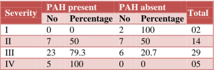
Normal echocardiography was seen in 24% cases in our study. In the analysis of echocardiographic findings, our study showed 70% of the patients had echocardioghaphic evidence of pulmonary arterial hypertension. Frequency of PAH in ECHO in mild, moderate, severe, and very severe COPD was 0/2 = 0%, 7/14 = 50%, 23/29 = 79.3%, and 5/5 = 100%, respectively. There was statistically significant correlation between severity of COPD and
PAH with correlation coefficient of +0.434 and p value <0.05 (Table 1).
Table 1: Comparison of PAH in echo according to severity of disease.
Severity PAH present PAH absent Total
No Percentage No Percentage
I 0 0 2 100 02
II 7 50 7 50 14
III 23 79.3 6 20.7 29
IV 5 100 0 0 05
Cor-pulmonale comprising of right ventricular enlargement was found in ECHO in 23/50= 46% of patients. Frequency of RV enlargement in ECHO in mild, moderate, severe, and very severe COPD was 0/2 = 0%, 4/14 = 28.6%, 14/29 = 48.3%, and 5/5 = 100%, respectively. Spearman’s correlation coefficient between severity of COPD and prevalence of RV enlargement was +0.395 and p value was 0.005. So, as the severity of COPD increased, prevalence of cor-pulmonale/RV enlargement also increased significantly (Table 2). Right atrial enlargement was present in 14% patients with frequency in mild, moderate, severe, and very severe COPD in 0/2 = 0%, 1/14 = 7.1%, 3/29 = 10.3%, and 3/5 = 60%, respectively. Prevalence of right atrial enlargement also correlated significantly with severity of COPD with correlation coefficient of +0.307 and p value of <0.05 (Table 3).
Table 2: Comparison of RV size in ECHO according to severity of disease.
Severity Normal Enlarged Total
No Percentage No Percentage
I 2 100 0 0 02
II 10 71.4 4 28.6 14 III 15 51.7 14 48.3 29
IV 0 0 5 100 05
Table 3: Comparison of RA size in ECHO according to severity of disease.
Severity Normal Enlarged Total
No Percentage No Percentage
I 2 100 0 0 02
II 13 92.9 1 7.1 14 III 26 89.7 3 10.3 29
IV 2 40 3 60 05
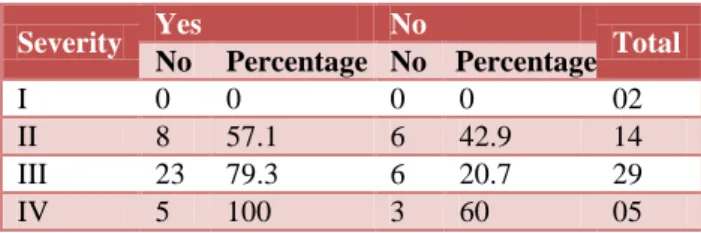
of tricuspid regurgitation also increased significantly (Table 4).
Table 4: Comparison of TR in ECHO according to severity of disease.
Severity Yes No Total
No Percentage No Percentage
I 0 0 0 0 02
II 8 57.1 6 42.9 14 III 23 79.3 6 20.7 29
IV 5 100 3 60 05
DISCUSSION
The cardiac manifestations of COPD are numerous. Impairment of right ventricular function and pulmonary blood vessels are well known to complicate the clinical course of COPD and co-relate inversely with survival. Pulmonary arterial hypertension (PAH) is the primary cardiovascular complication encountered in COPD. Chronic cor pulmonale is a strong predictor of death in COPD. Cor pulmonale is right ventricular enlargement secondary to pulmonary hypertension. Cor pulmonale can range clinically from mild changes in right ventricular function to frank right heart failure. Right ventricular dysfunction is reported to occur in up to 50% of the patients with moderate to severe COPD, and portends a higher mortality rate. Its recognition and treatment may lead to prolonged survival and improved quality of life.6
In this study, mean age was 59.52±12.53 years. Female: male sex ratio was 1:2.33. So, COPD is more common in males and smokers in 5th to 7th decade of life.
The study group was divided into subgroups depending upon the duration of the disease. The duration of the disease was determined on the basis of retrospective history and any medical records available. Majority of patients in the study group fell in the disease duration group of 4-8 years. The range of duration of disease was 4-20 years with the mean duration being 11.08±4.83 years. In study by Shreshta B et al, more than half of the COPD patients were in age group 60-75 years, followed by less number of patients (approximately 20.0%) in both 45-59 years and 75-89 years age groups.9
Most patients had moderate to severe disease at presentation. The study group was subdivided based on the severity of COPD according to GOLD guidelines. The group was divided into mild, moderate, severe, and very severe COPD with 2/50 = 4%, 14/50 = 28%, 29/50 = 58%, and 5/50 = 10% patients respectively. So, there were very few patients in the study group falling in Stage I (mild COPD) showing that the patients with COPD usually present late in the illness. In study by Suma et al[10], the number of patients with mild, moderate and severe COPD were 2/50 = 4%, 18/50 = 36% and 30/50 = 60% respectively.
On analysis by echocardiography, there is high prevalence of cardiac dysfunction in COPD patients with tricuspid regurgitation and pulmonary arterial hypertension being the most common cardiac manifestations of COPD. PAH in ECHO in our study was present in 35/50 = 70% patients. Frequency of PAH in ECHO in mild, moderate, severe, and very severe COPD was 0/2 = 0%, 7/14 = 50%, 23/29 = 79.3%, and 5/5 = 100%, respectively. Tiwari et al, 2015 found that PAH was present in 51.6% of COPD patients and prevalence in mild, moderate, severe and very severe patients was 17%, 52%, 60% and 78% respectively.11 The result of present study is comparable to previous studies. Statistical analysis of our study showed that the difference in the presence of PAH in different groups was statistically significant (p value<0.05, p = 0.002) and there is positive correlation (correlation coefficient+0.434) between severity of COPD and PAH i.e. with increase in severity of COPD, incidence of PAH increases. Thus the incidence of PAH is directly proportional to severity of COPD.
In this study of total 50 patients, RV was enlarged in ECHO in 23/50 = 46% of patients. Frequency of RV enlargement in ECHO in mild, moderate, severe, and very severe COPD was 0/2 = 0%, 4/14 = 28.6%, 14/29 = 48.3%, and 5/5 = 100%, respectively. Jain et al, found that right ventricular dilatation was present in 66.7% of COPD patients and prevalence in mild, moderate, severe and very severe patients was 66.6%, 66.6%, 60% and 100% respectively.12 Tiwari et al, found that cor plumonale was present in 23.7% of COPD patients and prevalence in mild, moderate, severe and very severe patients was 12%, 15%, 40% and 44% respectively.11 So the present study is comparable to the previous studies. The present study also showed a statistically significant correlation between right ventricular enlargement and severity of COPD with spearman’s correlation coefficient of +0.395 and p value 0.005. So as severity of COPD increases, incidence of RV enlargement also increases.
Out of total 50 patients, RA was enlarged in ECHO in 7/50 = 14% of patients. Frequency of RA enlargement in ECHO in mild, moderate, severe, and very severe COPD was 0/2 = 0%, 1/14 = 7.1%, 3/29 = 10.3%, and 3/5 = 60%, respectively. Jain et al, found that right atrial enlargement was present in 50% of COPD patients and prevalence in mild, moderate, severe and very severe patients was 41.6%, 53.3%, 60% and 100% respectively.12 Our study also showed a statistically significant correlation between right atrial enlargement and severity of COPD with spearman’s correlation coefficient of +0.307 and p value 0.03.
mild, moderate, severe and very severe patients was 58.3%, 60%, 60% and 100% respectively.12 Tiwari et al, observed that tricuspid regurgitation was present in 74.2% of COPD patients.11 These findings were comparable to our study. The present study showed a statistically significant correlation between tricuspid regurgitation and severity of COPD with spearman’s correlation coefficient of +0.387 and p value 0.005. So as severity of COPD increases, incidence of tricuspid regurgitation also increases.
CONCLUSION
The echocardiographic finding of pulmonary arterial hypertension, right ventricular enlargement, right atrial enlargement and tricuspid regurgitation positively correlated with severity of COPD as evidenced by echocardiographic finding of increased prevalence of these conditions with increasing severity of COPD. So, prevalence of cardiac dysfunction increases as the severity of COPD increases and echocardiography helps in analysis of cardiac complications of COPD. It is recommended that echocardiography should be done early in all cases of COPD to diagnose the cardiac complications of COPD, so that early interventions can be undertaken in order to improve quality of life and decrease mortality and morbidity in COPD patients.
Funding: No funding sources Conflict of interest: None declared
Ethical approval: The study was approved by the institutional ethics committee
REFERENCES
1. Chesnutt MS, Prendergast TJ, Tavan ET. Pulmonary disorders. In: Papadakis MA, Mcphee SJ, Rabow MW, editors. Current Medical Diagnosis and Treatment.53rd ed, New York: McGraw-Hill Education. 2014:234-314.
2. Siafakas N, Vermeire P, Pride N, Paoletti P, Gibson J, Howard P, et al. Optimal assessment and management of chronic obstructive pulmonary disease (COPD). Eur Respir J. 1995;8(8):1398-420. 3. Macnee W. Chronic bronchitis and emphysema. In:
Seaton A, Seaton D, Leitch A, editors. Crofton and
Douglas’s Respiratory Diseases. 5th edition. France:
Blackwell Science. 2002:616.
4. Anthonisen N, Connett JE, Kiley JP, Altose MD, Bailey WC. Effects of smoking intervention and the use of an inhaled anticholinergic bronchodilator on the rate of decline of FEV1. JAMA. 1994;272:1497-505.
5. Sin DD, Anthonisen NR, Soriano JB, Agusti AG. Mortality in COPD: role of comorbidities. Eur Respir J. 2006;28:1245-57.
6. Klinger JR, Hill NS. Right ventricular dysfunction in chronic obstructive pulmonary disease, Evaluation and management. Chest. 1991;99:715-23.
7. Luke SH, Julia G, David D, Michael B, John B. Chambers, Navroz D, et al. Echocardiographic assessment of pulmonary hypertension: standard operating procedure. Eur Respir Rev. 2012;21(125):239-48.
8. Daniels LB, Krummen DE, Blanchard DG. Echocardiography in pulmonary vascular disease. Cardiol Clin.2004;22:383-99.
9. Shrestha B, Dhungel S, Chokhani R. Echocardiography based cardiac evaluation in the patients suffering from chronic obstructive pulmonary disease. Nepal Med Coll J. 2009;11(1):14-8.
10. Suma KR, Srinath S, Praveen. Electrocardiographic and echocardiographic changes in chronic obstructive pulmonary disease (COPD) of different grades of severity. Journal Evolution Medical Dental Sci. 2015;4:5093-101.
11. Tiwari VK, Agarwal R, Kumar A, Kumar A, Kumar R. The cardiac evaluation in chronic obstructive pulmonary disease patients. Indian J Applied Res. 2015;15(11):434-5.
12. Jain J, Soni P, Apte S, Chanchlani R. A Study of correlation between echocardiographic changes with the duration and severity of chronic obstructive pulmonary disease. Journal Evolution Med Dental Sci. 2014;3(8):1997-2002.