Original Research Article
Tissue doppler imaging for estimation of left ventricular filling pressure
in patients with systolic and diastolic heart failure: a comparative
simultaneous doppler catheterization study
Manohar J. Suranagi, K. Subramanyam*, K. S. Subramani, K. H. Srinivasa
INTRODUCTION
Tissue Doppler imaging is a robust and reproducible echocardiographic tool which has permitted a quantitative assessment of both global and regional function and
timing of myocardial events.1,2 Most published studies
have examined the long-axis function of the heart by TDI from apical views.
In diastole, potentially important prognosticators include peak myocardial early diastolic velocity measured at the lateral mitral annulus (E′) as well as measurement of trans-mitral to TDI of early diastolic velocity ratio (E/E′).
ABSTRACT
Background: Noninvasive assessment of diastolic filling by Doppler echocardiography provides important information about left ventricular (LV) status in selected subsets of patients. This study was designed to assess whether the lateral mitral annular velocity as assessed by tissue Doppler imaging is associated with invasive measures of diastolic LV performance in patients with diastolic and systolic heart failure. Aim of the study was to compare the diagnostic accuracy of lateral mitral annular E/E′ as an estimate of LV filling pressure with invasive LVEDP measurement in subjects with systolic or purely diastolic heart failure.
Methods: Total 100 patients were studied, 50 patients with diastolic heart failure and 50 patients with systolic heart failure in patients undergoing diagnostic coronary angiogram. Detailed 2D Echocardiography, Trans mitral Doppler and Tissue Doppler velocities of lateral mitral annulus was obtained. The ratio of peak mitral velocity (E) to lateral mitral annular velocity (E′) by TDI (E/E′) was calculated.
Results: The ratio of E/E′ in diastolic group was 13.4±4.9 and in systolic group it was 13.7±5.2. The mean LVEDP in diastolic heart failure patients was 14.3±4.5 and 14.2±4.9 in systolic heart failure patients. The ratio of E/E′ showed a better correlation with LVEDP. E/E′ <8 accurately predicted normal LVEDP, and E/E′ >15 identified increased LVEDP ≥15mmHg.
Conclusions: E/E′ is a reliable estimate of LV filling pressures in subjects with systolic and diastolic heart failure. In subjects with diastolic heart failure, E/E′ seems helpful to identify those with truly elevated LV filling pressures. In patients with diastolic heart failure and normal E/E′, a search for other causes of symptoms (pulmonary disease, obesity and so forth) may be warranted.
Keywords: Diastolic heart failure, Left ventricular end diastolic pressure, Lateral mitral annular velocity, Peak mitral velocity, Receiver-operating characteristic curves
Department ofCardiology, Sri Jayadeva Institute of Cardiovascular sciences and research, Bangalore, Karnataka, India
Received: 21 December 2019
Revised: 02 January 2020
Accepted: 06 January 2020
*Correspondence:
Dr. K. Subramanyam,
E-mail: [email protected]
Copyright: © the author(s), publisher and licensee Medip Academy. This is an open-access article distributed under the terms of the Creative Commons Attribution Non-Commercial License, which permits unrestricted non-commercial use, distribution, and reproduction in any medium, provided the original work is properly cited.
These myocardial velocity measurements with TDI have been shown to be useful in various diseases, including heart failure (HF), hypertension, acute myocardial infarction (MI) and in patients undergoing stress
echocardiography for suspected coronary heart disease.3
Diastolic dysfunction is common in cardiac disease and contributes to the signs and symptoms of heart failure. Doppler echocardiography is widely used for the noninvasive assessment of diastolic filling of the left
ventricle (LV).4 Analysis of the mitral inflow velocity
curve has provided useful information for determination of filling pressures and prediction of prognosis in selected patients. However, mitral flow is dependent on multiple interrelated factors, including the rate and extent of ventricular relaxation, suction, atrial and ventricular
compliance, and left atrial pressure.4,5 These factors may
have confounding effects on the mitral inflow; thus, it has not been possible to determine diastolic function from the
mitral flow velocity curves in many subsets of patients.6,7
To overcome these limitations of the mitral inflow parameters, combinations of the mitral flow velocity curves with other Doppler parameters have been used. These include the pulmonary venous velocity curves, color M-mode propagation velocity, and the response of
the mitral inflow to altered loading conditions.8-17 Tissue
Doppler imaging (TDI) of mitral annular motion has been proposed to correct for the influence of myocardial relaxation on trans-mitral flows. This has been shown to be an excellent predictor of diastolic filling in subsets of patients.18-28
The present study compares the diagnostic accuracy of E/E′ with invasive LVEDP as an estimate of LV filling pressure in subjects with systolic and pure diastolic heart failure.
METHODS
Total 100 patients were studied between July 2007 to March 2008, 50 patients (22 coronary artery disease, 28 hypertensive) with diastolic heart failure (defined by heart failure signs and symptoms but with preserved ejection fraction) and 50 patients (48 coronary artery disease, 26 dilated cardiomyopathy) with systolic heart failure (heart failure signs and symptoms and reduced ejection fraction) in patients who underwent clinically indicated cardiac catheterization in the admitted patients to the hospital (Sri Jayadeva Institution of Cardiology, Bangalore, India.) and who were able provide written informed consent.
The Patients with systolic and diastolic heart failure aged between 21 to 80 years who underwent clinically indicated cardiac catheterization were included. Patients with mitral stenosis, Patients with prosthetic valves, Patients with arrhythmias/heart blocks during study, Patients with mitral annular calcification and Patients
Data are expressed as mean values±SD. Comparison between the diastolic and systolic variables were using non paired Student’s t test. For dichotomous variables, chi-square analysis was used for comparison. Statistical significance was set at p <0.05. Statistical relations between conventional Doppler echo/TDI variables and LV end-diastolic pressures were assessed by simple
linear regression analysis. Receiver-operating
characteristic (ROC) curves analysis was generated for the E/E′ ratio to predict a LV end-diastolic pressure ≥15 mm Hg. Linear regression analysis was used to assess statistical relations between E/E′ and hemodynamic variables.
Patients underwent Echocardiographic examination in the catheterization laboratory just before case was taken for coronary angiography. LV end diastolic pressure was estimated by pigtail catheter before doing left ventriculogram.
The 2D Echocardiography and evidence of impaired LV relaxation or filling, that is, abnormal age dependent isovolumic relaxation time, mitral E/A ratio, deceleration time and thus met the criteria for diastolic heart failure
(diastolic heart failure group).29 Fifty (50) patients had
systolic dysfunction with EF <50% (systolic heart failure group).
Images were taken ≤2 hours before cardiac
catheterization according to the guidelines of the American Society of Echocardiography using a PHILIPS
EnVisor machine.30 For TDI recordings from the apical
window, a 5-mm sample volume was located at the septal and lateral sites of the mitral annulus in the 4-chamber
view.31 Peak systolic (S′), early (E′), and late (A′)
diastolic velocities were obtained at each site and average values of these measurements were calculated for each patient. The mitral E/E′ ratio was subsequently derived
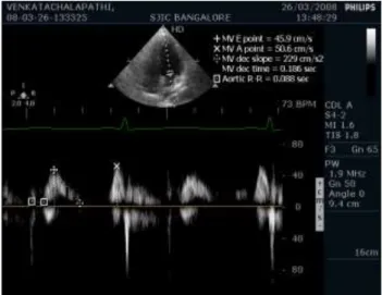
(Figure 2).31
E/E′=4.3, Measured LVEDP=8mmHg
Figure 2: TDI velocity tracings with the sample volume located at the lateral mitral annulus from which peak systolic (S′), peak early diastolic (E′), and peak late diastolic (A′) annular velocities are derived.
RESULTS
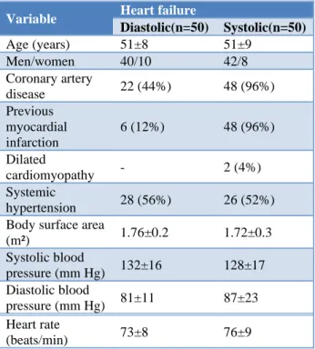
Coronary artery disease was the most common cause for heart failure in patients with systolic and diastolic heart failure; whereas dilated cardiomyopathy was only present in the systolic heart failure group (Table 1).
Table 1: Clinical data of the study participants.
Variable Heart failure
Diastolic(n=50) Systolic(n=50)
Age (years) 51±8 51±9
Men/women 40/10 42/8
Coronary artery
disease 22 (44%) 48 (96%)
Previous myocardial infarction
6 (12%) 48 (96%)
Dilated
cardiomyopathy - 2 (4%)
Systemic
hypertension 28 (56%) 26 (52%)
Body surface area
(m²) 1.76±0.2 1.72±0.3
Systolic blood
pressure (mm Hg) 132±16 128±17
Diastolic blood
pressure (mm Hg) 81±11 87±23
Heart rate
(beats/min) 73±8 76±9
Patients with systolic heart failure were in a worse New York Heart Association functional (NYHA) class than patients with diastolic heart failure. In patients with systolic heart failure, LV dimensions and volumes were increased, but ejection fraction and fractional shortening
were reduced compared with the diastolic heart failure group (Table 2).
Table 2: Baseline Echocardiographic Variables among participants.
Variable
Heart failure
p value
Diastolic (n=50)
Systolic (n=50)
LV diastolic diameter
(cm) 3.7±1.1 5.7±1.6 0.62
LV systolic diameter
(cm) 2.1±0.8 4.3±1.6 0.82
LV diastolic volume
(ml) 89±21 114±23 0.97
LV systolic volume
(ml) 37±10 74±17 0.66
LA diameter (cm) 2.8±1.5 3.2±1.4 0.15
Fractional shortening
(%) 32±8 18±9 0.60
Ejection fraction (%) 58±2.6 35±4.7 0.65
In the diastolic heart failure group, deceleration time and isovolumic relaxation time were prolonged, and the E/A ratio was reduced compared with the systolic heart failure group (Table 3).
Table 3: Conventional Doppler and TDI- derived variables.
Variable
Heart failure
p value
Diastolic (n=50)
Systolic (n=50)
E (cm/s) 73±13 84±11.5 0.001
A(cm/s) 71±19 39±6.4 0.79
E/A ratio 1.2±0.5 2.2±0.3 0.23
Deceleration time
(ms) 179±32 159±27 0.39
Isovolumic relaxation
time (ms) 77±15 72±11 0.15
Systolic mitral annular velocity (S′) (cm/s)
7.0±1.1 4.3±1.3 0.67
Early diastolic lateral mitral annular velocity (E′) (cm/s)
6.5±3.2 7.1±2.8 0.41
Late diastolic lateral mitral annular velocity (A′) (cm/s)
9.0±2.1 6.0±2.6 0.95
E/E′ 13.4±4.9 13.7±5.2 0.46
In the diastolic heart failure group, the LV end-diastolic pressure was similar to systolic group (14.3±4.5 Vs 13.7±4.3mm Hg). In patients with diastolic heart failure, E/E′ was significantly related to the LV end-diastolic pressure (r=0.79, p <0.0001) (Figure 3).
In patients with systolic heart failure, E/E′ also correlated significantly with LV end-diastolic pressure (r =0.79, p <0.0001) (Figure 4).
Figure 3: The relation between LV end-diastolic pressure (LVEDP) and E/E′ in the diastolic heart
failure group.
Figure 4: The relation between LV end-diastolic pressure (LVEDP) and E/E′ in the systolic heart
failure group.
In the diastolic heart failure group, an E/E′ >15 derived from ROC analysis, (Figure 5) identified patients with a LV end-diastolic pressure ≥15 mm Hg with a sensitivity of 87% and a specificity of 90% (AUC-area under the curve, 0.80) positive predictive value, 93%; negative
Figure 5: ROC curve analysis for prediction of LV end-diastolic pressure >15 mm Hg in the diastolic heart failure group. the area under the curve (AUC)
was 0.80 for E/E′.
In patients with systolic heart failure, E/E′ >15 derived from ROC analysis, (Figure 6) for the prediction of LV end-diastolic pressure ≥15 mm Hg had 87% sensitivity and 90% specificity (AUC-area under the curve, 0.78); positive predictive value, 90%; negative predictive value, 84%.
Figure 6. ROC curve analysis for prediction of LV end-diastolic pressure >15 mm Hg in the systolic heart
failure group. The area under the curve (AUC) was 0.78 for E/E′.
DISCUSSION
Study confirms that the mitral E/E′ ratio is a reliable estimate of LV filling pressures in subjects who have diastolic heart failure, as evidenced by heart failure symptoms but preserved LV systolic performance. Our results confirm previous work that indicates that E/E′ readily identifies elevated LV filling pressures in subjects
with reduced LV systolic performance.32,33 Results was
21.00 18.00 15.00 12.00 9.00 6.00 3.00
E/E'
21.00
18.00
15.00
12.00
9.00
6.00
LVEDP(
mmH
g)
R Sq Linear = 0.687
20.00 18.00 16.00 14.00 12.00 10.00 8.00 6.00
E/E'
21.00
18.00
15.00
12.00
9.00
6.00
LVEDP(
mmH
g)
R Sq Linear = 0.635
1.0 0.8
0.6 0.4
0.2 0.0
1 - Specificity
1.0 0.8 0.6 0.4 0.2 0.0
Sensitivi
ty
in both systolic and pure diastolic heart failure.34 E/E′ also
seems an appropriate tool to exclude elevated LV filling pressures in subjects presenting with heart failure symptoms. E/E′ is reproducibly obtained so that a widespread clinical application of this ratio seems feasible.
Within the last 30 years, the number of patients affected by chronic heart failure has increased dramatically in the
Western population.35 In 2/3 of such patients, systolic and
diastolic dysfunction coexist; whereas in 1/3, signs and symptoms are attributable to purely diastolic heart
failure.36 Elevated LV filling pressures have long been
identified as the hallmark of diastolic heart failure. However, even in patients with a reduced ejection fraction, exercise limitation is determined by the increase in filling pressures, irrespective of the degree of systolic
dysfunction.37 Thus, in patients with heart failure with or
without systolic dysfunction, noninvasive estimation of filling pressures is desirable.
Using echocardiography, LV filling pressures have been estimated by using equations that incorporate ≥1 mitral or
pulmonary vein-flow- derived variable.37,38 Rossvoll and
Hatle have shown that combined trans mitral and pulmonary vein flow variables provide valuable
information about filling pressures.38 However, the
feasibility of this approach has been questioned, mainly because mitral and pulmonary vein flow variables are known to change with age, heart rate, and loading
conditions.39 Those limitations seem to have been partly
overcome with the introduction of TDI.
The mitral annular E′ velocity decreases with worsening diastolic function, suggesting a role of E′ as an index of
LV relaxation.40 In addition, the mitral E/E′ ratio was
proposed as an estimate of LV filling pressures, and this ratio was properly validated in subjects who were admitted to an intensive care unit, most of whom had underlying cardiac disease and reduced systolic
performance.32 Those findings were confirmed by
Ommen et al, who studied 100 patients during left-sided
cardiac catheterization.33 In these previous studies,
however, patients with diastolic heart failure were not separately evaluated, and the diagnostic accuracy of E/E′ in this important diastolic heart failure patient’s population was not addressed.
Study has few limitations that are LVEDP was the only invasive measure of LV diastolic function. Relatively small sample of population who had LVEDP >15 mmHg may have weakened the potential correlation between LVEDP and E/E′.
CONCLUSION
Our results confirm that E/E′ is a reliable estimate of LV filling pressures in subjects with systolic and diastolic heart failure. In subjects with diastolic heart failure, E/E′ seems helpful to identify those with truly elevated LV
filling pressures. In patients with diastolic heart failure and normal E/E′, a search for other causes of symptoms (pulmonary disease, obesity and so forth) may be warranted.
Funding: No funding sources Conflict of interest: None declared
Ethical approval: The study was approved by SJICS ethical board
REFERENCES
1. Gorcsan J, Gulati VK, Mandarino WA, Katz WE.
Color-coded measures of myocardial velocity throughout the cardiac cycle by tissue Doppler imaging to quantify regional left ventricular function. Am Heart J. 1996;131:1203-13.
2. Yu CM, Lin H, Ho PC, Yang H. Assessment of left
and right ventricular systolic and diastolic synchronicity in normal subjects by tissue Doppler echocardiography and the effects of age and heart rate. Echocardiography. 2003;20:19-27.
3. Sanderson JE, Wang M, Yu CM. Tissue Doppler
imaging for predicting outcome in patients with
cardiovascular disease. Curr Opin Cardiol.
2004;19:458-63.
4. Nishimura RA, Tajik AJ. Evaluation of diastolic
filling of left ventricle in health and disease: Doppler echocardiography is the clinician’s Rosetta Stone. J Am Coll Cardiol. 1997;30:8-18.
5. Choong CY, Herrmann HC, Weyman AE, Fifer
MA. Preload dependence of Doppler-derived indexes of left ventricular diastolic function in humans. J Am Coll Cardiol. 1987 Oct 1;10(4):800-8.
6. Yamamoto K, Nishimura RA, Chaliki HP, Appleton
CP, Holmes DR, Redfield MM. Determination of left ventricular filling pressure by Doppler echocardiography in patients with coronary artery disease: critical role of left ventricular systolic
function. J Am Coll Cardiol. 1997 Dec
1;30(7):1819-26.
7. Oh JK, Appleton CP, Hatle LK, Nishimura RA,
Seward JB, Tajik AJ. The noninvasive assessment of left ventricular diastolic function with two-dimensional and Doppler echocardiography. J Am Soc Echocardio. 1997 Apr 1;10(3):246-70.
8. Brunazzi MC, Chirillo F, Pasqualini M, Gemelli M,
Franceschini-Grisolia E, Longhini C, et al. Estimation of left ventricular diastolic pressures
from precordial pulsed-Doppler analysis of
pulmonary venous and mitral flow. Am Heart J. 1994 Aug 1;128(2):293-300.
9. Keren G, Sherez JA, Megidish RE, Levitt BA,
Laniado SH. Pulmonary venous flow pattern--its relationship to cardiac dynamics. A pulsed Doppler
echocardiographic study. Circulation. 1985
Jun;71(6):1105-12.
10. Kuecherer HF, Kusumoto F, Muhiudeen IA,
patterns by transesophageal pulsed Doppler echocardiography: relation to parameters of left ventricular systolic and diastolic function. Am Heart J. 1991 Dec 1;122(6):1683-93.
11. Nishimura RA, Abel MD, Hatle LK, Tajik AJ.
Relation of pulmonary vein to mitral flow velocities by transesophageal Doppler echocardiography. Effect of different loading conditions. Circulation. 1990 May;81(5):1488-97.
12. Rossvoll O, Hatle LK. Pulmonary venous flow
velocities recorded by transthoracic Doppler ultrasound: relation to left ventricular diastolic pressures [see comments]. J Am Coll Cardiol. 1993;21:1687-96.
13. Yamamoto K, Nishimura RA, Burnett Jr JC,
Redfield MM. Assessment of left ventricular end-diastolic pressure by Doppler echocardiography: contribution of duration of pulmonary venous versus mitral flow velocity curves at atrial contraction. J Am Soc Echocardio. 1997 Jan 1;10(1):52-9.
14. Thomas JD, Garcia MJ, Greenberg NL. Application
of color Doppler M-mode echocardiography in the assessment of ventricular diastolic function: potential for quantitative analysis. Heart Vessels. 1997; 12(suppl):135-7.
15. Steen T, Rootwelt K, Risøe C, Andreassen AK,
Ihlen H. A new colour M-mode index of diastolic
filling compared with radionuclide
ventriculography. Inter J Cardiol. 1995 Jan 27;48(1):89-95.
16. Dumesnil JG, Gaudreault G, Honos GN, Kingma Jr
JG. Use of Valsalva maneuver to unmask left ventricular diastolic function abnormalities by Doppler echocardiography in patients with coronary artery disease or systemic hypertension. Am J Cardiol. 1991 Aug 15;68(5):515-9.
17. Hurrell DG, Nishimura RA, Ilstrup DM, Appleton
CP. Utility of preload alteration in assessment of left
ventricular filling pressure by Doppler
echocardiography: a simultaneous catheterization and Doppler echocardiographic study. J Am Coll Cardiol. 1997 Aug 1;30(2):459-67.
18. Aranda JJ, Weston MW, Puleo JA, Fontanet HL.
Effect of loading conditions on myocardial relaxation velocities determined by Doppler tissue imaging in heart transplant recipients. J Heart Lung Transplantation: Official Publication Inter Soc Heart Transplantation. 1998 Jul;17(7):693-7.
19. Garcia MJ, Rodriguez L, Ares M, Griffin BP, Klein
AL, Stewart WJ, et al. Myocardial wall velocity assessment by pulsed Doppler tissue imaging: characteristic findings in normal subjects. Am heart j. 1996 Sep 1;132(3):648-56.
20. Garcia MJ, Rodriguez L, Ares M, Griffin BP,
Thomas JD, Klein AL. Differentiation of
constrictive pericarditis from restrictive
cardiomyopathy: assessment of left ventricular diastolic velocities in longitudinal axis by Doppler tissue imaging. J Am Coll Cardiol. 1996 Jan
21. Nagueh SF, Middleton KJ, Kopelen HA, Zoghbi
WA, Quiñones MA. Doppler tissue imaging: a noninvasive technique for evaluation of left ventricular relaxation and estimation of filling
pressures. J AmColl Cardiol. 1997 Nov
15;30(6):1527-33.
22. Nagueh SF, Mikati I, Kopelen HA, Middleton KJ,
Quiñones MA, Zoghbi WA. Doppler estimation of left ventricular filling pressure in sinus tachycardia: a new application of tissue Doppler imaging. Circulation. 1998 Oct 20;98(16):1644-50.
23. Nagueh SF, Lakkis NM, Middleton KJ, Spencer III
WH, Zoghbi WA, Quinones MA. Doppler estimation of left ventricular filling pressures in
patients with hypertrophic cardiomyopathy.
Circulation. 1999 Jan 19;99(2):254-61.
24. Nixdorff U, Rupprecht HJ, Mohr-Kahaly S, Kremer
M, Bickel C, Meyer J. Tissue Doppler
echocardiography: a new method of evaluating perfusion-dependent myocardial function during PTCA. Inter J Cardiac Imag. 1997 Apr 1;13(2):99-103.
25. Sohn DW, Chai IH, Lee DJ, Kim HC, Kim HS, Oh
BH, et al. Assessment of mitral annulus velocity by Doppler tissue imaging in the evaluation of left ventricular diastolic function. J Am Coll Cardiol. 1997;30:474-80.
26. Nishimura RA, Schwartz RS, Tajik AJ, Holmes Jr
DR. Noninvasive measurement of rate of left ventricular relaxation by Doppler echocardiography.
Validation with simultaneous cardiac
catheterization. Circulation. 1993 Jul;88(1):146-55.
27. Yamamoto K, Nishimura RA, Redfield MM.
Assessment of mean left atrial pressure from the left ventricular pressure tracing in patients with cardiomyopathies. Am J Cardiol. 1996 Jul 1;78(1):107-10.
28. Davidson CJ, Fishman RF, Bonow RO. Cardiac
catheterization. In: Braunwald E, ed. Heart Disease: A Textbook of Cardiovascular Medicine. 5th ed. Philadelphia, Pa: WB Saunders; 1997:177-203.
29. Silva CE, Ferreira LD, Peixoto LB, Monaco CG, Gil
MA, Ortiz J, et al. Evaluation of segmentary contractility in Chagas' disease by using the integral of the myocardial velocity gradient (myocardial
strain) obtained through tissue Doppler
echocardiography. Arquivos brasileiros de
cardiologia. 2005 Apr;84(4):285-91.
30. Schiller NB, Shah PM, Crawford M, DeMaria A,
Devereux R, Feigenbaum H, et al.
Recommendations for quantification of the left ventricle by two-dimensional echocardiography. J Am Soc Echocardiogr. 1989;2:358-67.
31. Rivas-Gotz C, Manolios M, Thohan V, Nagueh SF.
Impact of left ventricular ejection fraction on estimation of left ventricular filling pressures using tissue Doppler and flow propagation velocity. Am J Cardiol. 2003;91:780-4.
Technique for Estimation of Left Ventricular End Diastolic Pressure in Severe Mitral Regurgitation. J Tehran Heart Cent. 2010;5(3):122-7.
33. Ommen SR, Nishimura RA, Appleton CP, Miller
FA, Oh JK, Redfield MM, et al. Clinical utility of Doppler echocardiography and tissue Doppler imaging in the estimation of left ventricular filling pressures: a comparative simultaneous Doppler-catheterization study. Circulation. 2000;102:1788-94.
34. Bruch C, Stypmann J, Grude M, Gradaus R,
Breithardt G, Wichter T. usefulness of tissue Doppler imaging for estimation of let ventricular filling pressures in patients with systolic and diastolic heart failure. Am J Cardial. 2005;95:892-5.
35. Ho KK, Anderson KM, Kannel WB, Grossman W,
Levy D. Survival after the onset of congestive heart failure in Framingham Heart Study subjects. Circulation. 1993;88:107-15.
36. Grossmann W. Diastolic dysfunction in congestive
heart failure. Semi Med Beth Israel Hosp. 1991;325:1557-64.
37. Mulvagh S, Quinones MA, Kleiman NS, Cheirif J,
Zoghbi WA. Estimation of left ventricular end-diastolic pressure from Doppler transmitral flow velocity in cardiac patients independent of systolic performance. J Am Coll Cardiol. 1992;20:112-9.
38. Appleton CP, Galloway JM, Gonzalez MS, Gaballa
M, Basnight MA. Estimation of left ventricular
filling pressures using two-dimensional and Doppler echocardiography in adult patients with cardiac disease. Additional value of analyzing left atrial size, left atrial ejection fraction and the difference in duration of pulmonary venous and mitral flow velocity at atrial contraction. J Am Coll Cardiol. 1993 Dec;22(7):1972-82.
39. Rakowski H, Appleton C, Chan KL, Dumesnil JG,
Honos G, Jue J, et al. Canadian consensus recommendations for measurement and reporting of diastolic dysfunction by echocardiography. J Am Soc Echocardio. 1996;9:736-60.
40. Naqvi TZ, Padmanabhan S, Rafii F, Hyuhn HK,
Mirocha J. Comparison of usefulness of left ventricular diastolic versus systolic function as a
predictor of outcome following primary
percutaneous coronary angioplasty for acute myocardial infarction. Am J Cardiol. 2006 Jan 15;97(2):160-6.
Cite this article as: Suranagi MJ, Subramanyam K, Subramani KS, Srinivasa KH. Tissue doppler imaging for estimation of left ventricular filling pressure in patients with systolic and diastolic heart failure: a comparative simultaneous doppler