Modeling Cell-Extracellular Matrix Interactions: Spatial and Conformational Control of Ligand Presentation on Self-Assembled Monolayers
Brian Mathew Lamb
A dissertation submitted to the faculty of the University of North Carolina at Chapel Hill in partial fulfillment of the requirements for the degree of Doctor of Philosophy in the
Department of Chemistry
Chapel Hill
2011
ABSTRACT Brian Mathew Lamb
Modeling Cell-Extracellular Matrix Interactions: Spatial and Conformational Control of Ligand Presentation on Self-Assembled Monolayers
(Under the direction of Asst. Prof. Muhammad N. Yousaf)
iii
ACKNOWLEDGMENTS
The success and completion of my studies depended on several people. I would be remiss if I did not first thank my advisor Asst. Professor Muhammad Yousaf. I am indebted to him for teaching me the ropes of publishing science. He enabled me to pursue areas of broad interest that rely on multi-disciplinary science and encouraged me to take initiative to reach my passions.
My parents, Dale and Terrie, are chiefly responsible for inciting my scientific curiosity and encouraging me to pursue the frontier of knowledge in chemistry. Their enduring support keeps me strong and I strive to live up to their high standards and expectations.
I owe Merissa Olmer and her family credit for helping me navigate the choppy waters of science. Their advice, and especially Merissa’s companionship, has kept me heading in the right direction. Furthermore it is from interacting with them that makes me strongly believe that in a world of competition, collaboration is the best way to accomplish nature’s challenges.
Special thanks to Dale Lamb, Linda Olmer, Merissa Olmer, and Andrew Miklos for editing the document.
v
Table of Contents
List of Figures ... vii
List of Schemes ... xiii
List of Abbreviations ... xiv
1. Introduction ... 1
1.1. Cell Behavior is a Consequence of Spatial Constraints ... 2
1.2. Self-Assembled Monolayers on Gold: An Ideal Interfacial Modeling Scaffold .. 4
1.3. Microfabrication and Surface Patterning ... 6
1.4. An Introduction to Switchable Chemistry and Quinonoid Chemistry ... 8
1.5. Background Summary ... 14
1.6. References ... 17
2. Synergistic Microfluidic Etching and Functionalization of Biomaterials ... 22
2.1. Patterning Visually Apparent Chemoselective SAMs ... 22
2.2. Multistep Surface Modification with Microfluidic Lithography ... 23
2.3. Cell Polarity During Migration Around ECM Obstacles ... 27
2.4. Summary - Visually Apparent SAMs for Monitoring Cell Migration ... 30
2.5. Materials and Methods - Visually Apparent SAMs ... 32
2.6. References ... 34
3. Surface Gradient Fabrication and Assays for Cell Orientation Behavior ... 37
3.1. Introduction ... 37
3.3. Surface Gradients Orient Cell Migration ... 45
3.4. Micrometer-scale Surface Gradients Orient Cell Polarity ... 47
3.5. Surface Gradients Orient Cell Division ... 51
3.6. Orientation of Single Cell Divisions in Confluent Monolayers... 55
3.7. Conclusions ... 59
3.8. Materials and Methods ... 60
3.9. References ... 62
4. Dynamic control of peptide conformation on electrode surfaces ... 68
4.1. Introduction ... 68
4.2. Switchable Hydroquinones Bind and Release Oxyamine Ligands... 69
4.3. Incorporating Switchable Chemistry into Surface Bound Peptides ... 70
4.4. Model Reaction: Huisgen Cycloaddition and Oxime Formation ... 76
4.5. Switchable Peptides on Hydroquinone/Azide SAMs ... 81
4.6. Conclusion ... 86
4.7. Materials and Methods ... 88
4.8 Spectroscopic Characterization...……….………...99
vii List of Figures
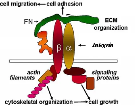
Figure 1.1 Cell adhesion to ECM is mediated by integrin receptors. Shown: general
αβ-integrin receptor binding to a cell adhesive protein fibronectin (FN). Binding initiates focal adhesion formation and actin polymerization
which drives cell migration and growth. ... 3
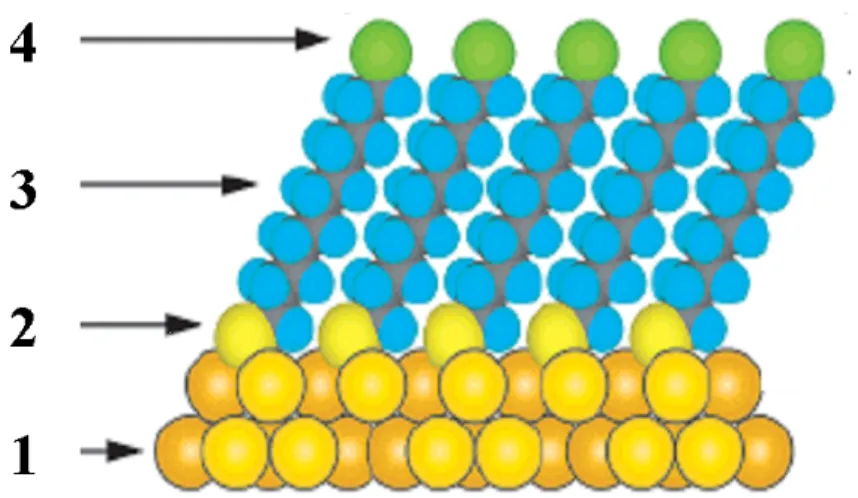
Figure 1.2 Design features of SAMs on gold. 1) Metal substrate (gold)has high
affinity for 2) ligand head group (thiol), creating the stable SAM. 3) Hydrophobic spacer enhances uniformity and prevents degradation. 4) Terminal functional group permits chemical modification, molecular
presentation, and determines macroscopic properties. ... 5
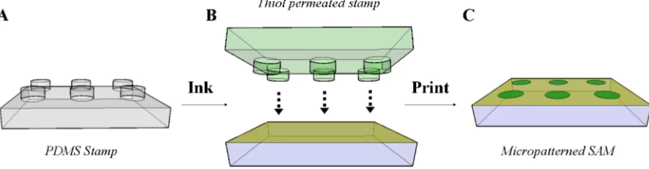
Figure 1.3 Surface patterning via microcontact printing. A) A PDMS stamp with
molded convex microfeatures is inked with a permeable alkanethiol. B) The stamp is dried and then depressed onto a surface (eg. gold) to micropattern alkanethiol exposure to the surface. C) Removal of the
PDMS stamp reveals a micropatterned SAM. ... 7
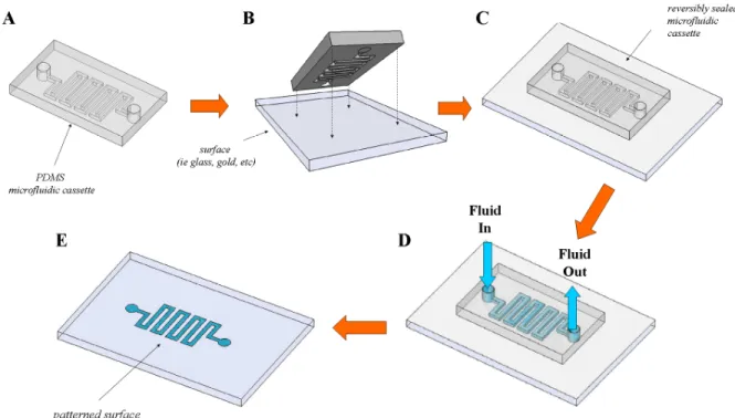
Figure 1.4 Utilization of PDMS microfluidic devices for patterned fluid delivery to
surfaces. A, B) A microfluidic device is sealed onto a surface. C) The device is connected to an external pump or other apparatus that introduces fluid and permits flow to occur. D) Fluid flows through the device,
patterning only regions of the surface exposed to microfluidic channels. E) The microfluidic device is removed from the surface, revealing the
chemically modified surface. ... 9
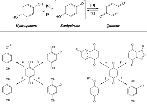
Figure 1.5 (Top) Redox-interconversion of hydroquinone and quinone (left)
Reactivity of HQ. a and b) Radical addition reactions occur at ‘C’ and ‘O’ positions. c) Electrophilic addition- dihalides (Br2), peroxyacids
(RCO3H), acrylates; d) Phenol reactivity with electrophilic reagents (eg. Halosilanes, hydroxyl protecting groups). (right) Reactivity of BQ. e) Diels-Alder with cyclopentadienes, dienes. f) 1,3-dipolarcycloaddition with alkylazides (R-N3). g)Oxime/hydrazone coupling products(Y = O, N, R= H, alkyl). h) reductive Michael addition of thiols, amines,
alkoxides, (Z = N, O, S). ... 12
Figure 1.6 Electrochemical, catalytic ligand immobilization strategy for SAMs on
gold. In pH 7.4 aqueous solution, oxime-immobilized benzoquinone monolayers can be electrochemically reduced, resulting in loss of an alcohol terminated ligand. Immobilizing the Arg-Gly-Asp (RGD) peptides to the surface allows integrin- mediated adhesion and release of
cells from the surface. ... 15
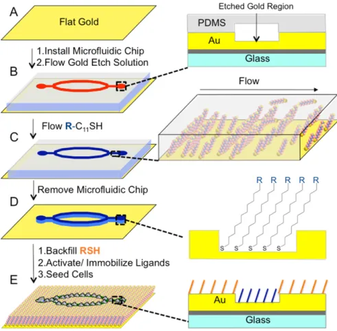
Figure 2.1 Schematic for the generation of patterned chemoselective SAMs on
the microfluidic cassette for 10 s to create visually apparent features. C) Rinsing with a solution of absolute ethanol and water, followed by flowing 1mM HQ-EG4 in ethanol for 60 s generates a partial SAM on the visually apparent regions. D) Removal of the PDMS device. E)
immersion of the substrate in 1 mM EG4 in ethanol solution for 8 h creates an inert SAM background resistant to protein and cell adhesion in non-etched regions. Depending on the identity of R-SH, several
chemoselective surface-functionalization methods are compatible with
the methodology. ... 24
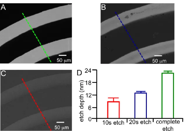
Figure 2.2 Controlling the amount of gold etch and therefore gold thickness by the
duration of triiodide solution in the microfluidic channels. Gold thickness was determined by linescan measurements of surface transparency with Metamorph imaging software. A) A 24 nm gold surface on a glass slide was completely etched showing a patterned gold and glass substrate. Gold was etched 24 nm. B) Partially etched surface for 20 s. Gold was etched 13 nm. C) Surface etched for 10 s. Gold was etched 8 nm. D) Bar graph
of etch results. ... 26
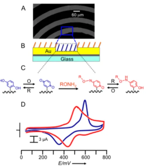
Figure 2.3 Electrochemical characterization of HQ SAMs on partially etched gold.
A) Brightfield micrograph of a partially etched surface. B) Sideview representation of a partially etched patterned SAM. C) The hydroquinone was activated by electrochemical oxidation to the benzoquinone and subsequently reacted with amino-oxy terminated ligands to generate a stable oxime-tethered biomolecule. D) Cyclic voltammetry of the surface showed diagnostic redox signals for the HQ-BQ and the Qox/AP product. Integrating the waves provided a quantitative measure of the amount of
oxime product on the partially etched regions. ... 28
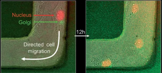
Figure 2.4 Multiwavelength time-lapse live-cell fluorescence microscopy of
transfected Rat2 fibroblast cells undergoing directed migration on partially etched electroactive RGD presenting SAM gold surfaces. The partially etched regions appear lighter in the micrographs. A) Initial position of cells on the etched region of the gold. Red = Nucleus. Green = Golgi. B) An image taken 12 hours later after cell migration had occurred
in the etched regions of the gold. ... 31
Figure 3.1 SPREAD methodology: inking PDMS microfluidic channels with a
gradient of alkanethiols. A) A PDMS microfluidic stamp is placed on a glass substrate. B) An alkanethiol (5mM R-SH, in EtOH) is flowed into the device, controllably permeating and diffusing into the walls of the microfluidic channels. C) Evacuation of the microfluidic stamp and timed diffusion further perpetuates the gradient, allowing control of gradient length. Inset) cross sectional perspective of alkanethiol gradient radiating
ix
Figure 3.2 Fabrication of surface gradients presenting cell adhesive RGD peptide. A)
Printing a stamp inked with 1 directly onto a gold substrate creates B) a gradient oxyamine terminated SAM. C) The remaining bare gold regions are backfilled with EG4 to form a SAM that is inert to non-specific cell attachment. Addition of ketone containing ligands results in the immobilization of ligands via an oxime linkage. (Bottom)
Characterization of a representative gradient generated by the gradient stamping method and reaction with a ketone-rhodamine. Fluorescence intensity profile of the SPREAD gradient on a gold surface showing a
linear slope extending ~200 µm. ... 42
Figure 3.3 Gradient length characterization of printed oxyamine-alkanethiols on gold
surfaces obtained using the SPREAD procedure in a microfluidic device. A) Diffusion length dependence on diffusion time of an ethanolic solution of 5 mM oxyamine alkanethiol. B) Gradient length dependence on
CH2Cl2 co-solvent inclusion in a 5 mM oxyamine alkanethiol solution
with 600 s diffusion time. ... 44
Figure 3.4 A “protect and print“ technique for creating chemoselective gradient
arrays. A) A PDMS microfluidic cassette is placed directly onto a bare gold surface. A solution of EG4 (1 mM, EtOH, 30 s) was flowed through the channels of this separate microfluidic device to generate a patterned EG4 SAM. After surface protection and removal of the first microfluidic stamp from the surface, B) a PDMS device SPREAD inked with 1 is then stamped onto the surface (15 s) to create a oxyamine-terminated SAM gradient on the bare gold surface. C) Alkanethiol transfer does not occur on the blocked EG4 SAM regions. This allows for spatial separation of the gradient features. D) The remainder of the surface is then backfilled with EG4. (E, F) Subsequent reaction with a ketone-functionalized
rhodamine shows an ordered gradient array. (scale bar = 80 µm). ... 46
Figure 3.5 Cell migration is directed by the SPREAD gradient presenting RGD
peptides. A) Cartoon showing a fibroblast cell migrating towards the higher RGD peptide density region of the gradient. B) A vector histogram showing the distribution of cell migration direction on the RGDS peptide gradient (n = 75). Cells consistently migrate towards the
higher density region of the gradient. ... 48
Figure 3.6 Cell adhesion and polarization on RGD SAM gradients created with
presenting gradient (scale bar = 30 µm). Cells adhered to the high peptide density region and polarized to the edge. The net cell polarity vector consistently pointed outward (n = 20, >80%) from the center of the
pattern for all radial gradient shapes. ... 50
Figure 3.7 Experimental assay for determining single cell orientation of cell division
preference on a gradient. (Top) Cartoon showing a single cell migrating on a gradient and then undergoing division. The nucleus-nucleus vector of cell division on Region A (higher density) and Region B (lower density) is determined by time-lapse microscopy and compared to the direction of the underlying gradient. (Bottom) Overlay images of a Rat2 cell undergoing cell division where the Nucleus and Golgi are
fluorescently stained to observe the plane of cell division. Scale bar 10
µm. ... 53
Figure 3.8 Inverted microscope configuration for imaging live cell fluorescence on
gold. A mixture of vacuum grease and 25 µm diameter microspheres are applied to the corners of the patterned substrate prior to placement
directly on top of a petri dish, serving as the imaging chamber. Light from the Hg/Xe arc lamp passes through excitation and emission filters
appropriate for the cellular fluorescence being observed (mCherry: Nikon G-2E/C, 540 nm/620 nm; GFP: Nikon B-2E, 470nm/540nm) and is
recorded on a CCD camera (Roper Scientific, Coolsnaps HQ). ... 54
Figure 3.9 Orientation of single cells undergoing cell division on cell adhesive RGD
peptide gradients. (Top) Observed cells undergoing cell division in region A and region B of the gradient. Cells divide perpendicular to the gradient in Region A and parallel to the gradient in Region B. (Bottom) Vector histograms of cell division orientation. A) Cells divide perpendicular to the gradient in Region A (n = 48). B) Cells divide parallel to the gradient in Region B (n = 76). C) A FAK knock-out cell line that is absent focal adhesion kinase shows no preference of cell division on Region A or B. D) With a uniform presentation of RGD peptides (no gradient) the Rat2
cells show random orientation of cell division. (scale bar 25 µm) ... 56
Figure 3.10 Cell division orientation as part of a confluent monolayer of cells on a
gradient. (Top) Illustration of the gradient and overlay image of a confluent layer of cells on the gradient. A) Cell division orientation on Region A is perpendicular to the gradient (n = 35). B) Cell division orientation is random in region B (n = 81). The interplay of cell-cell contact and the underlying gradient contribute to determining the
orientation of cell division. (scale bar 50 µm) ... 58
Figure 4.1 Oxyamine immobilization and release on hydriquinone terminated
xi
electrochemically reversible. Immobilization of amino-oxy terminated molecules to BQ results in immobilization via a stable oxime bond, or Qox. In acidic solution, AP is stable, but under neutral conditions
(aqueous pH 7.4) AP degrades to reproduce the HQ/BQ redox couple. ... 71
Figure 4.2 General methodology for immobilizing, activating, conjugating, and
releasing small molecules and peptides on SAMs. (Left to Right) Small molecules are ‘clicked’ onto a bifunctional SAM presenting HQ and azide moieties. An applied oxidizing potential activates HQ, converting it to BQ, promoting intramolecular cyclization and formation of Qox on the surface. An applied reducing potential releases the oxime bond,
converting the oxime to AP and a primary alcohol. ... 72
Figure 4.3 Cyclic voltammograms confirming surface reactivity by immobilized A)
alkynyl ferrocene and B) methoxylamine onto hydroquinone and azide
moieties. ... 78
Figure 4.4 UV-Vis characterization of intramolecular coupling of oxyamine and
benzoquinone, and subsequent release to for the alcohol product... 79
Figure 4.5 MALDI of alkynyl ferrocene immobilization with
tri-hydroxyacetophenone (THAP) matrix. (disulfide expected) ... 80
Figure 4.6 MALDI, RGDPTh immobilization- sinapic acid matrix (thiol expected) ... 83
Figure 4.7 Electrochemically controlled cyclization and linearization of a synthesized
PgRGDF-Oxy peptide containing alkyne and oxyamine functional groups. (Left to Right) Copper catalyzed azide/alkyne cycloaddition of PgRGDF-Oxy peptide onto the secondary azide installs an oxyamine terminated peptide on the surface. Oxidation of the HQ to BQ results in rapid cyclization. Reduction of Qox to AP results in cleavage of the N-O bond
and linearization of the RGDF peptide. ... 84
Figure 4.8 Cell adhesion visualization on cyclized and linearized RGD peptides using
dynamic chemistry. A) An adhered cell stained for cytoskeleton and vinculin (focal adhesions), illustrating greater cell spreading due to more focal adhesions between the cell and the high density cyclized RGD peptide. B) A stained cell on linearized RGD, showing more peripheral cytoskeletal filaments and focal adhesions. C) Data showing differences in cell adhesion diameter and migration rates (p < 0.05 n = 78 for cell
adhesion, p < 0.1 n = 55 for cell migration) ... 85
Figure 4.9 A dynamic hide and reveal strategy for biospecific RGDS ligand. A)
adhesion and migration. B) Pharmacological profile of cell integrin
xiii
List of Schemes
Scheme 3.1 Structures of molecules used for gradient patterning, surface
characterization, and cell experiments. ... 40
Scheme 4.1 a) i. t-butyllithium, tetrahydrofuran (THF), 0 oC; ii. 1-bromoheptene, HMPA, 3 h. b) MCPBA, bicarbonate, dichloromethane (DCM), 3 h, RT. c) i. bicarbonate, pthalimide, 100 oC, 24 h. ii.) mesityl chloride, 10 eq. triethylamine (Et3N), THF, 1 h RT. iii. N,N-dimethylformamide (DMF), sodium azide, 110 oC, 3 days. d) i. Hydrazine, methanol, 50 oC 12 h. ii. THF, succinic anhydride, 30 min, RT. e) DMF, HBTU, di-isopropyl-ethylamine, 9, 2 h, RT. f) DCM, trifluoroacetic acid, triethylsilane, 30 min, RT. ... 74
Scheme 4.2. Synthesis of an amine-terminated trityl protected TEG molecule (9). a) DMF, sodium hydride (2 eq) at 0 oC, then 4 eq. 1,6-dibromohexane at RT, 4 h; b) potassium pthalimide, DMF, 80 oC, 6 h; c) Hydrazine, MeOH, Et3N, 50 oC. ... 75
Scheme 4.3. Peptides and small molecules for synthesis and surface tailoring ... 77
Scheme 4.4 Model reaction: ‘Click-Electrochemical-Click-Release’... 95
List of Abbreviations
AP aminooxy-phenol
BQ benzoquinone
DAPI 4',6-diamidino-2-phenylindole
DCM dichloromethane (solvent)
DIEA di(isopropyl)ethylamine
DMF dimethylformamide (solvent)
ECM extracellular matrix
EG4 tetra(ethylene-glycol)undecane thiol
Et3N triethylamine
EtOH ethanol (solvent)
FADH2 flavin-adenine dinucleotide-H2
FITC fluorescein isothiocyanate
HBTU o-benzotriazole-N,N,N’,N’-tetramethyl-
uronium-hexafluoro-phosphate
HDT hexadecanethiol
HMPA hexamethylphosphoramide
HQ hydroquinone
HQ-EG4 hydroquinone terminated EG4
HQ-N3 bifunctional hydroquinone/azide terminated
material
MALDI matrix-assisted laser desorption ionization
MeOH methanol
xv
NADH nicotinamide adenine dinucleotide-H
µCP microcontact printing
µFL microfluidic lithography
n-BuLi n-butyllithium
NBS n-bromosuccinimide
NMR nuclear magnetic resonance
PBS phosphate buffered saline
PDMS poly(dimethylsiloxane)
Pg/Pth propargyl glycine and pthalimide-acetic acid
RGD Arginine-Glycine-Aspartic acid peptide
RT room temperature
SAM self-assembled monolayer
SEM scanning electron microscopy
SPREAD solute permeation and diffusion
SPR surface plasmon resonance
TEM transmission electron microscopy
TIPS triisopropylsilane
TFA trifluoroacetic acid
THF tetrahydrofuran (solvent)
THP tetrahydropyran
TLC thin-layer chromatography
1.
Introduction
The extracellular matrix (ECM) is a structural support network immediately surrounding a cells’ lipid membrane that is crucial for regulating cell behavior.1-3. The ECM composed of numerous proteins, carbohydrates, peptides, and small molecules presented on surfaces, such as connective tissue. A major challenge in studying cell behavior is the quantitative analysis of biochemical interactions mediated by physical parameters. As an example, in cell adhesion, although two separate substrate-bound molecules may have a high specificity for interaction, the number of interactions required to support a given histological parameter depends on the physical characteristics of the supporting medium. To analyze these processes, a solution-orthogonal (reagent free) platform for examining biochemical interactions at solution/surface interfaces is required.
2
restrict cell interactions within its microenvironment for calculating the pharmacological impact of geometric constraints.
The research presented in this dissertation is focused on engineering surfaces for modeling cell interactions with ECM. The goals of designing spatially restricted, biologically relevant microenvironments for studying cell behavior and incorporating dynamic, switchable chemistry into a surface for modulating molecular recognition were both sought and achieved. These efforts have been applied to examine the effects of switchable biomolecules and surface gradients on cell adhesion, migration, polarity, and replication.
1.1. Cell Behavior is a Consequence of Spatial Constraints
4
transmembrane receptors that mediate cell adhesion to the ECM.4 When integrin receptors recognize adhesive proteins or peptides, they promote the formation of focal adhesion complexes called plaques that initiate actin polymerization.4, 9 This results in cytoskeletal reorganization and ultimately facilitates normal cell adhesion and migration. 9-11 As a testament to both the importance of the technology and to the novelty of the biological finding, it was previously found that geometric constraints and available adhesive surface area are critical parameters for proper cell function. If the available surface area and volume a cell is exposed to is insufficient for a given cell histology, apoptotic pathways are initiated.12 These experiments point to the importance of geometric constraints for in vivo cell behavior and reiterate the impetus for studying geometry as a biochemical parameter.
1.2. Self-Assembled Monolayers on Gold: An Ideal Interfacial Modeling Scaffold
Figure 1.2 Design features of SAMs on gold. 1) Metal substrate (gold)has high affinity for 2)
6
electrochemically switchable, permitting solution-orthogonal functional group modulation. 16-20
The design features inherent in SAMs on gold are summarized in Figure 1.2. Alkanethiols and disulfides form reproducible SAMs gold surfaces via chemisorption. Both sulfur moieties are stable under ambient conditions, have generally orthogonal reactivity with most organic functional groups allowing for flexibility in molecular design, and furthermore are concealed from reactivity at the interface of the SAM with surrounding chemical environments.13 The incorporation of a long hydrophobic alkane tail promotes monolayer uniformity, maximizes Van der Waals interactions, minimizes free energy, and provides a monolayer tilting angle and is critical for creating an ordered monolayer. The packing efficiency due to a 12-carbon alkanethiol results in a crystalline carbon covering on the surface, preventing exposure of the gold substrate.14, 15, 21-23 Once these key features are in place, nearly any terminal functional group, which controls many of the macroscopic properties of the surface, can be incorporated into a monolayer at a desired mole fraction (χ) of surface coverage. 13, 24
1.3. Microfabrication and Surface Patterning
The conventional platform technologies for imparting micropatterns to polymers and surfaces, such as SAMs, are based on combinations of soft lithography and photolithography. Soft lithography relies on general microfabrication technology to create a template for polymer molding.25-27 This methodology allows relatively facile fabrication of both poly(dimethyl)siloxane (PDMS) microcontact printing stamps and microfluidic devices.
Figure 1.3 Surface patterning via microcontact printing. A) A PDMS stamp with molded
8
process used to imprint SAMs onto surfaces: 1.) ink, 2.) print, 3.) and surface rinsing. During the ‘print’ phase, the alkanethiol diffuses from the PDMS onto the surface and rapidly forms a SAM.28 This classic methodology has enabled rapid generation of SAM features between 10 µm and 1 mm for nearly twenty years.
PDMS microfluidic devices can be produced by an identical stamp manufacturingprotocol. The difference between microcontact printing stamps and microfluidic devices lies in comparison of the micropatterns molded into the PDMS. Concave long and connected web-like channels are molded into PDMS microfluidic stamps versus convex island features on the surface of microcontact printing stamps. Fluid flow is induced by an external vacuum or syringe pump that draws fluid into and out of the device through access channels. An illustration of the utilization of a microfluidic device to pattern surfaces is shown in Figure 1.4. Microfluidic devices are commonly utilized to produce chemical gradients via fluid diffusion in multi-channel devices.
Until now, no method has been available to produce gradient features with the convenience of µCP. The work in this dissertation demonstrates a new technique that simplifies the production of surface patterned gradients on SAMs. It combines alkanethiol permeation into PDMS microfluidic devices with the classic microcontact printing technique
1.4. An Introduction to Switchable Chemistry and Quinonoid Chemistry
Figure 1.4 Utilization of PDMS microfluidic devices for patterned fluid delivery to surfaces.
10
to changes in charge equilibrium.35 All of these examples, a few of many, illustrate the importance of switchable molecular ‘gates’ within living organisms, and underscore the importance of research dedicated to exploring and developing switchable biomaterials for applications at the frontier of biomedical research.33
There are many types of chemoselective chemical switches that are commonly observed and utilized in biochemistry and materials science. These include photochemical switches, such as photolabile ‘cage’ molecules or photoisomerization reactions,36-39 electrical switches, in which an applied electric field can modify molecular properties,40 and ligand-activated switches, such as allosteric inhibitors, that cause conformational changes in proteins or polymers.41
One of the focuses of this dissertation is electrochemical switches that respond to changes in the redox potential of their local chemical environment. Electrochemical switches are more numerous than any other type of switch in biology and advanced materials owing to the diversity of redox-active organic molecules and metals available. Some common redox switches include quinonoids, ferrocenes, porphyrins, tetrathiafulvalene, and cyclic bipyridines, many of which have been utilized in biochemical reactions or tailored for nanotechnology.30, 42, 43 These switches can be activated and controlled by oxidation and reduction reactions, either by oxidizing and reducing agents in solution or direct electrochemical modification via voltammetry.
of methodologies for their synthetic modification and ultimate incorporation into functional materials.
Hydroquinones (HQ or 1,4-dihydroxybenzenes) are redox-active and can be converted chemically or electrochemically to benzoquinones (BQ or p-benzoquinones). Both HQ and BQ forms have similar and orthogonal chemical reactivity. The mechanisms for the interconversion between these two forms are well-known and can be classified as one and two electron processes depending on the conditions of the reaction (Figure 1.5, top).44, 45 Mild oxidizing agents, such as iron(III), copper (II), dissolved molecular oxygen, or other organic oxidizing agents like NBS, convert HQ rapidly and in high yields into BQ. Hydroquinones can also be oxidized electrochemically in both aqueous and non-aqueous solutions by applying electrochemical oxidizing voltages. The conversion of BQ to HQ can also occur electrochemically or with chemical reducing agents such as lithium aluminum hydride and ascorbic acid. Although the redox-state can be manipulated relatively easily, this electrochemical reactivity can lead to both unexpected synthetic difficulty and novel chemoselectivity.
12
Figure 1.5 (Top) Redox-interconversion of hydroquinone and quinone (left) Reactivity of
HQ. a and b) Radical addition reactions occur at ‘C’ and ‘O’ positions. c) Electrophilic addition- dihalides (Br2), peroxyacids (RCO3H), acrylates; d) Phenol reactivity with
processes, HQ are also excellent radical scavengers. Oxidative radical addition or substitution reactions can readily occur at phenolic and aromatic reaction centers (carbon and oxygen), though selectivity may be difficult to control (BQ can undergo reductive radical addition in a similar manner).45, 48 In general, the reactivity of HQ tends to be less specific than that of BQ, owing to multiple reaction pathways for the same set of conditions (electrophilic substitution on the phenol vs. aromatic ring), which is both advantageous and problematic in designing valid syntheses.45
14
In addition to the reactivity of the alkene, the carbonyl groups can react with nucleophiles whose highest occupied molecular orbital matches the lowest unoccupied molecular orbital energy level of the quinone carbonyl.51 Aminooxy-terminated molecules, due to the α-effect of the neighboring oxygen atom, avoid traditional Michael addition pathways and react rapidly with BQ ketone groups to form an oxime conjugate (Qox)that is stable in acidic and neutral solutions. 19, 45 Hydrazines also react with BQ to form stable hydrazones, and are excellent intermolecular coupling agents for quinonoids in general.
Hydroquinone-terminated SAMs on conductive surfaces can be reversibly interconverted between HQ and BQ redox states.52 Chan and coworkers developed a HQ/BQ dependent catalytic surface for attaching and releasing aminooxy-containing peptides on SAMs on gold (Figure 1.6).19, 53 It was demonstrated that amino-oxy terminated ligands react chemoselectively with BQ on surfaces and in solution, forming a stable oxime-coupled product. The kinetics of Qox formation on could be assessed in real-time by using cyclic voltammetry (CV), which showed a shift in the redox potential as Qox formation occurred. At neutral pH, the oxyamino-phenol (AP) is unstable, and applied reducing potentials lead to loss of the aminooxy attached ligand. The released product was shown to be an alcohol, implicating N-O heteroatom bond cleavage in the release mechanism. Although the mechanism of this release has not been fully addressed, it is presumed that the oxyamino-phenol (AP, reduced form) is unstable in aqueous pH 7.4 environments, resulting in oxidative loss of the alcohol and reversion of AP to BQ.
1.5. Background Summary
Figure 1.6 Electrochemical, catalytic ligand immobilization strategy for SAMs on gold. In
pH 7.4 aqueous solution, oxime-immobilized benzoquinone monolayers can be
16
REFERENCES
(1) Choi, C. K.; Breckenridge, M. T.; Chen, C. S. Engineered materials and the cellular microenvironment: a strengthening interface between cell biology and bioengineering.
Trends Cell Biol. 2010, 20, 705-714.
(2) Medalia, O.; Geiger, B. Frontiers of microscopy-based research into cell-matrix adhesions. Curr. Opin. Cell Biol. 2010, 22, 659-668.
(3) Schwarz, U. S.; Bischofs, I. B. Physical determinants of cell organization in soft media.
Med. Eng. Phys. 2005, 27, 763-772.
(4) Alberts, B. In Molecular biology of the cell; Garland Science: New York, 2002.
(5) Bond, C. W.; Angeloni, N. L.; Harrington, D. A.; Stupp, S. I.; McKenna, K. E.; Podlasek, C. A. Peptide amphiphile nanofiber delivery of sonic hedgehog protein to reduce smooth muscle apoptosis in the penis after cavernous nerve resection. J. Sex. Med.
2011, 8, 78-89.
(6) Angeloni, N. L.; Bond, C. W.; Tang, Y.; Harrington, D. A.; Zhang, S.; Stupp, S. I.; McKenna, K. E.; Podlasek, C. A. Regeneration of the cavernous nerve by Sonic hedgehog using aligned peptide amphiphile nanofibers. Biomaterials 2011, 32, 1091-1101.
(7) Adams, D. N.; Kao, E. Y.; Hypolite, C. L.; Distefano, M. D.; Hu, W. S.; Letourneau, P. C. Growth cones turn and migrate up an immobilized gradient of the laminin IKVAV peptide. J. Neurobiol. 2005, 62, 134-147.
(8) Tashiro, K.; Sephel, G. C.; Weeks, B.; Sasaki, M.; Martin, G. R.; Kleinman, H. K.; Yamada, Y. A synthetic peptide containing the IKVAV sequence from the A chain of laminin mediates cell attachment, migration, and neurite outgrowth. J. Biol. Chem.
1989, 264, 16174-16182.
(9) Moissoglu, K.; Schwartz, M. A. Integrin signalling in directed cell migration. Biol. Cell.
2006, 98, 547-555.
(10) Hynes, R. O. Integrins: versatility, modulation, and signaling in cell adhesion. Cell
1992, 69, 11-25.
(11) Pierschbacher, M. D.; Ruoslahti, E. Cell attachment activity of fibronectin can be duplicated by small synthetic fragments of the molecule. Nature 1984, 309, 30-33. (12) Chen, C. S.; Mrksich, M.; Huang, S.; Whitesides, G. M.; Ingber, D. E. Geometric
18
(13) Love, J. C.; Estroff, L. A.; Kriebel, J. K.; Nuzzo, R. G.; Whitesides, G. M.
Self-assembled monolayers of thiolates on metals as a form of nanotechnology. Chem. Rev.
2005, 105, 1103-1169.
(14) Allara, D. L.; Nuzzo, R. G. Modification of properties of metals. Ger. Offen., 1983, 34. (15) Bain, C. D.; Troughton, E. B.; Tao, Y. T.; Evall, J.; Whitesides, G. M.; Nuzzo, R. G.
Formation of monolayer films by the spontaneous assembly of organic thiols from solution onto gold. J. Am. Chem. Soc. 1989, 111, 321-35.
(16) Yeo, W. S.; Yousaf, M. N.; Mrksich, M. Dynamic interfaces between cells and surfaces: electroactive substrates that sequentially release and attach cells. J. Am. Chem. Soc.
2003, 125, 14994-14995.
(17) Dillmore, W. S.; Yousaf, M. N.; Mrksich, M. A photochemical method for patterning the immobilization of ligands and cells to self-assembled monolayers. Langmuir 2004,
20, 7224-7231.
(18) Mrksich, M. A surface chemistry approach to studying cell adhesion. Chem. Soc. Rev.
2000, 29, 269-273.
(19) Chan, E. W.; Park, S.; Yousaf, M. N. An electroactive catalytic dynamic substrate that immobilizes and releases patterned ligands, proteins, and cells. Angew. Chem. Int. Ed
Engl. 2008, 47, 6267-6271.
(20) Hoover, D. K.; Chan, E. W.; Yousaf, M. N. Asymmetric peptide nanoarray surfaces for studies of single cell polarization. J. Am. Chem. Soc. 2008, 130, 3280-3281.
(21) Nuzzo, R. G.; Zegarski, B. R.; Dubois, L. H. Fundamental studies of the chemisorption of organosulfur compounds on gold(111). Implications for molecular self-assembly on gold surfaces. J. Am. Chem. Soc. 1987, 109, 733-40.
(22) Troughton, E. B.; Bain, C. D.; Whitesides, G. M.; Nuzzo, R. G.; Allara, D. L.; Porter, M. D. Monolayer films prepared by the spontaneous self-assembly of symmetrical and unsymmetrical dialkyl sulfides from solution onto gold substrates: structure, properties, and reactivity of constituent functional groups. Langmuir 1988, 4, 365-85.
(23) Schlenoff, J. B.; Li, M.; Ly, H. Stability and Self-Exchange in Alkanethiol Monolayers.
J. Am. Chem. Soc. 1995, 117, 12528-36.
(24) Ulman, A. Formation and Structure of Self-Assembled Monolayers. Chem. Rev. 1996,
96, 1533-1554.
(26) Xia, Y.; Whitesides, G. M. Soft lithography. Annu. Rev. Mater. Sci. 1998, 28, 153-184. (27) Gates, B. D.; Whitesides, G. M. Replication of vertical features smaller than 2 nm by
soft lithography. J. Am. Chem. Soc. 2003, 125, 14986-14987.
(28) Wilbur, J. L.; Kumar, A.; Enoch, K.; Whitesides, G. M. Microfabrication by microcontact printing of self-assembled monolayers. Adv. Mat. 1994, 6, 600-604. (29) Yoo, J. W.; Mitragotri, S. Polymer particles that switch shape in response to a stimulus.
Proc. Natl. Acad. Sci. U. S. A. 2010, 107, 11205-11210.
(30) Pease, A. R.; Jeppesen, J. O.; Stoddart, J. F.; Luo, Y.; Collier, C. P.; Heath, J. R.
Switching devices based on interlocked molecules. Acc. Chem. Res. 2001, 34, 433-444. (31) Aime, S.; Castelli, D. D.; Terreno, E. Chapter 10 - Lanthanide-loaded paramagnetic
liposomes as switchable magnetically oriented nanovesicles. Methods Enzymol. 2009,
464, 193-210.
(32) Leunissen, M. E.; Dreyfus, R.; Cheong, F. C.; Grier, D. G.; Sha, R.; Seeman, N. C.; Chaikin, P. M. Switchable self-protected attractions in DNA-functionalized colloids.
Nat. Mater. 2009, 8, 590-595.
(33) Ostermeier, M. Designing switchable enzymes. Curr. Opin. Struct. Biol. 2009, 19, 442-448.
(34) Milbradt, A. G.; Boulegue, C.; Moroder, L.; Renner, C. The two cysteine-rich head domains of minicollagen from Hydra nematocysts differ in their cystine framework and overall fold despite an identical cysteine sequence pattern. J. Mol. Biol. 2005, 354, 591-600.
(35) Bear, M. F.; Connors, B. W.; Paradiso, M. A.; Stuart, A. E. In Neuroscience : exploring
the brain; Lippincott Williams & Wilkins: Philadelphia, PA, 2007.
(36) Wu, Y. I.; Frey, D.; Lungu, O. I.; Jaehrig, A.; Schlichting, I.; Kuhlman, B.; Hahn, K. M. A genetically encoded photoactivatable Rac controls the motility of living cells. Nature
2009, 461, 104-108.
(37) Liu, D.; Xie, Y.; Shao, H.; Jiang, X. Using azobenzene-embedded self-assembled monolayers to photochemically control cell adhesion reversibly. Angew. Chem. Int. Ed
Engl. 2009, 48, 4406-4408.
(38) Milbradt, A. G.; Loweneck, M.; Krupka, S. S.; Reif, M.; Sinner, E. K.; Moroder, L.; Renner, C. Photomodulation of conformational states. IV. Integrin-binding RGD-peptides with (4-aminomethyl)phenylazobenzoic acid as backbone constituent.
20
(39) Peng, K.; Tomatsu, I.; Kros, A. Light controlled protein release from a supramolecular hydrogel. Chem. Commun. (Camb) 2010, 46, 4094-4096.
(40) Lahann, J.; Mitragotri, S.; Tran, T. N.; Kaido, H.; Sundaram, J.; Choi, I. S.; Hoffer, S.; Somorjai, G. A.; Langer, R. A reversibly switching surface. Science 2003, 299, 371-374.
(41) King, W. J.; Pytel, N. J.; Ng, K.; Murphy, W. L. Triggered drug release from dynamic microspheres via a protein conformational change. Macromol. Biosci. 2010, 10, 580-584.
(42) Saha, S.; Flood, A. H.; Stoddart, J. F.; Impellizzeri, S.; Silvi, S.; Venturi, M.; Credi, A. A redox-driven multicomponent molecular shuttle. J. Am. Chem. Soc. 2007, 129, 12159-12171.
(43) Boulas, P. L.; Gomez-Kaifer, M.; Echegoyen, L. Electrochemistry of supramolecular systems. Angew. Chem. , Int. Ed. 1998, 37, 216-247.
(44) Chambers, J. Q. Electrochemistry of Quinones. The Chemistry of the Quinonoid
Compounds 1988, 2, Chapter 12.
(45) Patai, S.; Rappoport, Z., Eds.; In The Chemistry of the Quinonoid Compounds; John Wiley & Sons: Chichester, New York, Brisbane, Toronto, Singapore, 1988; Vol. 2, pp 1711.
(46) Carpino, L. A.; Triolo, S. A.; Berglund, R. A. Reductive lactonization of strategically methylated quinone propionic acid esters and amides. J. Org. Chem. 1989, 54, 3303-3310.
(47) Zheng, A.; Shan, D.; Wang, B. A Redox-Sensitive Resin Linker for the Solid Phase Synthesis of C-Terminal Modified Peptides. J. Org. Chem. 1999, 64, 156-161. (48) Kumli, E.; Montermini, F.; Renaud, P. Radical addition to 1,4-benzoquinones: addition
at O- versus C-atom. Org. Lett. 2006, 8, 5861-5864.
(49) Curreli, M.; Li, C.; Sun, Y.; Lei, B.; Gundersen, M. A.; Thompson, M. E.; Zhou, C. Selective functionalization of In2O3 nanowire mat devices for biosensing applications.
J. Am. Chem. Soc. 2005, 127, 6922-6923.
(50) Maminon, C.; Gentili, J.; Barrett, R.; Nebois, P. Synthesis of N-benzylated indole-, indazole- and benzotriazole-4,7-diones. Tetrahedron 2007, 63, 739.
(51) England, W. B.; Kovacic, P.; Hanrahan, S. M.; Jones, M. B. Molecular orbital theory of supernucleophiles: complementary criteria and supporting evidence. J. Org. Chem
(52) Mubrahtu, T.; Berry, G. M.; Bravo, B. G.; Michelhaugh, S. L.; Soriaga, M. P. Substrate-mediated adsorbate-adsorbate interactions: effect of submonolayer coverage and coadsorbed iodine on the reversible redox of 2,5-dihydroxythiophenol chemisorbed at gold and platinum. Langmuir 1988, 4, 1147-1151.
2.
Synergistic Microfluidic Etching and Functionalization of Biomaterials
For more details see: Lamb et al, ChemBioChem 2008, 9, 2220-4
Biomaterials manufacturing, including polymers and surfaces, requires methods for performing multiple sequential surface modifications. Although standard methods for micropatterning surfaces traditionally rely on soft lithography and microcontact printing, these methods are not ideal for multi-step surface modifications that may be required for site-selective molding and chemical modification. This is due to the micrometer scale precision required for consecutive surface manipulations.
PDMS microfluidic devices can be reversibly sealed to most surfaces and allow multiple chemical reactions to be performed with high precision without removal of the device from the surface. I sought to apply this multi-step synergy to patterning visually apparent SAMs and for molding and functionalizing the surfaces of biodegradable polyesters. Both methods illustrate the elegant use of a microfluidic device for performing consecutive surface manipulations that are unable to be achieved by conventional micropatterning techniques.
2.1. Patterning Visually Apparent Chemoselective SAMs with a Microfluidic Device
repair, embryogenesis, and cancer metastasis.1, 2 Self-assembled monolayers (SAMs) of alkanethiolates on gold have been utilized as model systems to study and simulate cell interactions with ECM components in a controlled environment and several methods have been explored for patterning SAMs in specific geometries (see Chapter 1 for an overview).3, 4
SAM patterns are not visible by conventional light microscopy techniques. Thus, tracking and accurately predicting cell migration on complex patterns and geometries are difficult because the precise boundaries to which a migrating cell is confined cannot be visually verified. To ameliorate this problem, gold coated microscope slides have been etched down to the glass support to confine cells onto patterned geometries of protein by utilizing the protein adhesion resistant advantages of polyethyelene glycol SAMs on gold.5 While this method enables pattern visualization, cell adhesion to the surface is no longer a precise ligand-receptor mediated interaction and the great variety of biospecific ligand immobilization strategies developed for SAMs can no longer be utilized. I developed a microfluidic lithography (µFL) technique to enable consecutive partial etching of a gold surface with dilute triiodide solutions followed by SAM formation to produce model surfaces that enable vivid pattern recognition. These surfaces were applied to preliminary studies of in
vitro cell polarity orientation as cells migrate around easily discernable ECM obstacles.
2.2. Multistep Surface Modification with Microfluidic Lithography
24
Figure 2.1 Schematic for the generation of patterned chemoselective SAMs on partially
and allowed to etch for 10 s.6 Without removing the PDMS stamp, the channels were rinsed and a HQ SAM was formed by flowing hydroquinone tetra(ethylene-glycol) undecanethiol (0.2 mM, HQ-EG4) through the microfluidic device for 30 s. The choice of HQ-EG4 SAMs was made to provide both a chemoselective ligand immobilization method for examining integrin mediated ECM interactions and electrochemical characterization of overall SAM quality on the partially etched gold surfaces.7-9 During the period of alkanethiol exposure, an incomplete SAM forms on the gold surface exposed to fluid in the microchannels. The PDMS device is then removed from the gold surface, and the substrate is submersed in 1mM tetra(ethylene-glycol)undecanethiol (EG4) solution at room termparature for 8 h to render unpatterned regions of the gold surface inert to cell and protein adhesion and to order the HQ-EG4 SAMs. Experimental characterization enables us to conclude that the etching process does not affect the ability of the gold to form an ordered SAM, which completely prevents non-specific cell adhesion. Depending on the nature of the alkanethiol patterned in the etched region, several immobilization reactions can be employed to install a variety of ligands chemoselectively to the etched regions. When viewed under magnification on a light microscope, the ligand pattern localized in the partially etched regions is readily visible as a bright pattern against the darker unetched gold surface (Figure 2.2).
26
of HQ-EG4 SAMs on freshly evaporated gold and partially etched surfaces were first compared to determine whether the chemical environment surrounding the hydroquinone SAMs were identical. The partially etched surface shows two distinct redox peaks (HQ/BQ) at 570 and 290 mV, which correlates with standard oxidation and reduction peaks for this molecule on unmodified gold (Figure 2.3). Chemoselective immobilization of amino-oxy terminated molecules to BQ on the surface resulted in two redox peak shifts to 483 mV and 342 mV, for the oxidation and reduction peaks respectively. These redox peaks confirm the ability of the HQ SAM to tether biomolecules via oxime formation. The quality of the SAM was further verified using cyclic voltammetry to calculate the maximum density of electroactive molecule on the partially etched regions.7, 10 As a sample for calculation, the total area etched and therefore total area of installed electroactive hydroquinone was 0.375 cm2. The total current for the hydroquinone SAMs on this partially etched area was calculated to be 4.05 µC with the equation Q = nFAΓ (Q represents total charge, n = mols of
electrons (2), F = Faraday’s constant, Γ = molecules per surface area). An HQ SAM on unmodified gold of should theoretically have a total of ~1 x 10¹⁴ (3.75 x 10¹³ for 0.375 cm2) electroactive molecules on the surface if a full monolayer is present on the surface. We found experimentally on the partially etched surface a density of 3.38 x 10¹³ molecules on the etched surface, closely approximating the density of a SAM on unmodified gold.
2.3. Cell Polarity During Migration Around ECM Obstacles
28
Figure 2.3 Electrochemical characterization of HQ SAMs on partially etched gold. A)
(90%) was used during µFL (1 mM R-SH in EtOH, 1 min exposure). After patterning and backfilling with EG4, the surface was activated with a 750 mV oxidation potential for 10 s convert the HQ to the reactive BQ. Coupling a synthetic cell adhesion peptide, amino-oxy terminated Arg-Gly-Asp (RGD, the minimum adhesion peptide for fibronectin8), provided a surface for examining integrin mediated cell adhesion. Cells seeded to the surface only adhered to the partially etched regions of the gold where RGD-presenting SAMs were localized.
With cell adhesion onto the patterned regions confirmed, a preliminary study was undertaken to determine the correlation between cell polarity and direction of migration. The role of cell polarity, approximated by the relative positional vector of the concentrated Golgi and nucleus in directed cell migration, is a fundamental question in cell motility. We were especially interested in this inquiry because of the dual roles cytoskeletal proteins, such as actin and microtubules, exhibit both in organizing the intracellular compartments and for adhering cells to surfaces. It was sought to use the µFL methodology to determine the relative orientations of the Golgi and nucleus when the cell changes direction and migrates around corners.
30
the nucleus to Golgi vector could be measured in live cells as they predictably change their direction of migration.
Using the µFL technique, an experimental assay was designed to monitor in real-time cell polarity as a cell responds to navigational cues within its ECM. Visually apparent right-angle patterns were designed to force migrating cells to change their direction of migration as obstacles are encountered. To enable measurement of the orientation of cell polarity vectors, a genetically modified Rat2 fibroblast cell line was acquired encoding mCherry-labelled H2-histone and GFP-α-tubulin. These genetic modifications enable live-cell high resolution microscopy of the cell nucleus and Golgi, allowing measurements of cell polarity.17 Upon cell seeding, the cells adhered only to the partially etched regions of gold because the RGD ligands were localized in these areas only. Cell migration was observed on the partially etched surface over a period of 12 h (Figure 2.4). The cells stayed confined to the etched regions and the Golgi was concentrated toward the leading edge of migration. Although only preliminary data were obtained, our results suggest that the polarity vector (nucleus to Golgi) reorients after completion of a directional turn around corners.
2.4. Summary - Visually Apparent SAMs for Monitoring Cell Migration
Figure 2.4 Multiwavelength time-lapse live-cell fluorescence microscopy of transfected Rat2
fibroblast cells undergoing directed migration on partially etched electroactive RGD presenting SAM gold surfaces. The partially etched regions appear lighter in the
32
ability to pattern partially etched gold surfaces, install and pattern a variety of alkanethiolates provides a synergistic methodology that enables the observation and study of a variety of cell adhesion, migration, and growth dynamics on molecularly tailored gold surfaces. Microfabrication methods to generate layered microfluidic devices will allow for the etching and/or patterning of alkanethiolates in isolated regions to generate complex but addressable geometries and patterns on the surface.
2.5. Materials and Methods - Visually Apparent SAMs
Synthesis of alkanethiols and peptides. The undecane thiols terminated with tetra(ethylene
glycol), hydroquinone–tetra(ethylene glycol), and of RGD–oxyamine were synthesized as reported previously.7, 18, 19
Electrochemistry. All electrochemical measurements were made by using a bioanalytical
Systems Epsilon potentiostat. An Ag/AgCl electrode served as the reference, the gold monolayer acted as the working electrode, and a platinum wire served as the counter electrode. The electrolyte was HClO4 (1 M) and the scan rate was 100 mVs-1. All measurements were made in a standard electrochemical cell.
Microfabrication. The microchips were fabricated by using soft lithography.20 Patterns were
The prepolymer was then cured for 1 h at 75oC. The PDMS was removed from the master and access holes were punched into the PDMS to allow fluid flow.
Preparation of monolayers. Gold substrates were prepared by electron beam deposition of
titanium (6 nm) and gold (24 nm) on 24 mm 100 mm glass microscope slides. The slides were cut into 1 cm2 pieces and washed with absolute ethanol before use.
Chemical gold etch. A PDMS microfluidic stamp was reversibly sealed to a gold substrate.
To chemically etch the gold surface, a solution containing potassium iodide (18 mM) and iodine (4.3 mM) was flowed into microchannels for various lengths of time (Figure 2.2) to partially etch the gold layer. By controlling the duration of the etch conditions, it was possible to control the amount of gold etched. Once the gold had been etched, flowing water and then ethanol (30 s each) were used to clean the microchannels.
SAM Formation. For the patterned and electroactive surface characterization, a 1 mM
solution of HQ-EG4 in ethanol was flowed into the channels for 30 s to install a SAM on the etched regions. The microfluidic device was then removed and the remaining bare gold regions were backfilled with EG4 for 10 min. Cyclic voltammetry was used to determine the density of electroactive HQ-EG4 on the etched regions. For cell biology studies, a mixed SAM that contained HQ-EG4 (10%) and EG4 (90%) was flowed through the microchannels (1 mM total, 1 min). The microfluidic cassette was then removed, and the remaining bare gold regions were backfilled with EG4 (1 mM) for 8 h. The surface was electrochemically oxidized and the BQ surface was treated with amino-oxy-RGD (1 mM, 2 h) to install the peptide on the surface for subsequent biospecific cell adhesion and migration studies.
Oxyamine coupling reaction. The surface was activated with the application of 750 mV for
34
REFERENCES
(1) Nelson, W. J.; Ridley, A. J.; Schwartz, M. A. Adaptation of core mechanisms to generate cell polarity. Nature 2003, 422, 766-774.
(2) Ridley, A. J.; Schwartz, M. A.; Burridge, K.; Firtel, R. A.; Ginsberg, M. H.; Borisy, G.; Parsons, J. T.; Horwitz, A. R. Cell migration: integrating signals from front to back.
Science 2003, 302, 1704-1709.
(3) Love, J. C.; Estroff, L. A.; Kriebel, J. K.; Nuzzo, R. G.; Whitesides, G. M.
Self-assembled monolayers of thiolates on metals as a form of nanotechnology. Chem. Rev.
2005, 105, 1103-1169.
(4) Whitesides, G. M.; Kriebel, J. K.; Love, J. C. Molecular engineering of surfaces using self-assembled monolayers. Sci. Prog. 2005, 88, 17-48.
(5) Kandere-Grzybowska, K.; Campbell, C.; Komarova, Y.; Grzybowski, B. A.; Borisy, G. G. Molecular dynamics imaging in micropatterned living cells. Nat. Methods 2005, 2, 739-741.
(6) Hu, Z.; Ritzdorf, T. Cyanide- and thiourea-free electrochemical etching of gold for microelectronics applications. J. Electrochem. Soc. 2007, 154, D543-D549.
(7) Chan, E. W.; Yousaf, M. N. Immobilization of ligands with precise control of density to electroactive surfaces. J. Am. Chem. Soc. 2006, 128, 15542-15546.
(8) Chan, E. W.; Park, S.; Yousaf, M. N. An electroactive catalytic dynamic substrate that immobilizes and releases patterned ligands, proteins, and cells. Angew. Chem. Int. Ed
Engl. 2008, 47, 6267-6271.
(9) Chan, E. W.; Yousaf, M. N. A photo-electroactive surface strategy for immobilizing ligands in patterns and gradients for studies of cell polarization. Mol. Biosyst 2008, 4, 746-753.
(10) Hong, H.; Park, W. A study of adsorption kinetics and thermodynamics of ω-mercaptoalkylhydroquinone self-assembled monolayer on a gold electrode.
Electrochim. Acta 2005, 51, 579-587.
(11) Ueda, M.; Graf, R.; MacWilliams, H. K.; Schliwa, M.; Euteneuer, U. Centrosome positioning and directionality of cell movements. Proc. Natl. Acad. Sci. U. S. A. 1997,
94, 9674-9678.
(12) Gomes, E. R.; Jani, S.; Gundersen, G. G. Nuclear movement regulated by Cdc42, MRCK, myosin, and actin flow establishes MTOC polarization in migrating cells. Cell
36
(13) Hoover, D. K.; Chan, E. W.; Yousaf, M. N. Asymmetric peptide nanoarray surfaces for studies of single cell polarization. J. Am. Chem. Soc. 2008, 130, 3280-3281.
(14) Thery, M.; Racine, V.; Piel, M.; Pepin, A.; Dimitrov, A.; Chen, Y.; Sibarita, J. B.; Bornens, M. Anisotropy of cell adhesive microenvironment governs cell internal organization and orientation of polarity. Proc. Natl. Acad. Sci. U. S. A. 2006, 103, 19771-19776.
(15) Etienne-Manneville, S.; Hall, A. Integrin-mediated activation of Cdc42 controls cell polarity in migrating astrocytes through PKC. Cell 2001, 106, 489-498.
(16) Czuchra, A.; Wu, X.; Meyer, H.; van, H., Jolanda; Schroeder, T.; Geffers, R.; Rottner, K.; Brakebusch, C. Cdc42 is not essential for filopodium formation, directed migration, cell polarization, and mitosis in fibroblastoid cells. Mol. Biol. Cell 2005, 16, 4473-4484.
(17) Uetrecht, A. C.; Bear, J. E. Golgi polarity does not correlate with speed or persistence of freely migrating fibroblasts. Eur. J. Cell Biol. 2009, 88, 711-717.
(18) Chan, E. W.; Yousaf, M. N. Site-selective immobilization of ligands with control of density on electroactive microelectrode arrays Chemphyschem 2007, 8, 1469-1472. (19) Westcott, N. P.; Yousaf, M. N. Synergistic microfluidic and electrochemical strategy to
activate and pattern surfaces selectively with ligands and cells. Langmuir 2008, 24, 2261-2265.
3.
Surface Gradient Fabrication and Assays for Cell Orientation Behavior
For more details, see Lamb et al, Langmuir 2010, 26(15), 12817-12823.
3.1. Introduction
Directed cell and tissue migration in space and time is crucial for many fundamental biological processes including wound healing, metastasis, inflammation, and development.1, 2 For these phenomena, the precise timing and movement of cells and tissue are guided by a complex interplay of soluble molecules, ECM immobilized ligands, and physicomechanical forces.3, 4 The ability for cells to compute, interpret, and then decide cellular function based on information from the dynamic microenvironment underlies the very nature of cellular systems biology. Cell behavior is often guided by gradients of soluble factors (chemotaxis) and adhesive ECM (haptotaxis), both of which provide function-specific and navigational information.
38
that allow for rapid and inexpensive printing of chemoselective gradients in patterns and radial geometric patterns. This capability would significantly enhance studies of complex cell behavior on model substrates by enabling a routine and facile manufacture of biologically relevant SAM gradients with geometric patterning control. Such a methodology would greatly simplify the technological requirements for studying biological phenomena at surface interfaces, and would immediately enable a greater number of chemists and biologists to investigate cell guidance behaviors elicited by molecular surface gradients.
A new methodology is presented utilizing controlled solute permeation and diffusion (SPREAD) of alkanethiols into PDMS microfluidic devices to print chemical gradients onto surfaces.22 It is demonstrated that chemoselective alkanethiols can permeate and diffuse into a PDMS microfluidic device and then be printed directly onto a gold surface to form a gradient SAM. Cell polarity, cell migration, and cell division orientations were found to be influenced by the produced surface gradients presenting an immobilized cell adhesive Arg-Gly-Asp (RGD) peptide. The SPREAD technique and its combined applications are flexible and capable of scale-up for performing cell based assays of fundamental cell behaviors (adhesion, polarization, migration, growth, differentiation).
3.2. Microcontact printing well-defined SAM gradients
Due to its ubiquitous use in soft lithography applications, PDMS has been investigated for organic solvent permeation and diffusion for a number of applications.25-27
Based on experimental findings in previous publications,28 we sought to control the diffusion of alkanethiols into microfluidic devices and simplify the creation of SAM surface gradients. A three-step experimental procedure was devised to ink the microfluidic device with a chemical gradient (Figure 3.1). In PDMS microfluidic devices, alkanethiols are extracted from solution within into the side-walls of the surrounding polymer matrix. By controlling both the concentration of alkanethiol solution flowed through the microchannels and diffusion time within the PDMS, reproducible alkanethiol gradients emanating from the channels of the microfluidic device are produced. The gradient can be stamped onto a gold surface to form a gradient SAM, as with µCP (See Chapter 1.3).
40
Scheme 3.1 Structures of molecules used for gradient patterning, surface characterization,
Figure 3.1 SPREAD methodology: inking PDMS microfluidic channels with a gradient of
42
Figure 3.2 Fabrication of surface gradients presenting cell adhesive RGD peptide. A)
oxyamine-alkanethiol capable of chemoselective ligand immobilization.30 As a technical note, a SPREAD inked PDMS stamp can be used repeatedly to print patterned gradient SAMs to many additional gold surfaces, but best reproducibility is attained by using freshly permeated stamps for a single printing. This is due to the minute quantities of R-SH permeating the PDMS microfluidic device, as each subsequent stamping consumes a significant amount of the total ink (alkanethiol) remaining within the stamp.
To characterize the gradients produced by the SPREAD inking and printing technique, ketone-functionalized rhodamine (3) was reacted to the oxyamine terminated gradient SAMs.12 Visualization of the oxime conjugated surface with fluorescence microscopyclearly shows a SAM gradient with a linear slope (Figure 3.1). As a control, the surface was exposed to non-functionalized rhodamine, which lacks the ketone group, and no fluorescence was observed. Furthermore, when the oxyamine SAM surface is first reacted with acetone and then the ketone-rhodamine, no fluorescence is observed.
44
Figure 3.3 Gradient length characterization of printed oxyamine-alkanethiols on gold
accommodating other solutes and materials (e.g. alkylphosphonates-ITO, alkylsilanes-glass) To illustrate the flexibility of the SPREAD strategy to generate high-throughput and multiplex surfaces for cell behavior assays, we generated an array of SAM gradients using a “protect and print” method (Figure 3.4). This strategy relies on patterning a protective inert monolayer onto a gold surface to inhibit the subsequent printing of alkanethiol gradients onto the protected features. Using a microfluidic lithography (µFL) strategy, a high density EG4 monolayer can be patterned directly onto a gold surface.28, 31, 32 The EG4 molecule in ethanol does not significantly permeate the PDMS cassette over the short time interval of µFL, and the protective monolayer can be formed in as little as 30 s when a solution of 1 mM EG4 in ethanol is flowed through the microfluidic device. After selective surface protection, a SPREAD inked PDMS stamp of alkanethiol (1) is printed onto the surface bisecting the protected features, which immediately generates a SAM gradient exclusively onto the unprotected bare gold surface regions. Backfilling the remaining surface with EG4 creates a patterned chemoselective gradient array for high-throughput assays of cell behavior.
To visualize the array, a fluorescent ketone-functionalized rhodamine was immobilized. Characterization by fluorescence microscopy revealed the high fidelity of the protection process (Figure 3.4). Using this simple general method, it is possible to make microarrays of various shapes and sizes, including radial gradient microarrays. The level of control provided by the “protect and print” strategy to generate inert and chemoselective gradient arrays rapidly and inexpensively is important for performing cell behavior assays.
46
Figure 3.4 A “protect and print“ technique for creating chemoselective gradient arrays. A) A
To determine whether cell adhesive gradients could influence cell migration, gradients of dimensions 200 µm in width and 5 cm in length were produced (Figure 3.5A). These dimensions are chosen to accommodate single cells (average diameter 20-40 µm) and to ensure that only the effects elicited by a haptotactic gradient in one dimension are observed. Furthermore, this experimental setup should eliminate the effect of geometric boundaries to allow examination of cell behavior elicited solely by the gradients presented on the surface.
To observe the underlying effects of adhesive ligand gradients on cell migration, a ketone functionalized RGD peptide (4, Scheme 1) was immobilized to gradient microarray surfaces. The RGD peptide sequence is found in ECM proteins, including fibronectin and virtonectin, and is known to facilitate cell adhesion via cell integrin receptors.33 Cell migration was assayed on the fabricated gradients to determine whether migration preferences existed (Figure 3.5B). The results show that after cells adhere to the RGD peptide gradient they consistently migrate toward the higher RGD density. Fibroblasts bound to the highest density RGD tended to migrate parallel to high density regions, only migrating to low density regions when cell-cell contacts force cells to override the patterned surface guidance cues. As controls, when the surface does not present RGD or presents scrambled peptide (RDG), cells do not adhere, thus showing that the surface is inert to non-specific cell attachment. These data confirm that surface gradients manufactured via the SPREAD technique are haptotactic.
3.4. Micrometer-scale Surface Gradients Orient Cell Polarity
48
Figure 3.5 Cell migration is directed by the SPREAD gradient presenting RGD peptides. A)
fundamental cellular process strongly influenced by environmental factors and is critical for a range of biological processes.34, 35 In order to migrate, cells must polarize and establish cellular asymmetry, inducing reorganization of the cytoskeleton and organelles responsible for directed migration. This process typically results in the alignment of the nucleus and Golgi/centrosome in the direction of polarization, called the polarization vector. Cells often polarize in response to external cues from the extracellular matrix.36-38 The role of the underlying adhesive environment is critical to establishing cell polarity, but is not well understood. Swiss 3T3 fibroblast cells were confined to small patterns (50 µm width x 150 µm gradient length) where the cells could attach and become polarized. After 16 h, cells were fixed and stained for polarity via immunofluorescence staining.
50
Figure 3.6 Cell adhesion and polarization on RGD SAM gradients created with SPREAD