Modulation of Interleukin-8 Protein Expression by Micro-RNA 181 in Human Pulpal Fibroblasts
Johnah C. Galicia
A thesis submitted to the faculty at the University of North Carolina at Chapel Hill in partial fulfillment of the requirements for the degree of Master of Science in the School of Dentistry
(Endodontics).
Chapel Hill 2014
Approved by:
Asma Khan Peifeng Lim
ii
iii
ABSTRACT
Johnah C. Galicia: Modulation of Interleukin-8 Protein Expression by Micro-RNA 181 in Human Pulpal Fibroblasts
(Under the direction of Asma A. Khan)
MicroRNAs (miRNA) regulate the synthesis of cytokines in response to Toll-like
receptor (TLR) activation. miRNA-181 is differentially expressed in pulpitis, which represents
an immune reaction to bacteria and is also the most common cause of emergency room visits due
to dental pain. We employed an in-vitro model to determine the role of miRNA in pulpitis,
which has been shown in several studies to overexpress Interleukin-8 (IL-8). Primary human
dental pulp fibroblasts (HDPF) were stimulated with the TLR-2/4 agonist P. gingivalis
lipopolysaccharide. An inversely proportional relationship between IL-8 and miRNA-181a was
observed, which was validated by in-silico identification of a miR-181a binding site on the
3’UTR of IL-8 and by dual-luciferase assays. This is the very first report demonstrating
miR-181a regulation of IL-8. Considering that both mir-181 and IL-8 have been implicated in various
iv
To my mother who did not only invest in her children’s education, but also in kindness, love, gratefulness and joyful memories that held her family together. I miss you mama and see you in
v
ACKNOWLEDGEMENTS
To my thesis committee members especially to my mentor, Dr. Asma Khan, my heartfelt
gratitude to you all;
To Dr. Salvador Nares, Dr. Afsar A. Naqvi, Dr. Ching-Chang Ko and Dr. Jezrom Self-Fordham
for their invaluable help and support;
To Dr. Eric Rivera, my co-residents and the Endodontic faculty and staff;
To the National Institute for Dental and Craniofacial Research grant numbers R01DE021052 and
T90DE021986;
To the UNC School of Dentistry’s Next Generation of Oral Health Researchers (NextGen);
To Dr. Richard Pestell (Jefferson Medical College, Philadelphia, PA) and Dr. David A. Scott
vi
TABLE OF CONTENTS
LIST OF TABLES ………...viii
LIST OF FIGURES ………...ix
INTRODUCTION AND REVIEW OF LITERATURE ……….1
Discovery, Biogenesis and Function of microRNA ………....1
microRNA and the Immune Response ………....3
mir-181 family and their role in immune response ……….4
Cytokines and mir-181 in inflammation ……….7
Immunocompetence of the dental pulp ………...8
PURPOSE ………...9
SPECIFIC AIMS ……….9
HYPOTHESIS ………9
MATERIALS AND METHODS ………....9
vii
HDPF Stimulation ……….11
Quantitative Real-time PCR ……….12
ELISA ………...12
Luciferase assay ………12
MiRNA-181 transfection in HDPF and Macrophages ………..13
Statistical Analysis ………13
RESULTS ………...14
DISCUSSION ………...19
CONCLUSION AND FUTURE DIRECTIONS ………..22
viii
LIST OF TABLE
ix
LIST OF FIGURES
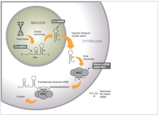
Figure 1- Biogenesis of micro-RNA ………...1
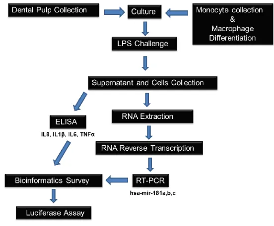
Figure 2- Flow diagram of the methods employed in the study ………...10
Figure 3 – Human dental pulp tissue and fibroblasts in culture ………11
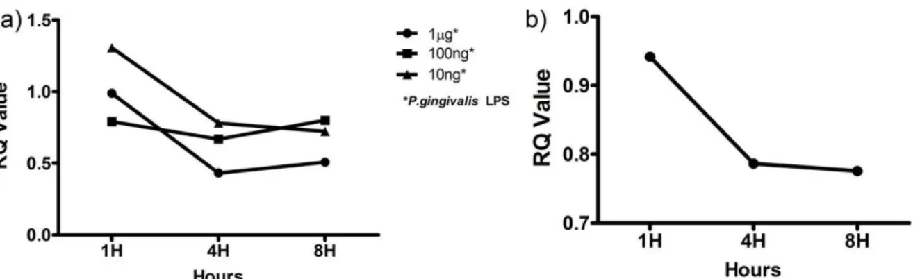
Figure 4 - Mir-181a and -181-b expression upon LPS challenge………..14
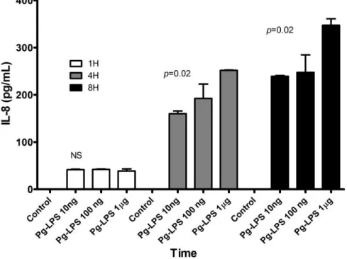
Figure 5 - Interleukin-8 response to LPS stimulation ………...15
Figure 6 - Relationship between level of IL-8 protein and hsa-mir181a and -181b ……….16
Figure 7 – Complementarity between mir-181 family and IL8 and Luciferase assay …………..17
Figure 8 – IL-8 response on LPS challenge with or without mir-181 mimic transfection ……...18
1
INTRODUCTION AND REVIEW OF LITERATURE
Discovery, Biogenesis and Function of MicroRNAs
MicroRNAs (miRNA) were discovered by a group of developmental biologists at
Harvard University while studying the development of the nematode C. elegans. The report
published by this group centered on a short sequence of 22 nucleotides, thus the term ‘micro’,
which complemented with lin-14 messenger RNA (mRNA) (Lee et al., 1993). As a consequence
of this pairing, the translation of lin-14 mRNA to LIN-14 protein that was essential to the
nematode’s development was impaired. This discovery led to hundreds of subsequent discoveries
that catapulted miRNAs to the vertex of gene regulation research. At present, over 1000 miRNAs
have been identified in humans, which potentially target over 30% of the human genome
(Griffiths-Jones et al., 2008).
The biogenesis of miRNAs involves several steps that start in the cell nucleus and
terminate in the cytoplasm by a coordinated process of cropping and dicing (Figure 1) (Kim,
2005). The step begins by transcription of intergenic or intragenic DNA sequences by RNA
polymerase II that forms a one to three kilobase (kb) length sequence known as primary miRNA
(Cai et al., 2004; Lee et al., 2004). Next, the enzyme Drosha and the RNA-binding protein Pasha
(also known as DiGeorge syndrome critical region 8) cleave the primary miRNA into a shorter
sequence of 70-100 nucleotide stem-loop structure called pre-miRNA (Denli et al., 2004;
Gregory et al., 2004; Han et al., 2004; Landthaler et al., 2004).
Exportin-5 transports pre-mirRNA to the cytoplasm (Bohnsack et al., 2004; Lund et al.,
2
oligonucleotide by an enzyme named Dicer (Bernstein et al., 2001; Hutvagner et al., 2001). The
double strand then separates into two single strands. One of the strands transforms into a mature
miRNA molecule incorporated into RNA induced silencing complex or RISC. This complex can
complement with mRNA, inhibiting protein translation (Knight and Bass, 2001).
Figure 1. Biogenesis of micro-RNA (source: Life Technologies).
The miRNA-mRNA interaction depends on the degree of complementarity between their
sequences. If there is a close to one hundred percent complementarity between miRNA and
mRNA, mRNA can be cleaved and degraded; otherwise, mRNA translation is just repressed
(Kim, 2005). Some miRNAs are produced at unusually high or low concentrations and
consequently may bind more or less extensively to their mRNA targets. This dysregulation has
been implicated in tumorigenesis where a disturbance in protein levels, identified as tumor
3
The degree of miRNA activity can also be altered if the concentration of the target mRNA
changes during a physiological event like differentiation or development, or as the result of
changes in the surrounding environment (Arvey et al., 2010). This fascinating area of
miRNA-mRNA interaction has spurred studies on how disturbances in this interaction may be associated
with pathological conditions like cancer (Genovesi et al., 2011; Kim et al., 2013; Li et al., 2011)
and hepatitis (Janssen et al., 2013). The role of miRNAs in the regulation of gene expression
makes them the key players in almost every biological function, both physiological and
pathological (O'Connell et al., 2010).
micro-RNAs and the Immune Response
Toll-like receptors (TLR) are a class of proteins that recognize conserved pathogen
structures, trigger innate immune responses and prime antigen-specific adaptive immunity
(Eskan et al., 2008; Hajishengallis et al., 2006; Hirao et al., 2009; Kawai and Akira, 2010;
Medzhitov et al., 1997). TLRs are a group of proteins expressed in the transmembrane region or
in the cytoplasm of cells and recognize specific microbial patterns (Kopp and Medzhitov, 1999).
TLR4 for example recognizes bacterial lipopolysaccharide (LPS) (Medzhitov et al., 1997). Upon
microbial stimulation, TLR4 sends a cascade of downstream signals that trigger the cell to
produce cytokines and /or chemokines that initiate the inflammatory process (Shimazu et al.,
1999).
MiRNAs have recently emerged as an important post-transcriptional regulators of
inflammation, metabolism and healing (van Rooij et al., 2012). As key regulators of
4
TLR4 pathway activation (Xie et al., 2013b) and in various TLR-mediated immune responses to
bacterial infection where miRNAs either suppressed the inflammatory response or reduced
inflammatory triggers (Case et al., 2011; Liu et al., 2009).
Our recent microarray report showed differential expression of miRNAs in human dental
pulp tissues that were clinically diagnosed to have pulpal inflammation or pulpitis (Zhong et al.,
2012), a relatively common and painful dental disease that represents an immune response to
bacterial infection (Love and Jenkinson, 2002). Pulpitis and its sequela, periapical periodontitis
have been directly linked to the pathogenicity of the oral microflora (Adachi et al., 2007; Huang
et al., 1999; Kakehashi et al., 1966; Love and Jenkinson, 2002; Siqueira et al., 2009). With the
preceding information at hand, it elicits an interest to investigate the potential association
between pulpal infection and changes in miRNA levels that may translate directly on how
inflammatory triggers are stimulated or repressed.
mir-181 Family and their Role in Immune Response
The multiple validated gene targets of the mir-181 family (Table 1) underscore their role
in regulating immune response and inflammation. A two to five fold increase in mir-181b and c
expression was reported in inflamed periodontal tissues compared with healthy gingival tissues
(Xie et al., 2011). In another similar study, mir-181b was found to be four times overexpressed in
chronic periodontitis tissues compared with normal periodontal tissues (Lee et al., 2011). This
increase in expression in diseased periodontal tissues seems to provide a link between infection
5 Table 1. MiRNA-181 validated target genes
Like pulpitis, periodontitis is a microbial disease that is directly correlated with the
preponderance of a group of gram negative anaerobes called the red complex species
(Ximenez-Fyvie et al., 2000). The root canal system, like the deep pockets in periodontitis, is an anaerobic
environment. In fact, there is an overlap in the identified species between periodontitis that
miRNA Target gene Gene product function
GO Term (Accession, Ontology)
mir-181a DUSP5/6
Regulation of T Cell Receptor (TCR)-signalling and activation threshold; alteration of T helper (Th) subset differentiation
GO:0070373, Biological Process
IL-6
Acute and chronic inflammation and the maturation of
B cells; T helper (Th)17 differentiation GO:0019981, Molecular Function
STAT1 Cytokine-mediated signaling pathway; LPS-mediated signaling pathway
TGFB1
Adaptive immune response; positive regulation of collagen biosynthesis, chemotaxis, fibroblasts migration, and odontogenesis
GO:0034713, Molecular Function
TLR-4
Toll-like receptor-4 (TLR) plays a fundamental role in
pathogen recognition and activation of innate immunity GO:0035662, Molecular Function
mir-181b CCL8 Immune response, inflammatory response, chemokine activity, phospholipase activator
IL-6
Negative regulation of cytokine secretion; negative regulation of collagen biosynthetic process; positive regulation of acute inflammatory response; response to cold, heat and mechanical stimuli; T Helper (Th)17 differentiation
GO:0005138, Molecular Function
MMP9
Cell response to IL-1, LPS; macrophage differentiation; response to heat and mechanical stimuli; positive regulation of apoptosis and angiogenesis
GO:0004229, Molecular Function
TGFB1
Adaptive immune response; positive regulation of collagen biosynthesis, chemotaxis, fibroblasts migration, and ondontogenesis; T helper (Th)17 differentiation, T regulatory (Treg) differentiation
GO:0034713, Molecular Function
mir-181c IL-2 Cytokine produced by T-cells in response to antigen or mitogen stimulation GO:0005134, Molecular Function
SOCS1 Cytokine mediated signaling pathway; negative regulator of JAK-STAT pathway; LPS response
mir-181d
MMP9
Cell response to IL-1, LPS; macrophage differentiation; response to heat and mechanical stimuli; positive regulation of apoptosis and angiogenesis
6
progressed from gingivitis and apical periodontitis that resulted from pulpitis (Siqueira et al.,
2009).
The difference in mir-181 family members identified in the two periodontitis studies
above (Lee et al., 2011; Xie et al., 2011) can be attributed to subject variability in terms of
disease stage or severity at the time the periodontal samples were collected
In terms of pain, which remains a diagnostic dilemma in endodontics and beyond,
circulating levels of miR-181a and c are significantly lower in patients diagnosed with chronic
regional pain syndrome (Orlova et al., 2011). miR-181a has been demonstrated to be a key
regulator of AMPA-type glutamate receptors in neurons (Saba et al., 2012). miRNAs are known
to modulate the expression and function of neuronal ions channels (Carrillo et al., 2011;
Favereaux et al., 2011; Saba et al., 2012). These include the neuronal ion channel Cav1.2, a
calcium channel implicated in chronic pain and NaV1.8, a sodium channel implicated in
inflammatory pain and thermal hyperalgesia (Akopian et al., 1999; Favereaux et al., 2011; Laird
et al., 2002; Zhao et al., 2011).
Extending outside the area of endodontics, circulating levels of mir-181b was found to be
lower in patients with sepsis and in animal models of septic shock (Sun et al., 2012). In sepsis
patients, each increase of one unit of miR-181b in blood is associated with an approximately
50% decrease in the odds of having sepsis. Inan animal modelof sepsis, thesystemic
administration of miR181-b reduces leukocyte influx in the vascular endothelium and decreases
mortality. This response was attributed to the nuclear factor κβ (NF-κβ) targeting of mir-181b,
7 Cytokines and mir-181 in Inflammation
Among the cytokines produced during inflammation, Interleukin-8 (IL-8) has gained
significant attention due to its essential involvement in acute response to infection (Harada et al.,
1994). IL-8 is a potent chemoattractant that is increased in pulpitis (Huang et al., 1999) and in
gingival crevicular fluid collected from teeth with acute pulpitis (Karapanou et al., 2008). Its
essential involvement and causative role in acute inflammation by recruiting and activating
neutrophils have been firmly established (Gura, 1996; Harada et al., 1994).
In our study that compared normal and infected human dental pulps, we reported the
differential expression of 36 miRNAs (Zhong et al., 2012). In another in-vivo study examining
infected tissues surrounding the apices of teeth with infected root canal systems, 24 miRNAs
were differently expressed (Chan et al., In press). The mir-181 family, which regulates a wide
range of gene targets, was differentially expressed in both of these tissues. They have also been
recently implicated in TLR-induced in-vivo increase in cytokine levels (Xie et al., 2013b).
However, there have been no reports as yet on the role of these miRNAs in regulating the
immune response in primary human dental pulpal fibroblasts (HDPF).
Both IL-8 and mir-181 family have been associated with other inflammatory conditions
such as rheumatoid arthritis and neuroinflammation and inflammatory responses of various cell
types like astrocytes and macrophages (Hutchison et al., 2013; Seitz et al., 1992; Sun et al., 2012;
8 Immunocompetence of the Dental Pulp
The role of bacteria in pulpal disease is well established (Kakehashi et al., 1966). These
microorganisms invade the dentinal tubules of the susceptible tooth and eventually the dental
pulp through a carious lesion, which is the most common etiology for pulpal inflammation and
necrosis (Love and Jenkinson, 2002). As the carious lesion progresses deeper into the
pulp-dentin interface, the microflora shifts from a Gram-positive to a more Gram-negative, anaerobic
bacteria (Ozok et al., 2012).
Pulpal fibroblasts, the predominant resident cells of the dental pulp, express TLRs (Hirao
et al., 2009) that, upon microbial infection, stimulates pulp cells to produce cytokines and
chemokines (Adachi et al., 2007; Nagaoka et al., 1996; Park et al., 2004; Tokuda et al., 2001).
Other cells types like endothelial cells in the dental pulp’s blood vessels and the resident
macrophages also express TLRs (Gallego et al., 2011; Hijiya et al., 2002). The dental pulp,
therefore, is an immunocompetent tissue.
Inflammation of the pulp and periapical tissues is commonly associated with pain, and
approximately 90% of dental emergency visits with pain as the chief complaint are attributable to
activation of pulpal or periapical nociceptors (Hasselgren and Calev, 1994). The prevalence of
periapical disease in the United States is estimated to be about 4.1% (Buckley and Spangberg,
1995). Despite the prevalence of endodontic disease and the great discomfort associated with it,
the fundamental molecular aspects of its pathogenesis are still not fully understood. The current
endodontic literature on pulpal immune response to microbial infection continues to expand;
9
PURPOSE
The purpose of this study is to determine how pathogens modulate the expression of
mir-181 family and to investigate this expression with the production of IL-8.
SPECIFIC AIMS
1) To characterize the expression of mir-181 family in primary HDPF;
2) To investigate if their expression level is influenced by TLR agonists;
3)To establish the regulatory mechanism of mir-181 with IL-8 in HDPF;
4) To determine if this regulatory mechanism is similar in macrophages, a resident immune cell
in the dental pulp.
HYPOTHESIS
Mir-181 expression is influenced by bacterial stimulation that translates into modulation
of IL-8 production in HDPF.
MATERIALS AND METHODS
10
Figure 2. Flow diagram of the methods employed in this study.
Dental Pulp Tissue Collection and Culture
The study was performed with the approval of and compliance to the guidelines set by
the Institutional Regulatory Board of the UNC Office of Human Research Ethics. Dental pulp
tissue from a clinically normal tooth of three healthy donors was extirpated immediately after
extraction for orthodontic reason (Figure 3a). The pulp tissue was obtained under informed
consent.
HDPF cultures were established using a previously published protocol by Adachi et al
(Adachi et al., 2007). In brief, minced pieces of pulp tissues were explanted into 35-mm culture
11
USA), supplemented with 10% fetal bovine serum (FBS) (Sigma, St. Louis, MO, USA), 1 mM
sodium pyruvate (Gibco), and 50 IU/mL penicillin/50 3g/mL streptomycin (Gibco) at 37°C in a
humidified atmosphere of 5% CO2. Confluent primary cultures were harvested and subcultured.
Morphologically fibroblastic cells obtained by this method were used as HDPF for experiments
at passages 4 to 6 (Figure 3b).
HDPF Stimulation
HDPFs were seeded in 6-well culture plates and incubated until a confluent monolayer of
approximately 106 cells was established. The cells were then challenged with 1μg, 100ng or 10
ng of Porphyromonas gingivalis (P. gingivalis) W83 LPS (Pg-LPS) (Dr. David A. Scott,
University of Louisville, KY, USA) at 1, 4 and 8 hours. Culture supernatants were collected and
stored at -80 oC until use. Adherent HDPF were lysed for RNA extraction. Among the bacteria
implicated in endodontic disease, P. gingivalis was utilized in this study because its presence in
infected root canals has been repeatedly documented and its characteristics fit the nature of a root
canal environment (Dougherty et al., 1998; Gomes et al., 1996; Kurihara et al., 1995)
Figure 3. Human dental pulp tissue and fibroblasts in culture .a) Dental pulp extirpated from a donor patient; b, c) Cultured primary HDPF at different confluence (x10).
12 Quantitative Real-time PCR
Total RNA was extracted from LPS-challenged HDPF and assessed for mir-181 a, b and
c by real-time PCR using Taq-Man primers and probe sets (Applied Biosystems, Foster City, CA,
USA). Normalization was performed using RNU-6B primer (Ucar et al., 2012). Relative
quantification (RQ) values were analyzed using Excel spreadsheet from Microsoft (Redmond,
WA). The experiments were carried out in three independent experimental set-ups.
ELISA
IL-1β, IL-6, IL-8 and TNF-α were measured by enzyme-linked immunosorbent assay
(ELISA) using commercially available kits (R&D Systems, Minneapolis, MN, USA) according
to the manufacturer’s instructions. The absorbance was read at 450 nm.
Luciferase Assay
IL8 3’UTR (~1.38 kb) was amplified by primers (Forward- GCACTCGAG
TGTGTGGGTCTGT TGTAGGG; Reverse – ATGCGGCCGCTGACTGTGGAGTTTTGGCTG
T) using pGL3-IL8 construct (kind gift of Dr. Richard Pestell Jefferson Medical College,
Philadelphia, PA) as template. The amplified fragment (~950 bp) was digested by XhoI and NotI
and cloned into psiCHEKTM2 vector (Promega). The transfection was performed as described
previously (Naqvi et al., 2013). Briefly, actively growing Human Embryonic Kidney 293 cells
(HEK cells) were seeded in 96-well plate at a density of 2 × 10⁴ cells per well in complete
DMEM. The next day, cells were co-transfected using Lipofectamine 2000 (Invitrogen) with 80
ng of IL8 construct and different concentration (2pmol and 5pmol) of miR-181a-5p mimics
13
BioSystems, Sunnyvale, CA, USA). The fold change in luciferase activity was calculated as
previously described (Naqvi et al., 2013).
MiRNA-181 Transfection in HDPF and Macrophages
Transfections with mir-181a were performed as described previously (Naqvi et al., 2013).
HDPFs and differentiated macrophages were transfected with mir-181a mimic (5'AACAUUCA
ACGCUGUCGGUGAGU) or inhibitor (5' AACAUUCAACGCUGUCGGUGAGU) (Qiagen)
using HiPerfect reagent (Qiagen) according to manufacturer’s instructions. Briefly, 1x10⁶ cells
were seeded in six-well plates and transfected with miRNA mimics or inhibitor. 50nM was used
for HDPFs while 50nM and 200nM were used for macrophages. Scrambled miRNAs were used
as controls. After 36 hours of incubation with the mimics or antagomirs, LPS was added to
HDPFs and was incubated for another one, four and eight hours before the supernatants were
collected. Macrophages supernatants were collected at two and 20 hours post-incubation.
Protein detection using ELISA was performed. Transfection efficiency of miRNAs was
determined bySiGLO Green and SiGLO Red siRNA (Thermo Scientific) that served as
surrogates for mir-181a. A transfection efficiency of over 80% was observed by dividing the
number of fluorescent cells with total number of cells.
Statistical Analysis
One-way ANOVA with Tukey’s post-hoc analysis was employed where statistical
difference is noted in the study using Graph Pad (La Jolla, CA, USA). Relative quantification
14
RESULTS
Expression of Mir-181-a, -b and IL-8 in HDPF is Influenced by P. gingivalis LPS
To examine if the TLR response affect the expression of miR-181 family in HPDF, cells
were challenged with Pg LPS. Figure 4a shows that mir-181a expression in HDPF is influenced
by P. gingivalis in a time- and dose-dependent manner. Compared with the one-hour time point,
the expression of mir-181a decreased at 4-hours. At the 8-hour time point, the expression level
either remained relatively similar (10 ng ml-1 LPS) or increased slightly (100 ng ml-1 and 1 μg
ml-1 LPS). This result is not surprising as a time- and dose-dependent stimulation by P.
gingivalis LPS has been shown in other cell types (Herath et al., 2013; Tabeta et al., 2000).
Mir-181b expression was only noted upon challenge with 1μg ml-1P. gingivalis LPS (Figure 4b)
while mir-181 c was not detected in this study. 1μg ml-1of P. gingivalis LPS was used in the
subsequent experiments as this dose had been widely used in other studies (Hajishengallis et al.,
2006) and had shown the most consistent result in our experiments.
15
Induction of pro-inflammatory cytokines is a key feature of TLR signaling. We
monitored the levels of IL-1β, IL-6, IL-8 and TNF-α in the supernatants of LPS stimulated
HPDF. Among the cytokines assayed in this study, only IL-8 was detectable in the cell culture
supernatant (Figure 5). IL-1β, IL-6 and TNF-α were all below minimum detectable dose (7.5
pg/mL); therefore, IL-8 was chosen to be further investigated in this study.
Interestingly, IL-8 was secreted in a time- and dose-dependent manner compared with
LPS-free controls, which was below the minimum detectable dose of the assay.
16
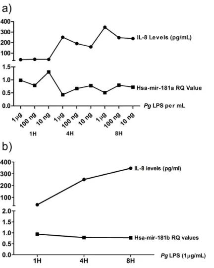
IL-8 and mir-181a and b Show an Inversely Proportional Relationship
From the dose and time dependent data we observed an inversely proportional
relationship between mir-181a gene expression and IL-8 protein level (Figure 6a). The same
relationship was observed between IL-8 and mir-181b (Figure 6b).This relationship suggests a
possible modulatory role of mir-181a and miR-181b on IL-8 expression.
17 IL-8 is Modulated by mir-181a
To investigate the possible mechanistic role of miR-181mediated regulation of IL8, we
scanned the 3’UTR for miR-181 binding site. Bioinformatics analysis identified a novel miRNA
binding region spanning 346-368 nts of IL8 3’UTR, as shown in Figure 7a. Importantly, the seed
sequence is conserved in all the four miRNAs of miR-181 family. To validate functional
miRNA-target interaction, dual luciferase assays were performed. HEK293 cells transfected with
miR-181a show reduced luciferase activity compared with the control miRNA mimic (Figure 7b).
Increasing the miR-181amimic concentration to 5 pmol had no further significant impact on the
luciferase activity (Figure 7b). Thus, miR-181a directly regulates IL8 levels by interacting with
the 3’UTR.
*
a b
Figure 7.a) Complementarity between IL-8 and mir-181 family sequences. mirSVR score: -0.0308; PhastCons 0.5158. b) dual luciferase assay demonstrating the downregulation of IL-8 by mir-181a. *P<0.05 compared with control mimic using one-way ANOVA.
18 Mir-181a Directly Binds to 3’UTR of IL-8
The luciferase assay results indicated that mir-181a directly binds to the 3’ UTR of IL-8,
thus capable of negatively regulating the activity of IL-8. In this experiment, we examined if
overexpression of mir-181a would have a negative impact on IL-8 expression. Mir-181a mimics
were transfected in both HDPFs and macrophages. Mir-181a antagonists were also transfected to
determine if suppressing this miRNA’s activity would increase the IL-8 protein levels upon LPS
stimulation. As shown in the figure below (Figure 8), LPS challenge resulted in significantly
increased IL-8 secretion in HDPFs. Interestingly, while the miRNA mimics significantly
increased IL-8 upon LPS stimulation, the antagomirs significantly reduced IL-8 levels. This
result is counterintuitive with the luciferase assay results. However, this is not surprising as a
previous study has shown similar differences in luciferase and miRNA overexpression
experiments (Tserel et al., 2011).
19
The results obtained from HDPFs above were also replicated in macrophages below
(Figure 9), albeit without LPS challenge, indicating that overexpression of mir-181a in cells,
independent of their type, function as a positive regulator of IL-8 while antagonizing this
miRNA down regulates IL-8.
At two hours, no changes were detected in both doses of the mimics and the antagomirs;
however, significant differences were observed at 20 hours, with the higher doses elucidating a
higher IL-8 response. Control mimics – the mock controls – did not show any difference
compared with the test samples. Modest IL-8 levels were obtained in this experiment as the
macrophages were not challenged with LPS, thus no TLR stimulation ensued to trigger the
release of cytokines.
20
DISCUSSION
Mir-181 is a critical micro-RNA that was largely thought to regulate lymphocyte
development and homeostasis, among other important functions (Henao-Mejia et al., 2013).
However, recent studies have also revealed its role in immunoregulation (Guo et al., 2013; Xie et
al., 2013b). In this study, we have shown that mir-181 family is also expressed in non-leukocytic
cells and that expression of both interleukin-8 and mir-181a and b are influenced by a TLR
agonist in a time- and dose-dependent manner. Moreover, the inversely proportional relationship
between mir-181a and IL-8 suggests that miR-181a directly binds to the 3'UTR of IL-8, an
important inflammatory component of the pulpal immune response, and modulates its levels.
The findings above are important additions to our current understanding on the regulatory
network governing the response of various cell types to microbial insults. Fibroblasts are
primarily structural in function but their ability to respond to infection as first or second line of
defense (after epithelial cells) is critical in limiting the spread of infection and in wound healing
(McDougall et al., 2006).The presence of TLRs in HDPF confers immunocompetency on these
cells (Hirao et al., 2009), giving merit to the importance of studying how their response is
regulated. As part of the initial responders to microbial entry during the carious process, the
capability of HDPF to recruit professional immune cells to the site of infection can be a
double-edged sword that may halt or propagate the infectious process (Gura, 1996).
The polymicrobial etiology of pulpitis and the clinical implications associated with it
make this disease an ideal model of immunoregulation. As a more gram negative, anaerobic
21
the conserved microbial patters through toll-like receptors (Hirao et al., 2009). Consequently,
various inflammatory mediators are expressed that initiate and enhance the inflammatory process.
We mimicked this response by utilizing an in-vitro model using HDPF stimulated with Pg-LPS.
Our results agree with previous studies on the immunocompetence of pulpal fibroblasts (Adachi
et al., 2007; Hirao et al., 2009; Nagaoka et al., 1996; Tokuda et al., 2001).
In this study, the detectable levels of IL-8 in Pg-LPS stimulated cells may recruit
circulating immune cells (e.g. neutrophils) to the site of infection as shown in-vivo by Izumi et al
(Izumi et al., 1995) and Olgart et al (Olgart et al., 1974). In their study, bacteria induced
inflammatory changes in the dental pulp characterized by infiltration of immune cells, activation
of the complement system due to the development of a local immune reaction, and the
accumulation of arachidonic acid metabolism with the destruction of cellular components.
The conflicting data obtained in miRNA overexpression in both HDPFs (Figure 8) and
macrophages (Figure 9) in comparison with the luciferase assay results (Figure 7b) create an area
for further investigation. This difference is interesting because there are multiple predicted or
validated mRNA targets for a single miRNA that could have a wide-ranging effect on the overall
immune response.
Mir-181 family for example does not only complement to IL-8 as shown in this study, but
it also targets other mRNAs whose protein products are essential for the downstream signaling of
IL-8 such as TLR-4, STAT1 and SOCS1 (Table 1). Therefore, it is quite unexpected that
overexpressing mir-181 to block IL-8 mRNA resulted in positive regulation of IL-8 and
22
the switching of miRNA from repression to activation resulting in mRNA translational
up-regulation instead of down-up-regulation in cells under cell cycle arrest (Vasudevan et al., 2007;
2008). With the in-vitro model that we have employed in this study, transfection with miRNA
mimics and antagonists may have caused the HDPFs and the macrophages to undergo cell cycle
arrest as shown in another study (Tserel et al., 2011). Therefore, depending on the cellular
environment, mir-181 can either up- or down-regulate the target gene expression. At this point
however, this extrapolation is purely speculative. Additional functional studies are definitely
encouraged to further investigate the applicability of mir-181 mimics to down-regulate
inflammatory response.
We confirmed for the very first time the regulation of IL-8 by mir-181a, a micro-RNA
that has been shown to be differentially expressed in inflamed pulp (Zhong et al., 2012).
Micro-RNAs posttranscriptionally regulate gene expression by targeting specific mMicro-RNAs for
degradation or translational repression (Pauley and Chan, 2008).Their cytoplasmic levels
directly influence the protein bioavailability of their targeted genes. We have shown in this study
that the level of mir-181a is inversely proportional with secreted IL-8 level (Figure 5). We
confirmed the repression of IL-8 by mir-181a using Luciferase assay (Figure 6b) and in-silico
alignment analysis (Figure 6a). This pattern of negative regulation by micro-RNAs has been
shown in other studies (Perry et al., 2008). Paradoxically, overexpression of mir-181a by
transfection of mimics into HDPF and macrophages resulted in a significant increase of IL-8 and
23
CONCLUSION AND FUTURE DIRECTIONS
Developing a treatment modality that can be used to directly dampen inflammation and
promote healing in various endodontic procedures like direct pulp capping, apical microsurgery,
root resorption treatment, post-trauma management, apexification and apexogenesis sounds very
encouraging. The results of this study can be, in theory, used to develop an miRNA based
anti-inflammatory therapy not only in endodontics but in other anti-inflammatory conditions as well
where regulation of inflammatory markers by miRNA were reported (Case et al., 2011; Guo et
al., 2013; Henao-Mejia et al., 2013; Hutchison et al., 2013; Sun et al., 2012; Ucar et al., 2012).
With the understanding that IL-8, a potent pro-inflammatory cytokine, can be directly
regulated with a micro-RNA, a targeted therapy to control inflammation is imminent in the near
future. This miRNA based therapeutics is already in large scale clinical trials in other fields
(Janssen et al., 2013).
Another advancing field in miRNA research is miRNA diagnostics. At present, novel
miRNA-based prognostic and diagnostic tools are being explored for cancers with poorer
prognosis (Larne et al., 2013; Stenvang et al., 2008; Szafranska-Schwarzbach et al., 2011). As
the field of miRNA research expands, it is predicted that the utility of these short sequenced
nucleotide will have longer and broader applications in a myriad of conditions (van Rooij et al.,
2012).
A critical barrier to progress in treatment of pulpitis is the incomplete understanding of
the regulatory network governing this disease. This study provides evidence that the dental pulp
24
microbial insults and modulating inflammatory response. Furthermore, the pathogenesis of
pulpitis and its immunoregulation can serve as a model for other microbial diseases that provide
less opportunity for ex-vivo or in-vitro studies due to their inaccessibility or critical location like
25
REFERENCES
Adachi T, Nakanishi T, Yumoto H, Hirao K, Takahashi K, Mukai K et al. (2007). Caries-related bacteria and cytokines induce CXCL10 in dental pulp. Journal of dental research 86(12): 1217-1222.
Akopian AN, Souslova V, England S, Okuse K, Ogata N, Ure J et al. (1999). The tetrodotoxin-resistant sodium channel SNS has a specialized function in pain pathways. Nature neuroscience
2(6):541-548.
Arvey A, Larsson E, Sander C, Leslie CS, Marks DS (2010). Target mRNA abundance dilutes microRNA and siRNA activity. Molecular systems biology 6(363.
Bernstein E, Caudy AA, Hammond SM, Hannon GJ (2001). Role for a bidentate ribonuclease in the initiation step of RNA interference. Nature 409(6818):363-366.
Bohnsack MT, Czaplinski K, Gorlich D (2004). Exportin 5 is a RanGTP-dependent dsRNA-binding protein that mediates nuclear export of pre-miRNAs. RNA 10(2):185-191.
Buckley M, Spangberg LS (1995). The prevalence and technical quality of endodontic treatment in an American subpopulation. Oral surgery, oral medicine, oral pathology, oral radiology, and endodontics 79(1):92-100.
Cai X, Hagedorn CH, Cullen BR (2004). Human microRNAs are processed from capped, polyadenylated transcripts that can also function as mRNAs. RNA 10(12):1957-1966.
Carrillo ED, Escobar Y, Gonzalez G, Hernandez A, Galindo JM, Garcia MC et al. (2011). Posttranscriptional regulation of the beta2-subunit of cardiac L-type Ca2+ channels by MicroRNAs during long-term exposure to isoproterenol in rats. Journal of cardiovascular pharmacology 58(5):470-478.
Case SR, Martin RJ, Jiang D, Minor MN, Chu HW (2011). MicroRNA-21 inhibits toll-like receptor 2 agonist-induced lung inflammation in mice. Experimental lung research 37(8): 500-508.
Chan LT, Zhong S, Naqvi AR, Self-Fordham J, Nares S, Bair E et al. (In press). MicroRNAs: New Insights into the Pathogenesis of Endodontic Periapical Disease. Journal of endodontics.
Denli AM, Tops BB, Plasterk RH, Ketting RF, Hannon GJ (2004). Processing of primary microRNAs by the Microprocessor complex. Nature 432(7014):231-235.
26
Eskan MA, Benakanakere MR, Rose BG, Zhang P, Zhao J, Stathopoulou P et al. (2008). Interleukin-1beta modulates proinflammatory cytokine production in human epithelial cells.
Infection and immunity 76(5):2080-2089.
Favereaux A, Thoumine O, Bouali-Benazzouz R, Roques V, Papon MA, Salam SA et al. (2011). Bidirectional integrative regulation of Cav1.2 calcium channel by microRNA miR-103: role in pain. The EMBO journal 30(18):3830-3841.
Gallego C, Golenbock D, Gomez MA, Saravia NG (2011). Toll-like receptors participate in macrophage activation and intracellular control of Leishmania (Viannia) panamensis. Infection and immunity 79(7):2871-2879.
Genovesi LA, Carter KW, Gottardo NG, Giles KM, Dallas PB (2011). Integrated analysis of miRNA and mRNA expression in childhood medulloblastoma compared with neural stem cells.
PloS one 6(9):e23935.
Gomes BP, Lilley JD, Drucker DB (1996). Clinical significance of dental root canal microflora.
J Dent 24(1-2):47-55.
Gregory RI, Yan KP, Amuthan G, Chendrimada T, Doratotaj B, Cooch N et al. (2004). The Microprocessor complex mediates the genesis of microRNAs. Nature 432(7014):235-240.
Griffiths-Jones S, Saini HK, van Dongen S, Enright AJ (2008). miRBase: tools for microRNA genomics. Nucleic acids research 36(Database issue):D154-158.
Guo XK, Zhang Q, Gao L, Li N, Chen XX, Feng WH (2013). Increasing expression of microRNA 181 inhibits porcine reproductive and respiratory syndrome virus replication and has implications for controlling virus infection. Journal of virology 87(2):1159-1171.
Gura T (1996). Chemokines take center stage in inflammatory ills. Science 272(5264):954-956.
Hajishengallis G, Tapping RI, Harokopakis E, Nishiyama S, Ratti P, Schifferle RE et al. (2006). Differential interactions of fimbriae and lipopolysaccharide from Porphyromonas gingivalis with the Toll-like receptor 2-centred pattern recognition apparatus. Cellular microbiology 8(10): 1557-1570.
Han J, Lee Y, Yeom KH, Kim YK, Jin H, Kim VN (2004). The Drosha-DGCR8 complex in primary microRNA processing. Genes & development 18(24):3016-3027.
Harada A, Sekido N, Akahoshi T, Wada T, Mukaida N, Matsushima K (1994). Essential involvement of interleukin-8 (IL-8) in acute inflammation. Journal of leukocyte biology
56(5):559-564.
27
Henao-Mejia J, Williams A, Goff LA, Staron M, Licona-Limon P, Kaech SM et al. (2013). The MicroRNA miR-181 Is a Critical Cellular Metabolic Rheostat Essential for NKT Cell Ontogenesis and Lymphocyte Development and Homeostasis. Immunity 38(5):984-997.
Herath TD, Darveau RP, Seneviratne CJ, Wang CY, Wang Y, Jin L (2013). Tetra- and penta-acylated lipid A structures of Porphyromonas gingivalis LPS differentially activate TLR4-mediated NF-kappaB signal transduction cascade and immuno-inflammatory response in human gingival fibroblasts. PloS one 8(3):e58496.
Hijiya N, Miyake K, Akashi S, Matsuura K, Higuchi Y, Yamamoto S (2002). Possible involvement of toll-like receptor 4 in endothelial cell activation of larger vessels in response to lipopolysaccharide. Pathobiology : journal of immunopathology, molecular and cellular biology
70(1):18-25.
Hirao K, Yumoto H, Takahashi K, Mukai K, Nakanishi T, Matsuo T (2009). Roles of TLR2, TLR4, NOD2, and NOD1 in pulp fibroblasts. Journal of dental research 88(8):762-767.
Huang GT, Potente AP, Kim JW, Chugal N, Zhang X (1999). Increased interleukin-8 expression in inflamed human dental pulps. Oral surgery, oral medicine, oral pathology, oral radiology, and endodontics 88(2):214-220.
Hutchison ER, Kawamoto EM, Taub DD, Lal A, Abdelmohsen K, Zhang Y et al. (2013). Evidence for miR-181 involvement in neuroinflammatory responses of astrocytes. Glia
61(7):1018-1028.
Hutvagner G, McLachlan J, Pasquinelli AE, Balint E, Tuschl T, Zamore PD (2001). A cellular function for the RNA-interference enzyme Dicer in the maturation of the let-7 small temporal RNA. Science 293(5531):834-838.
Izumi T, Kobayashi I, Okamura K, Sakai H (1995). Immunohistochemical study on the immunocompetent cells of the pulp in human non-carious and carious teeth. Archives of oral biology 40(7):609-614.
Janssen HL, Reesink HW, Lawitz EJ, Zeuzem S, Rodriguez-Torres M, Patel K et al. (2013). Treatment of HCV infection by targeting microRNA. The New England journal of medicine
368(18):1685-1694.
Kakehashi S, Stanley HR, Fitzgerald RJ (1966). The effects of surgical exposures of dental pulps in germfree and conventional laboratory rats. Journal - Southern California Dental Association
34(9):449-451.
28
Kawai T, Akira S (2010). The role of pattern-recognition receptors in innate immunity: update on Toll-like receptors. Nature immunology 11(5):373-384.
Kim SJ, Ha JW, Zhang BT (2013). Constructing higher-order miRNA-mRNA interaction networks in prostate cancer via hypergraph-based learning. BMC systems biology 7(47.
Kim VN (2005). MicroRNA biogenesis: coordinated cropping and dicing. Nature reviews Molecular cell biology 6(5):376-385.
Knight SW, Bass BL (2001). A role for the RNase III enzyme DCR-1 in RNA interference and germ line development in Caenorhabditis elegans. Science 293(5538):2269-2271.
Kopp EB, Medzhitov R (1999). The Toll-receptor family and control of innate immunity.
Current opinion in immunology 11(1):13-18.
Kurihara H, Kobayashi Y, Francisco IA, Isoshima O, Nagai A, Murayama Y (1995). A microbiological and immunological study of endodontic-periodontic lesions. J Endod
21(12):617-621.
Laird JM, Souslova V, Wood JN, Cervero F (2002). Deficits in visceral pain and referred hyperalgesia in Nav1.8 (SNS/PN3)-null mice. The Journal of neuroscience : the official journal of the Society for Neuroscience 22(19):8352-8356.
Landthaler M, Yalcin A, Tuschl T (2004). The human DiGeorge syndrome critical region gene 8 and Its D. melanogaster homolog are required for miRNA biogenesis. Current biology : CB
14(23):2162-2167.
Larne O, Martens-Uzunova E, Hagman Z, Edsjo A, Lippolis G, den Berg MS et al. (2013). miQ--a novel microRNA based diagnostic and prognostic tool for prostate cancer. International journal of cancer Journal international du cancer 132(12):2867-2875.
Lee RC, Feinbaum RL, Ambros V (1993). The C. elegans heterochronic gene lin-4 encodes small RNAs with antisense complementarity to lin-14. Cell 75(5):843-854.
Lee Y, Kim M, Han J, Yeom KH, Lee S, Baek SH et al. (2004). MicroRNA genes are transcribed by RNA polymerase II. The EMBO journal 23(20):4051-4060.
Lee YH, Na HS, Jeong SY, Jeong SH, Park HR, Chung J (2011). Comparison of inflammatory microRNA expression in healthy and periodontitis tissues. Biocell : official journal of the Sociedades Latinoamericanas de Microscopia Electronica et al 35(2):43-49.
29
Liu G, Friggeri A, Yang Y, Park YJ, Tsuruta Y, Abraham E (2009). miR-147, a microRNA that is induced upon Toll-like receptor stimulation, regulates murine macrophage inflammatory responses. Proceedings of the National Academy of Sciences of the United States of America
106(37):15819-15824.
Love RM, Jenkinson HF (2002). Invasion of dentinal tubules by oral bacteria. Critical reviews in oral biology and medicine : an official publication of the American Association of Oral Biologists 13(2):171-183.
Lu J, Getz G, Miska EA, Alvarez-Saavedra E, Lamb J, Peck D et al. (2005). MicroRNA expression profiles classify human cancers. Nature 435(7043):834-838.
Lund E, Guttinger S, Calado A, Dahlberg JE, Kutay U (2004). Nuclear export of microRNA precursors. Science 303(5654):95-98.
McDougall S, Dallon J, Sherratt J, Maini P (2006). Fibroblast migration and collagen deposition during dermal wound healing: mathematical modelling and clinical implications. Philosophical transactions Series A, Mathematical, physical, and engineering sciences 364(1843):1385-1405.
Medzhitov R, Preston-Hurlburt P, Janeway CA, Jr. (1997). A human homologue of the Drosophila Toll protein signals activation of adaptive immunity. Nature 388(6640):394-397.
Nagaoka S, Tokuda M, Sakuta T, Taketoshi Y, Tamura M, Takada H et al. (1996). Interleukin-8 gene expression by human dental pulp fibroblast in cultures stimulated with Prevotella intermedia lipopolysaccharide. Journal of endodontics 22(1):9-12.
Naqvi AR, Fordham JB, Khan A, Nares S (2013). MicroRNAs responsive to Aggregatibacter actinomycetemcomitans and Porphyromonas gingivalis LPS modulate expression of genes regulating innate immunity in human macrophages. Innate immunity.
O'Connell RM, Rao DS, Chaudhuri AA, Baltimore D (2010). Physiological and pathological roles for microRNAs in the immune system. Nature reviews Immunology 10(2):111-122.
Olgart L, Brannstrom M, Johnson G (1974). Invasion of bacteria into dentinal tubules. Experiments in vivo and in vitro. Acta odontologica Scandinavica 32(1):61-70.
Orlova IA, Alexander GM, Qureshi RA, Sacan A, Graziano A, Barrett JE et al. (2011). MicroRNA modulation in complex regional pain syndrome. Journal of translational medicine
9(195.
30
Park SH, Hsiao GY, Huang GT (2004). Role of substance P and calcitonin gene-related peptide in the regulation of interleukin-8 and monocyte chemotactic protein-1 expression in human dental pulp. International endodontic journal 37(3):185-192.
Pauley KM, Chan EK (2008). MicroRNAs and their emerging roles in immunology. Annals of the New York Academy of Sciences 1143(226-239.
Perry MM, Moschos SA, Williams AE, Shepherd NJ, Larner-Svensson HM, Lindsay MA (2008). Rapid changes in microRNA-146a expression negatively regulate the IL-1beta-induced inflammatory response in human lung alveolar epithelial cells. J Immunol 180(8):5689-5698.
Saba R, Storchel PH, Aksoy-Aksel A, Kepura F, Lippi G, Plant TD et al. (2012). Dopamine-regulated microRNA MiR-181a controls GluA2 surface expression in hippocampal neurons.
Molecular and cellular biology 32(3):619-632.
Seitz M, Dewald B, Ceska M, Gerber N, Baggiolini M (1992). Interleukin-8 in inflammatory rheumatic diseases: synovial fluid levels, relation to rheumatoid factors, production by mononuclear cells, and effects of gold sodium thiomalate and methotrexate. Rheumatology international 12(4):159-164.
Shimazu R, Akashi S, Ogata H, Nagai Y, Fukudome K, Miyake K et al. (1999). MD-2, a molecule that confers lipopolysaccharide responsiveness on Toll-like receptor 4. The Journal of experimental medicine 189(11):1777-1782.
Siqueira JF, Jr., Rocas IN, Alves FR, Silva MG (2009). Bacteria in the apical root canal of teeth with primary apical periodontitis. Oral surgery, oral medicine, oral pathology, oral radiology, and endodontics 107(5):721-726.
Stenvang J, Silahtaroglu AN, Lindow M, Elmen J, Kauppinen S (2008). The utility of LNA in microRNA-based cancer diagnostics and therapeutics. Seminars in cancer biology 18(2):89-102.
Sun X, Icli B, Wara AK, Belkin N, He S, Kobzik L et al. (2012). MicroRNA-181b regulates NF-kappaB-mediated vascular inflammation. The Journal of clinical investigation 122(6):1973-1990.
Szafranska-Schwarzbach AE, Adai AT, Lee LS, Conwell DL, Andruss BF (2011). Development of a miRNA-based diagnostic assay for pancreatic ductal adenocarcinoma. Expert review of molecular diagnostics 11(3):249-257.
Tabeta K, Yamazaki K, Akashi S, Miyake K, Kumada H, Umemoto T et al. (2000). Toll-like receptors confer responsiveness to lipopolysaccharide from Porphyromonas gingivalis in human gingival fibroblasts. Infection and immunity 68(6):3731-3735.
31
Tserel L, Runnel T, Kisand K, Pihlap M, Bakhoff L, Kolde R et al. (2011). MicroRNA expression profiles of human blood monocyte-derived dendritic cells and macrophages reveal miR-511 as putative positive regulator of Toll-like receptor 4. The Journal of biological chemistry 286(30):26487-26495.
Ucar A, Gupta SK, Fiedler J, Erikci E, Kardasinski M, Batkai S et al. (2012). The miRNA-212/132 family regulates both cardiac hypertrophy and cardiomyocyte autophagy. Nature communications 3(1078.
van Rooij E, Purcell AL, Levin AA (2012). Developing microRNA therapeutics. Circulation research 110(3):496-507.
Vasudevan S, Tong Y, Steitz JA (2007). Switching from repression to activation: microRNAs can up-regulate translation. Science 318(5858):1931-1934.
Vasudevan S, Tong Y, Steitz JA (2008). Cell-cycle control of microRNA-mediated translation regulation. Cell Cycle 7(11):1545-1549.
Xie W, Li M, Xu N, Lv Q, Huang N, He J et al. (2013a). MiR-181a regulates inflammation responses in monocytes and macrophages. PloS one 8(3):e58639.
Xie W, Li Z, Li M, Xu N, Zhang Y (2013b). miR-181a and inflammation: miRNA homeostasis response to inflammatory stimuli in vivo. Biochemical and biophysical research communications
430(2):647-652.
Xie YF, Shu R, Jiang SY, Liu DL, Zhang XL (2011). Comparison of microRNA profiles of human periodontal diseased and healthy gingival tissues. International journal of oral science
3(3):125-134.
Ximenez-Fyvie LA, Haffajee AD, Socransky SS (2000). Comparison of the microbiota of supra- and subgingival plaque in health and periodontitis. Journal of clinical periodontology 27(9): 648-657.
Yi R, Qin Y, Macara IG, Cullen BR (2003). Exportin-5 mediates the nuclear export of pre-microRNAs and short hairpin RNAs. Genes & development 17(24):3011-3016.
Zhao C, Dreosti E, Lagnado L (2011). Homeostatic synaptic plasticity through changes in presynaptic calcium influx. The Journal of neuroscience : the official journal of the Society for Neuroscience 31(20):7492-7496.