
R E S E A R C H
Open Access
Does the sex ratio of singleton births after
frozen single blastocyst transfer differ in
relation to blastocyst development?
Hua Lou
1, Na Li
1, Xiaoke Zhang
1, Ling Sun
1, Xingling Wang
1, Dayong Hao
1and Shihong Cui
2*Abstract
Purpose:To investigate the associations between blastocyst development and the sex ratio (male:female) among singleton live births resulting from single-blastocyst frozen embryo transfer (FET) cycles.
Methods:Patients with singleton live births following the first autologous single FET of non- preimplantation genetic testing (PGT) blastocysts in a single reproductive medicine department between January 2015 and February 2019 were included in this retrospective study. The primary outcome measure was the singleton sex ratio.
Multivariable logistic regression models were used to estimate the associations between blastocyst quality and singleton sex ratio after adjustment for some potential confounders.
Results:There were 638 high-quality and 572 poor-quality single blastocyst FETs, and the blastocysts were conceived via 855 IVF and 355 ICSI treatments. A total of 1210 singleton live births were assessed. High-quality single blastocyst FET resulted in a significantly higher sex ratio than did poor-quality single blastocyst FET (60% vs. 49.7%,P< 0.001). The infertility cause was not associated with sex ratio among singleton live births (P= 0.537). The results of a multivariate analysis revealed that a high-quality blastocyst has a 150% higher probability of being male than a poor-quality blastocyst (adjusted odds ratio (aOR) 1.57; 95% CI 1.24–2,P <0.001). Among the three blastocyst morphological parameters, Grade B trophectoderm was significantly associated with a higher sex ratio than Grade C (aOR 1.71; 95% CI 1.33–2.21.P< 0.001). Neither expansion degree nor inner cell mass degree were significantly associated with the singleton sex ratio.
Conclusions:A single high-quality blastocyst FET has a higher chance of resulting in a male infant than a female infant. The results demonstrate that grade B trophectoderm confers benefits in improving the implantation potential of male blastocysts.
Keywords:Blastocyst, Frozen-thawed transfer, Inner cell mass, Trophectoderm, Sex ratio
© The Author(s). 2020Open AccessThis article is licensed under a Creative Commons Attribution 4.0 International License, which permits use, sharing, adaptation, distribution and reproduction in any medium or format, as long as you give appropriate credit to the original author(s) and the source, provide a link to the Creative Commons licence, and indicate if changes were made. The images or other third party material in this article are included in the article's Creative Commons licence, unless indicated otherwise in a credit line to the material. If material is not included in the article's Creative Commons licence and your intended use is not permitted by statutory regulation or exceeds the permitted use, you will need to obtain permission directly from the copyright holder. To view a copy of this licence, visithttp://creativecommons.org/licenses/by/4.0/. The Creative Commons Public Domain Dedication waiver (http://creativecommons.org/publicdomain/zero/1.0/) applies to the data made available in this article, unless otherwise stated in a credit line to the data.
* Correspondence:[email protected]
2Department of Obstetrics and Gynecology, the Third Affiliated Hospital of
Introduction
Ideally, human-assisted reproductive technology (ART) aims to achieve a single live birth. Following improvements in blastocyst culture systems and freez-ing technology [1, 2], single blastocyst transfer (SBT) is now considered the most effective means of avoid-ing multiple pregnancies subsequent to ART [3–6]. Gradually, SBT has become the preferred transfer strategy throughout the world [7–10]. However, blastocyst transfer has some potential limitations, in-cluding adverse effects such as a male-biased imbal-ance in the sex ratio and an increased incidence of monozygotic twinning (MZT) [11–13].
The sex ratio at birth is often calculated as the propor-tion of males among all live births or the number of male births per 100 female births [14]. Dean et al. re-ported a significantly higher sex ratio at birth among in-fants born after SBT (54.1%) than among those born after cleavage-stage transfer cycles (49.9%) [15]. Another study determined that the higher sex ratio was inde-pendent of the fertilization method [16]. However, the details of the relationship between the characteristics of blastocysts and the sex ratio at birth remain unclear.
Blastocyst grading prior to embryo transfer usually oc-curs on day 5 or day 6 of culture. A detailed blastocyst scoring system is required to identify the blastocysts with the highest implantation potential. Static morph-ology remains the most commonly used embryo selec-tion method worldwide. This system is used to grade blastocysts according to three different variables: blasto-coel expansion, inner cell mass and trophectoderm [17]. Several studies have attempted to identify the individual contributions of these parameters to the implantation potential or live birth rate. In particular, trophectoderm morphology may be predictive of pregnancy outcomes related to single blastocyst transfer [18].
Ebner et al. reported that the embryo sex ratio was skewed significantly in relation to morphology. Male blastocysts had a 2.53-fold higher odds of receiving a trophectoderm quality score of A than female blastocysts [19]. All previous related studies were based on morpho-logical parameters and embryo sex. To our knowledge, no previous study has explored the relationship between blastocyst development and the sex ratio of singleton live births after single-blastocyst frozen embryo transfer (FET) cycles. The objective of this study was to investi-gate this association and fill this gap in the literature.
Materials and methods
Study subjects
Patients with singleton live births following the first au-tologous single FET of non- preimplantation genetic testing (PGT) blastocysts at the Third Affiliated Hospital of Zhengzhou University between January 2015 and
February 2019 were included in this retrospective study. Only the first FET cycle of each patient was analyzed to avoid repeated measures bias. Patients who underwent PGT, used donor oocytes or donor sperm were ex-cluded. The study sample identification and collation process is illustrated in Fig.1. The data collection proto-col for this study was approved by our Institutional Re-view Board.
Ovarian stimulation protocol
Each female patient underwent a conventional ovarian hyperstimulation procedure involving a gonadotrophin-releasing hormone agonist or antagonist. The physician adjusted the starting dose according to the patient’s age, body mass index (BMI) and ovarian reserve. Ovarian fol-licle development was monitored based on serum estra-diol and transvaginal ultrasonographic measurements. Oocytes were retrieved transvaginally 36–38 h after human chorionic gonadotrophin (Serono, Aubonne, Switzerland) administration when at least 40% of the folli-cles had reached or exceeded an average diameter of 18 mm as determined by ultrasound. The follicles were aspi-rated using a single-lumen needle attached to a syringe under transvaginal ultrasound guidance. The oocytes were then inseminated via conventional in vitro fertilization (IVF) or intracytoplasmic sperm injection (ICSI).
Laboratory protocol
Embryos were placed into the incubator (K-MINC-1000, Cook, United States) and cultured at 6% CO2, 5% O2
and 37 °C. G-1™plus (Vitrolife, Sweden) was used to culture the embryos from the pronucleate stage to day 3. Morpho-logical evaluation of the cleavage-stage embryos was per-formed on day 2 and day 3 based on the number of blastomeres, rate of fragmentation, multinucleation of the blastomeres and early compaction [20]. The two best-quality cleavage embryos were chosen for fresh cycle transfer or cryopreservation on day 3, with surplus embryos being cul-tured to the blastocyst stage for possible cryopreservation.
Blastocyst morphology was evaluated before cryo-preservation according to the classification devised by Gardner and Schoolcraft [21] on day 5 or day 6 after in-semination. As a general rule, the inner cell mass quality should be at least B to optimize cryosurvival. High-quality blastocysts were defined as those with a blasto-coel grade > B3, inner cell mass grade A/B and troph-ectoderm grade A/B (AA, AB, BA, BB). Poor-quality blastocysts were defined as those with a blastocoel grade > B3, inner cell mass grade A/B and trophecto-derm grade C (AC, BC) [22].
Japan). The media employed were vitrification and warming kits. For vitrification, the blastocysts were first equilibrated in solution I [7.5% v/v ethylene glycol (EG) and 7.5% v/v dimethyl sulfoxide (DMSO)] at room temperature for 10 min and then placed into vitrification solution II (15% v/v EG, 15% v/v DMSO and 0.5 M su-crose) for 1 min. Subsequently, blastocysts were individu-ally loaded onto Cryotops in a volume of < 0.1μl and quickly plunged into liquid nitrogen. Warming was per-formed by placing the Cryotop in the thawing solution (1.0 M sucrose) for 1 min at 37 °C. Then, the blastocyst was moved to the dilution solution (0.5 M sucrose) for 3 min at room temperature, followed by two steps in wash-ing solution at room temperature for 5 min each. The blastocyte was then transferred into a 50-μL droplet of culture medium (G2; Vitrolife) under mineral oil.
Blasto-cysts were transferred 1–2 h postwarming.
Embryo transfer and clinical outcomes
FET was performed after preparation via hormone re-placement therapy (HRT) or during a natural cycle.
Vaginal or oral progesterone (Crinone, Merck Serono, Switzerland) was provided for luteal support. On day 6 of progesterone administration, a single vitrified blasto-cyst was selected for transfer based on morphology grad-ing. All transfer procedures were directed by ultrasound guidance as previously described [25]. Only single blas-tocysts were selected for transfer. No blastocyst transfer was performed if the endometrial thickness was < 7 mm. A clinical pregnancy was confirmed by the ultrasono-graphic visualization of an intrauterine gestational sac with fetal heart activity at 4 weeks after blastocyst trans-fer. In this study, the primary outcome measure was the singleton sex ratio. The sex ratio at birth was calculated as the proportion of males among all live births.
Statistical analysis
All data analyses were performed using the SPSS 25.0 statistical software package. Continuous data are pre-sented as the mean and standard deviation (SD). Differ-ences between two categorical variables were analyzed using the chi-square test depending on the data
distribution. A multivariate logistic regression analysis with adjustment for major covariates (age, body mass index, type of fertilization and day of embryo transfer) was used to assess whether the sex ratio of singleton live births was affected by various morphological parameters used for grading. The data are reported as adjusted odds ratios (ORs) and 95% confidence intervals (95% CIs). A
P value < 0.05 was set as the threshold of statistical significance.
Results
Demographics and basic characteristics
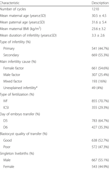
The final study dataset consisted of n= 1210 validated singleton live births. An analysis of the collected demo-graphic and clinical data revealed that there were 638 (52.7%) high-quality and 572(47.3%) poor-quality single blastocyst FETs, and blastocysts were conceived via 855 (70.7%) IVF and 355 (29.3%) ICSI treatments. The ana-lyzed cycles yielded 667 male infants and 543 female in-fants. The sex ratio was 55.1% (667/1210). The baseline characteristics of the included treatment cycles are de-scribed in Table1.
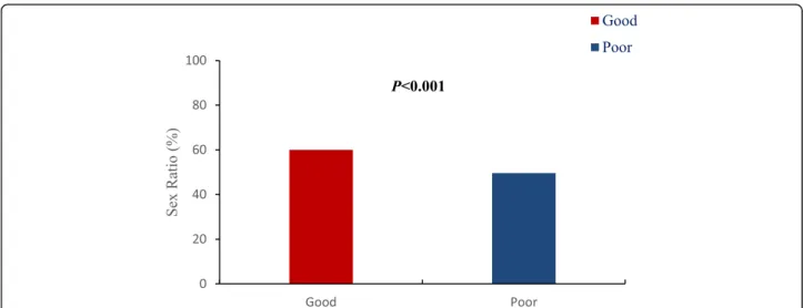
In the present study we checked the possible influence of patient characteristics and the embryo culture condi-tion on the sex ratio. We found that high-quality single blastocyst FET resulted in a significantly higher sex ratio than did poor-quality single blastocyst FET (60% vs. 49.7%, P< 0.001, Fig. 2.). Maternal age, maternal BMI, main infertility cause, type of fertilization or day of em-bryo transfer had no significant influence on the sex ra-tio as listed in Table2.
After adjustment for the potential confounder factors, we identified significant associations of the sex ratio among singleton births with the morphological charac-teristics of blastocysts. The results of a multivariate ana-lysis revealed that a high-quality blastocyst has a 150% higher probability of being male than a poor-quality blastocyst (aOR1.57; 95% CI 1.24–2,P <0.001). Particu-larly, when the trophectoderm was of Grade B, the blas-tocysts showed a 170% higher probability of being male than when the trophectoderm was of Grade C (aOR1.71; 95% CI 1.33–2.21, P <0.001). Neither expansion degree or inner cell mass degree were not significantly associ-ated with the singleton sex ratio (Table3).
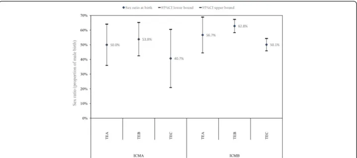
To further assess the effect of the trophectoderm grades on the sex ratio among singleton live births, we stratified the data according to inner cell mass grade, as shown in Fig. 3. In both the inner cell mass Grade A and B groups, Grade B trophectoderm showed the high-est sex ratio. In the inner cell mass Grade B group, when the trophectoderm was of Grade B, the blastocysts showed a 175% higher probability of being male than when the trophectoderm was of Grade C (odds ratio1.75; 95% CI1.34–2.29,P< 0.001).
Discussion
Our study results indicate that implantation with high-quality blastocysts, rather than poor-high-quality blastocysts, during single-blastocyst FET cycles is associated with an increased sex ratio among live births. We further deter-mined that only the trophectoderm grade was associated with a significantly higher sex ratio among singleton live births. Similar observations were not observed for the blastocoel expansion and inner cell mass grades. There-fore, our findings demonstrate that a grade B trophecto-derm is associated with a higher probability that a male infant will be born from a single-blastocyst FET cycle.
Single blastocyst transfer was designed to avoid the complications associated with multiple pregnancies and is thus becoming the preferred type of ART. The use of extended blastocyst culture conditions may favor the Table 1The characteristics of 1210 patients included the sex ratio among singleton births following the first single blastocyst frozen embryo transfer (FET) cycles performed from January 2015 to February 2019
Characteristic Description
Number of cycles 1210
Mean maternal age (years±SD) 30.5 ± 4.5
Mean paternal age (years±SD) 31.6 ± 5.4
Mean maternal BMI (kg/m2) 23.6 ± 3.2
Mean duration of infertility (years±SD) 3.3 ± 2.6
Type of infertility (%)
Primary 541 (44.7%)
Secondary 669 (55.3%)
Main infertility cause (%)
Female factor 661 (54.6%)
Male factor 307 (25.4%)
Mixed factor 193 (16%)
Unexplained infertility* 49 (4%)
Type of fertilization (%)
IVF 855 (70.7%)
ICSI 355 (29.3%)
Day of embryo transfer (%)
D5 783 (64.7%)
D6 427 (35.3%)
Blastocyst quality of transfer (%)
Good 638 (52.7%)
Poor 572 (47.3%)
Singleton livebirths (%)
Male 667 (55.1%)
Female 543 (44.9%)
Note: Data are presented as the mean ± SD and proportion (%). BMI = body mass index; D = day; IVF = in vitro fertilization; ICSI = intracytoplasmic sperm injection; *Unexplained infertility is rarely used as a diagnostic indicator for IVF in China because it is a controversial diagnosis
selection of male blastocysts for transfer, as male em-bryos are thought to exhibit greater preimplantation de-velopmental rates. In this study, the sex ratio among single-blastocyst FET cycles was 55.1%, which was con-sistent with the ratios reported in previous studies. For example, a large nationwide longitudinal birth cohort study of 103,099 pregnancies in Japan reported a skewed sex ratio in favor of males after blastocyst transfer rela-tive to spontaneous conception (OR: 1.095; 95% CI: 1.001–1.198). However, the sex ratios in the non-ART treatment and cleavage groups were slightly lower than or equivalent to the ratio in the spontaneous conception group [13]. A meta-analysis of 13 studies also suggested
Fig. 2Sex ratio among singleton births following single blastocyst frozen embryo transfer (FET) cycles with different blastocyst qualities
Table 2Sex ratio among singleton live births stratified by different demographic characteristics and the embryo culture condition
Characteristic Singletons (n) Male (n) Sex ratio(%) P-value
Blastocyst quality <0.001
High 638 383 60.0
Poor 572 284 49.7
Maternal age 0.261
<35 971 543 55.9
≥35 239 124 51.9
Maternal BMI 0.173
<18.5 47 32 68.1
18.5 ~ 24.9 772 425 55.1
≥25 391 210 53.7
Type of infertility 0.887
Primary 541 297 54.9
Secondary 669 370 55.3
Cause of infertility 0.537
Female factor 661 363 54.9
Male factor 307 178 58
Combined factor 193 102 52.8
Unexplained 49 24 49
Type of fertilization 0.191
IVF 855 461 53.9
ICSI 355 206 58.0
Day of frozen embryo 0.774
D5 783 434 55.4
D6 427 233 54.6
Note: The sex ratio at birth is calculated as the proportion of males among all livebirths.P< 0.05 was considered statistically significant. CI = confidence interval; BMI = body mass index; D = day; IVF = in vitro fertilization; ICSI = intracytoplasmic sperm injection
Table 3Multivariate logistic regression model of sex ratio among singleton births following single blastocyst frozen embryo transfer (FET) cycles in relation to blastocyst quality and morphology
Variable aOR(95% CI) Pvalue
Good 1.57 (1.24 ~ 2) < 0.001
Poor Reference
Expansion degree
4 Reference
5 0.96 (0.61 ~ 1.49) 0.843
6 1.63 (0.89 ~ 2.99) 0.111
Inner cell mass
A 0.71 (0.49 ~ 1.01) 0.059
B Reference
Trophectoderm
A 1.4 (0.91 ~ 2.16) 0.128
B 1.71 (1.33 ~ 2.21) < 0.001
C Reference
that blastocyst transfer was associated with a male-biased sex ratio when compared with cleavage-stage em-bryo transfer (OR: 0.89, 95% CI: 0.86–0.93) [26]. This observation may be attributable to the more frequent se-lection of male embryos for transfer, as male embryos develop more rapidly in vitro and thus may appear more viable at the blastocyst stage [27, 28]. We were inter-ested in determining the mechanism underlying the ob-served phenomenon of sex deviation in offspring after blastocyst transfer. Accordingly, we retrospectively ana-lyzed the potential effects of morphological parameters on the sex ratio after single-blastocyst FET.
Our study is the first to correlate various blastocyst par-ameter grades with the sex ratio among singleton live births after single-blastocyst FET cycles. We observed a significant increasing trend in the sex ratio in the context of high-quality versus poor-quality blastocyst transfer, suggesting that the transfer of more advanced blastocysts may increase the proportion of male infants. We note that male blastocysts may grow more rapidly and receive better morphological scores than female blastocysts. These dis-crepancies may at least partially account for the skewed sex ratio at birth. Mohamad et al. reported a greater likeli-hood of euploidy among blastocysts with good morph-ology scores and among embryos that progressed more rapidly to the blastocyst stage [29]. Therefore, differences in the observed sex ratios of live births subsequent to single-blastocyst FET cycles may also be attributable to blastocyst selection and transfer strategies.
We did not find that delayed blastulation had a signifi-cant effect on the newborn sex ratio, as similar values were observed for the groups that received day 5 and
day 6 embryo transfers. Our results are consistent with those of a study published by El-Toukhy et al., which re-ported similar live birth rates after the transfer of vitri-fied embryos on day 5 and day 6 [30]. Desai N et al. used a morphokinetics approach combined with preim-plantation genetic screening test models to determine that embryos exhibiting delayed blastulation were more likely to exhibit aneuploidy [31]. Blastocyst morphology, which is based on blastocoel expansion, the trophecto-derm and the inner cell mass, must also be considered when evaluating the outcomes of FET. Initially, we ob-served an association between the degree of blastocoel expansion and the sex ratio among singleton live births in a small sample. However, we did not observe any as-sociation between these variables. Samer et al. reported that sex-related differences in development are highly significant, such that male embryos were 2.6-times more likely to produce a grade 5 or 6 blastocyst than female embryos [32]. Our study differed from that study be-cause we focused on the sex ratio at birth rather than in the embryos. The functions of blastocoel expansion in-volve hatching from the zona pellucida, adhesion to and invasion of the endometrium and establishment of ma-ternal communication [32, 33]. In a recent study, Jing Zhao et al. confirmed that the degree of blastocoel ex-pansion was a better predictor than the trophectoderm or inner cell mass grade in terms of the likelihood of live birth after both single-blastocyst fresh transfer and FET cycles [34]. The degree of blastocoel expansion may be an essential factor in a successful pregnancy and should be prioritized when selecting a frozen blastocyst for transfer [35].
Fig. 3Sex ratio among singleton births following single blastocyst frozen embryo transfer (FET) cycles by different grades of trophectoderm (TE) and inner cell mass (ICM).Note: Sex ratio adjusted for age, BMI, type of fertilization, day of embryo transfer.P< 0.05 was considered statistically significant. CI = confidence interval; TE = trophectoderm; ICM = inner cell mass
Some previous studies have emphasized the import-ance of the inner cell mass with respect to pregnancy es-tablishment [36, 37]. Irani et al. applied the same embryo-grading criteria used in the analysis to 477 preimplantation-genetic-testing-confirmed single-euploid blastocyst FET cycles. These authors determined that the overall blastocyst quality and inner cell mass grade were the most effective predictors of a successful pregnancy and suggested the use of both parameters to fa-cilitate the selection of high-quality euploid blastocysts for IVF transfer [29]. In contrast, Ahlström et al. emphasized the higher predictive strength of the trophectoderm grade relative to the inner cell mass grade when selecting the optimal blastocyst for both fresh and frozen thawed cycles [38]. In our study, however, we did not find that the inner cell mass quality had a significant effect on the newborn sex ratio after single-blastocyst FET cycles.
In our analysis, the trophectoderm grade was identified as the most important factor affecting the newborn sex ratio after a single-blastocyst FET cycle. A significant as-sociation was observed between grade B trophectoderm and sex ratio among infants conceived via the transfer of blastocysts. In other words, the effect of blastocyst morphology on the sex ratio is only obvious when blas-tocysts with grade B trophectoderm are selected. The importance of the trophectoderm during the critical im-plantation phase might be ascribed to its role as a pro-moter of hatching and endometrial invasion [39, 40]. According to Thomas et al., the observed correlations of trophectoderm performance with infant sex and the rates of implantation, pregnancy and pregnancy loss in-dicate that this measure will eventually be prioritized over the inner cell mass score [19]. Interestingly, we also demonstrated that after controlling for the inner cell mass, trophoblast grade B was positively correlated with the highest sex ratio among singleton births. Disparities in morphological scores between male and female embryos are unlikely to reflect any difference in competence. How-ever, these variations clearly emphasize the tendency of male embryos to reach the final stages of blastocyst devel-opment more rapidly than female embryos [31].
This study has several strengths. First, all cycles and cryopreservation procedures were completed at a single institution according to a strict cycle protocol. Embryol-ogists scored several standardized transfer parameters using a consistent system according to the classification devised by Gardner and Schoolcraft [21]. We also in-cluded only single-blastocyst FET cycles that resulted in singleton live births to enable us to control the within-transfer characteristics that are known to affect out-comes. Our aim was to isolate morphological blastocyst parameters as the exposure while regulating several other potential confounders to the greatest extent possible.
However, our study also has some limitations. First, this was a retrospective study of a relatively small number of patients treated at a single center. In the future, a study with a large sample size is needed for validation. Second, further analyses of blastocyst grading according to mor-phological parameters should investigate dynamic param-eters by time-lapse microscopy, which allows the assessment of embryo morphodynamic patterns through-out preimplantation development. Third, our strategy re-quired that the two best-quality cleavage embryos be chosen for fresh cycle transfer or cryopreservation on day 3, with surplus embryos being cultured to the blastocyst stage for possible cryopreservation. The result was that the blastocyst used for culture was a suboptimal embryo. Therefore, we had very few good embryos (AA) and a large percentage of fair BB and poor BC embryos, and the strategy required an inner cell mass grade of B or higher for optimal cryosurvival. Therefore, we were only able to evaluate the outcomes associated with an inner cell mass grade of A or B. Fourth, blastocyst transfer is more advan-tageous than cleavage-stage embryo transfer in terms of successful implantation从and pregnancy rates. We prefer high-quality blastocyst transfer to obtain a better clinical pregnancy rate. Although we determined that the transfer of high-quality blastocysts may skew the sex ratio in favor of male embryos, it is difficult to change perceptions re-garding popular blastocyst transfer strategies.
Conclusion
In summary, we demonstrated that the morphological criteria associated with higher-quality blastocysts result in a higher sex ratio after single-blastocyst FET. How-ever, we found that only the trophectoderm grade was significantly associated with the sex ratio among off-spring. Our findings may elucidate the underlying deter-minants of the skewed sex ratio after blastocyst transfer. Clinicians should be aware of the effects of certain pro-tocols on sex distribution, given recent trends towards the increased use of blastocyst transfer. In the future, we aim to identify additional markers of embryonic implant-ation potential, with the aim of maintaining the equilib-rium of the sex ratio among offspring without affecting the pregnancy rate.
Abbreviations
ART:Assisted reproductive technology; FET: Frozen embryo transfer; BMI: Body mass index; IVF: In vitro fertilization; ICSI: Intracytoplasmic sperm injection; HRT: Hormone replacement therapy; OR: Odds ratio; 95% CI: 95% confidence interval; ICM: Inner cell mass; TE: Trophectoderm
Acknowledgments
Authors’contributions
Study design: HL, SHC, and DYH. Data acquisition and analysis: NL, XKZ, and LS. Drafting of the manuscript and interpretation: HL. Revision of the manuscript: XLW and SHC. The authors read and approved the final manuscript.
Funding
This work was supported by grant 2018020198 from Henan Medical Science and Technology Research Project, China.
Availability of data and materials
The datasets used and/or analyzed during the current study are available from the corresponding author on reasonable request.
Ethics approval and consent to participate
The study was approved by the Institutional Review Board and Ethics Committee of the Third Affiliated Hospital of Zhengzhou University.
Competing interests
The authors declare that they have no competing interests.
Author details
1Reproductive Center, the Third Affiliated Hospital of Zhengzhou University,
Zhengzhou 450052, Henan Province, China.2Department of Obstetrics and Gynecology, the Third Affiliated Hospital of Zhengzhou University, Zhengzhou 450052, Henan Province, China.
Received: 10 March 2020 Accepted: 16 June 2020
References
1. Penzias A, Bendikson K, Butts S. ASRM standard embryo transfer protocol template: a committee opinion. Fertil Steril. 2017;107:897–900. 2. Papanikolaou EG, Kolibianakis EM, Tournaye H, Venetis CA, Fatemi H,
Tarlatzis B, et al. Live birth rates after transfer of equal number of blastocysts or cleavage-stage embryos in IVF. A Syst rev meta-analysis. Hum Reprod Update. 2008;23:91–9.
3. Zhu D, Zhang J, Cao S, Heng BC, Huang M, Ling X, et al. Vitrified-warmed blastocyst transfer cycles yield higher pregnancy and implantation rates compared with fresh blastocyst transfer cycles–time for a new embryo transfer strategy? Fertil Steril. 2011;95:1691–5.
4. De C, Colman R, de Sutter P, Tilleman K. Blastocyst transfer for all? Higher cumulative live birth chance in a blastocyst-stage transfer policy compared to a cleavagestage transfer policy. Facts Views Vis Obgyn. 2019;11(2):169–76. 5. Chen H, Lv J-Q, Wu X-M, Yu X, Xi H-T, Zhu C-F, et al. Blastocyst-stage versus
cleavage-stage embryo transfer in the first frozen cycles of OHSS-risk patients who deferred from fresh embryo transfer. Gynecol Endocrinol. 2015;31(0951–3590):1–4.
6. Zhu L, Xi Q, Zhang H, Li Y, Ai J, Jin L. Blastocyst culture and
cryopreservation to optimize clinical outcomes of warming cycles. Reprod Biomed Online. 2013;27:154–60.
7. Yong H, Ryu S, Kyung E, Seung S, Yoon H, et al. A retrospective study of single frozen-thawed blastocyst transfer. Clin Exp Reprod Med. 2016;432:106–11. 8. Marissa S, Boulet L, Kissin S, Warner D, Jamieson L, Denise J. Elective single
embryo transfer trends and predictors of a good perinatal outcome--United States,1999 to 2010. Fertil Steril. 2013;997:1937–43.
9. Maheshwari A, Hamilton M, Bhattachry S. Should we be promoting embryo transfer at blastocyst stage? Reprod Biomed Online. 2016;32:142–6. 10. Pedro M, Ptersen A, Gf P, FM PP. Should single embryo transfer be used in
patients with any kind of infertility factor? Preliminary outcomes. JBRA Assisted reprod. 2019;233:200–4.
11. Supramaniam PR, Mittal M, Ohuma EO, Lim LN, McVeigh E, Granne I, et al. Secondary sex ratio in assisted reproduction:an analysis of 1376454 treatment cycles performed in the UK. Human Reproduction Open. 2019;4:1–9. 12. Pin-Yao L, Fu-Jen H, Fu-Tsai K, Li-Jung W, Young CS, Kuo-Chung L.
Comparison of the offspring sex ratio between cleavage stage embryo transfer and blastocyst transfer. Taiwan J Obstet Gynecol. 2010;491:323–6. 13. Hattori H, Kitamura A, Takahashi. The risk of secondary sex ratio imbalance
and increased monozygotic twinning after blastocyst transfer: data from the Japan environment and Children's study. Reprod Biol Endocrinol RB&E. 2019;171:1–8.
14. Jacobsen R, Møller H, Mouritsen A. Natural variation in the human sex ratio. Hum Reprod Update. 1999;14:3120–5.
15. Dean JH, Chapman MG, Sullivan EA. The effect on human sex ratio at birth by assisted reproductive technology (ART) procedures - an assessment of babies born following single embryo transfers, Australia and New Zealand,2002–2006. BJOG. 2010;117:1628–34.
16. Fernando D, Halliday JL, Breheny S, Healy DL. Outcomes of singleton births after blastocyst versus nonblastocyst transfer in assisted reproductive technology. Fertil Steril. 2012;97:579–84.
17. Gardner DK, Lane M. Stevens Jea. Blastocyst score affects implantation and pregnancy outcome: towards a single blastocyst transfer. Fertil Steril. 2000; 73(6):1155–8.
18. Chen X, Zhang J, Wu X, Cao S, et al. Trophectoderm morphology predicts outcomes of pregnancy in vitrified-warmed single-blastocyst transfer cycle in a Chinese population. J Assist Reprod Genet. 2014;31:1475–81. 19. Thomas E, Katja T, Mayer Richard B, Peter O. Quantitative and qualitative
trophectoderm grading allows for prediction of live birth and gender. J Assist Reprod Genet. 2016;331:49–57.
20. Papanikolaou EG. Live birth rate is significantly higher after blastocyst transfer than after cleavage-stage embryo transfer when at least four embryos are available on day 3 of embryo culture. A randomized prospective study. Hum Reprod. 2005;20(11):3198–203.
21. Schoolcraft WB, Gardner DK, Lane M, Schlenker T, Hamilton F, Meldrum DR, et al. Blastocyst culture and transfer: analysis of results and parameters affecting outcome in two in vitro fertilization programs. Fertil Steril. 1999;72: 604–12.
22. Charlène H, Sarah C, Sarah B, Cedrin-Durnerin EA. Impact of post-warming culture duration on clinical outcomes of vitrified good-quality blastocyst transfers: a prospective randomized study. Fertil Steril. 2018;1107:1290–7. 23. Kuwayama M. Highly efficient vitrification for cryopreservation of human oocytes and embryos: the Cryotop method2007. Theriogenology. 2007;67: 73–80.
24. Mukaida T, Takahashi K, Kasai M. Blastocyst cryopreservation: ultrarapid vitrification using cryoloop technique. Reprod Biomed Online. 2003;6:221–5. 25. Mamdoh E, Abou-Setta Ahmed M, Almushait Mona A, et al. Ultrasound
guidance during embryo transfer: a prospective, single-operator, randomized, controlled trial. Fertil Steril. 2008;904:1187-90.
26. Jinli D, Tailang Y, Yi Z, Danni Z, Yang J. The effect of blastocyst transfer on newborn sex ratio and monozygotic twinning rate: an updated systematic review and meta-analysis. Reprod Biomed Online. 2018;373:1187–90. 27. Luna M, Duke M, Copperman A, Grunfield L, Sandler B, Barritt J. Blastocyst
embryo transfer is associated with a sex-ratio imbalance in favor of male offspring. Fertil Steril. 2007;87:519–23.
28. Hentemann MA, Briskemyr S, Bertheussen K. Blastocyst transfer and gender: IVF versus ICSI. J Assist Reprod Genet. 2009;26:433–6.
29. Irani M, Reichman D, Robles A, Melnick A, Davis O, Zaninovic N, et al. Morphologic grading of euploid blastocysts influences implantation and ongoing pregnancy rates. Fertil Steril. 2017;107:664–70.
30. Elgindy E, Elsedeek MS. Day5 expanded blastocysts transferred on same day have comparable outcome to those left for more extended culture and transferred on day 6. J Assist Reprod Genet. 2012;29:1111–5.
31. Desai N, Ploskonka S, Goodman L, Attaran M, Jeffrey M, Jeffrey G, et al. Delayed blastulation,multinucleation, and expansion grade are independently associated with live-birth rates in frozen blastocyst transfer cycles. Fertil Steril. 2016;106:1–9.
32. Alfarawati S, Fragouli E, Colls P, Stevens J, Gutierrez-Mateo C, Schoolcraft WB, et al. The relationship between blastocyst morphology, chromosomal abnormality, and embryo gender. Fertil Steril. 2011;95:520–4.
33. Parks JC, McCallie BR, Janesch AM, Schoolcraft WB, Katz-Jaffe MG. Blastocyst gene expression correlates with implantation potential. Fertil Steril. 2011;95: 1367–72.
34. Jing Z, Yi Y, Xi H, Lunquan S, Yanping L. Blastocoele expansion: an important parameter for predicting clinical success pregnancy after frozen-warmed blastocysts transfer. Reprod Biol Endocrinol RB&E. 2019;171:15. 35. Du QY, Wang EY, Huang Y, Guo XY, Xiong YJ, Yu YP, et al. Blastocoele
expansion degree predicts live birth after single blastocyst transfer for fresh and vitrified/warmed single blastocyst transfer cycles. Fertil Steril. 2016;105: 910–9.
36. Subira J, Craig J, Turner K, Bevan A, Ohuma E, McVeigh E, et al. Grade of the inner cell mass, but not trophectoderm, predicts live birth in fresh blastocyst single transfers. Hum Fertil (Camb). 2016;19:254–61.
37. Licciardi F, McCaffrey C, Cheongeun O, et al. Birth weight is associated with inner cell mass grade of blastocysts. Fertil Steril. 2015;103(2):382–7. 38. Hlströ A, Westin C, Wikland M, Hardarson T. Prediction oflive birth in
frozen-thawed single blastocyst transfer cycles by prefreeze and post-thaw morphology. Hum Reprod. 2013;28:1199–209.
39. Ahlstrom A, Westin C, Reismer E, Wikland M, Hardarson T. Trophectoderm morphology: an important parameter for predicting live birth after single blastocyst transfer. Hum Reprod Biol. 2011;26:3289–96.
40. Norwitz ER, Schust DJ, Fisher SJ. Implantation and the survival of early pregnancy. N Engl J Med. 2001;345:1400–8.
Publisher’s Note
![Tetraaquabis[2,3 bis(5 methyl 1,3,4 thiadiazol 2 ylsulfanylmethyl)quinoxaline]nickel(II) bis(perchlorate) acetone disolvate](data:image/gif;base64,R0lGODlhAQABAIAAAP///wAAACH5BAEAAAAALAAAAAABAAEAAAICRAEAOw==)