R E S E A R C H
Open Access
Global transcriptome analysis of
Bacillus cereus
ATCC 14579 in response to silver nitrate stress
Malli Mohan Ganesh Babu
1, Jayavel Sridhar
2and Paramasamy Gunasekaran
1,2*Abstract
Silver nanoparticles (AgNPs) were synthesized usingBacillus cereus strains. Earlier, we had synthesized
monodispersive crystalline silver nanoparticles usingB. cereusPGN1 and ATCC14579 strains. These strains have showed high level of resistance to silver nitrate (1 mM) but their global transcriptomic response has not been studied earlier. In this study, we investigated the cellular and metabolic response ofB. cereusATCC14579 treated with 1 mM silver nitrate for 30 & 60 min. Global expression profiling using genomic DNA microarray indicated that 10% (n = 524) of the total genes (n = 5234) represented on the microarray were up-regulated in the cells treated with silver nitrate. The majority of genes encoding for chaperones (GroEL), nutrient transporters, DNA replication, membrane proteins, etc. were up-regulated. A substantial number of the genes encoding chemotaxis and flagellar proteins were observed to be down-regulated. Motility assay of the silver nitrate treated cells revealed reduction in their chemotactic activity compared to the control cells. In addition, 14 distinct transcripts overexpressed from the ‘empty’intergenic regions were also identified and proposed as stress-responsive non-coding small RNAs.
Keywords:silver nitrate stress, silver nanoparticles, transcriptomics,Bacillus cereus,sRNA
Background
Metal nanoparticles exhibit unique electronic, magnetic, catalytic and optical properties that are different from those of bulk metals. Nanoparticles are synthesized using several physical and chemical methods such as laser irra-diation, micelle, sol-gel method, hydrothermal and pyro-lysis. Attempts are being made to develop nontoxic and environmental friendly methods for the production of metal nanoparticles using biological systems. The use of bacteria, fungi and yeast for the synthesis of metallic nanoparticles is rapidly gaining importance due to the success of microbial production of nanometals [1]. Heavy metals are essential as trace elements and they are found in high concentrations in marine environments, trial effluents including mining and electroplating indus-tries. Untreated effluents from these industries have an adverse impact on the environment.
Metal ions play important roles in microbial metabo-lism. Some metal ions are essential as cofactor in the metabolic reactions, others are oxidized or reduced to
derive metabolic energy, while heavy metal ions such as Ag+, Cd2+, Hg2+, Co2+, Cu2+, Ni2+, Zn2+cause toxic effects. To counter the toxic effects, microorganisms have evolved adaptive mechanisms to survive under metal ionic stress [2]. Bioremediation approach is getting more attention because of its economical and environ-mental friendly aspects. Metal contaminated industrial sites are bioremediated by stimulating indigenous micro-bial communities. Bacteria belonging to different genera such asBacillus,Pseudomonas, Escherichiaand Desulfo-vibriohave been shown to accumulate and reduce var-ious heavy metals [3-5]. Ionic silver (Ag+) is known to be effective against wide range of microorganisms and has been traditionally used in therapeutics [6]. Basically, silver ions are charged atoms (Ag+), whereas silver nano-particles are zerovalent crystals of nanosize (nm). The crystallized nanoparticles have been used as a source of
Ag+ions in many commercial products, such as food
packaging, odour resistant textiles, household appliances and medical devices. Despite growing concerns, little is known about the potential impacts of silver nanoparticles on human health and environment. Microbial resistance to silver is most likely to occur in environments where silver is routinely used; for example, burns units in
* Correspondence: [email protected]
1Department of Genetics, Centre for Excellence in Genomic Sciences, School
of Biological Sciences, Madurai Kamaraj University, Madurai - 625 021, Tamil Nadu, India
Full list of author information is available at the end of the article
hospitals, catheters (silver-coated) and dental setting (amalgams contain 35% silver). In spite of the fact that silver is known to exhibit bactericidal effect, its impact on the transcriptome and cellular physiology have not been studied [7-9].
Microorganisms have evolved adaptive mechanisms to face the challenges under silver ionic stress condition.
B. cereusefficiently precipitates silver as discrete colloidal aggregates at the cell surface and occasionally in the cyto-plasm, thus the organism has the ability to reduce 89% of the total Ag+ and remove from the solution [10].
Simi-larly, B. licheniformis [11,12], B. cereus PGN1 [13],
B. subtilis[14] were shown to accumulate silver nanopar-ticles with well defined size and shape, within the cyto-plasm. Inside the cell, the toxic effects of heavy metals include nonspecific intracellular complexation with parti-cularly vulnerable thiol groups. Previous studies reported that several heavy metals were toxic to cellular processes. In Gram-negative bacteria, heavy metal ions can bind to glutathione and the resulting products tend to react with molecular oxygen to form oxidized bis-glutathione, releasing the metal cation and hydrogen peroxide. Some metal ions structurally mimic physiologically important molecules. Some metals are reduced intracellularly by both enzymatic and non-enzymatic reactions. This pro-cess may inadvertently cause damage to many cellular components, including DNA and proteins. In addition, metal stress is associated with oxidase activity, biofilm formation, motility, oxidative stress or sulphur assimila-tion in various microorganisms [12,15]. However, the response exhibited byB. cereusat transcript level under silver ionic stress has not yet been studied.
The transcriptional response ofBacillusspp. to envir-onmental perturbations can be large and complex, invol-ving multiple transcription factors and their regulons. DNA microarrays ofBacillusspp. were already employed to study the global response under acid/base [16], perox-ide [17], salt [18,19], organic/inorganic acid shocks [20], metal ions [21], superoxide radicals [22] and bile salts [23] stress conditions. Previously, some effector proteins in B. subtilisagainst multiple metal ion stresses were identified using DNA microarrays, but they were not stu-died for the global response against the metal ion stress. The availability of complete genome sequence ofB.
cer-eusATCC14579 [NC_004722] [24] facilitates to design
genome arrays which could be used for the analysis of global transcriptome in response to different stress conditions.
Recent studies have identified non-coding small RNAs (sRNAS) to play vital role in response to a variety of stress conditions. But very few small RNAs were reported inB. cereusATCC14579 [25]. To search for additional sRNAs expressing in response to silver metal stress, we have included those 900‘empty’intergenic regions in the
genomic microarray to detect transcripts arising from ‘empty’intergenic regions ofB. cereus. In this study, we performed DNA microarray for genome-wide transcrip-tional analysis ofB. cereusATCC 14579 in response to silver nitrate.
Results and Discussion
Effect of silver nitrate on the viability ofB. cereus
The effect of silver nitrate induced stress on the growth of
B. cereusATCC14579 was studied by challenging the cul-ture with silver nitrate. Figure 1 shows the viability of
B. cereusupon treatment with 1 mM silver nitrate. Expo-sure to silver nitrate decreased viability of the cells. Within 120 min exposure to silver nitrate, the viable cell number was decreased by two log scale i.e. from 108to 106cfu/ml. These results suggested that silver nitrate treatment signif-icantly affected the cell viability and growth ofB. cereus
ATCC14579.
Characteristics of silver nanoparticles formed inB. cereus
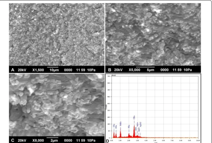
Scanning electron microscopy (SEM) analysis of the cells treated for 60 min with silver nitrate revealed the presence of silver nanoparticles within the cells (Figure 2A-C). Energy dispersive X-ray microanalysis (EDX) was done for qualitative analysis of the thin sections from the selected preparations. Elemental analysis of the silver nanoparticles was performed using EDX in SEM. The EDX spectrum of the silver nanoparticles synthesized byB. cereusis shown in Figure 2D. The vertical axis displays the number of x-ray counts whilst the horizontal axis displays energy (keV). The peaks between 3.00 - 3.40 keV correspond to the
binding energy of AgLa, AgLband AgLb2with ~ 50 - 60
counts, while the peaks near binding energies of 0.3 keV and 0.52 keV belongs to carbon and oxygen respectively. The carbon and oxygen peaks in the EDX analyses can be attributed to the surrounding residual material and/or the carbon tape used for SEM grid preparation. Throughout, the scanning range of binding energies, some peaks belonging to Na, Cl and P were also detected [26,27]. SEM with EDX analysis of the colloids in the cell pellet indi-cated the presence of silver material in nano size diameters bound within the cell wall of the bacteria. These particles are in the monodispersed size range between 4- 5 nm and spherical shape, which is comparable with silver nanopar-ticles synthesised by other bacteria [13].
Microarray experiments and their efficacy
Transcriptome analysis was carried out with microarray to study the effect of silver nitrate stress response on the global gene expression in B. cereus. These experi-ments were conducted with a custom-designed 8 × 15 K DNA microarray consisting of oligo probes for coding DNA sequences (CDS’s) and intergenic regions (IGR’s). The signal intensities obtained from the labelled cRNA Ganesh Babuet al.Journal of Nanobiotechnology2011,9:49
http://www.jnanobiotechnology.com/content/9/1/49
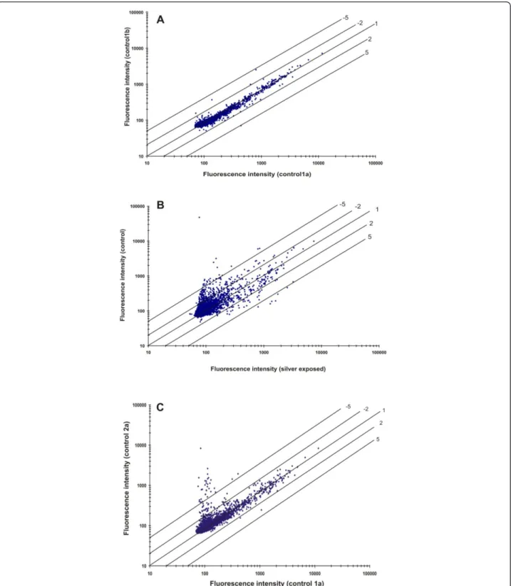
of the control cells (30 min) were presented in scatter plot to study the efficacy of the microarray experiments (Figure 3A). Virtually, majority of the spots lie on or close to the 45° line suggesting no difference in gene expression between the biological duplicates. However, some of the genes showed low-intensity signals suggest-ing a high standard deviation because of background signal (i.e., intensities less than 100 arbitrary units or twice the detection limit are considered as not signifi-cantly expressed).
The scatter plot of signal intensities obtained from the
cells grown with and without 1 mM AgNO3revealed a
clear difference in gene expression profile (Figure 3B). The genes that are up-regulated during silver nitrate stress condition showed signal intensity with at least one fold increase (shown by the upper and lower diago-nal lines in Figure 3B). There was also more than a five-fold difference (lower or higher) in signal intensity as indicated by the diagonal lines. The hybridization signal intensity obtained from the control cells at 30 and 60 min (1a and 2a) showed majority of ORFs lying close to the diagonal and few others at the low intensity (Figure 3C). These hybridizations results suggested that over all precision analysis of the microarray using various statis-tical parameters is of greater accuracy [28].
Response to silver nitrate stress at transcript level
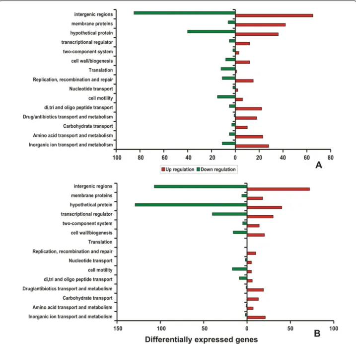
Genes showing differential expression in the cells exposed with silver nitrate at 30 and 60 min was compared with the controls. Expression of genes involved in basic cellular processes was classified based on the Clusters of Ortholo-gous Groups (COG). Most of the genes that showed down-regulation during silver nitrate stress conditions were identified to fall under the COG functional classes of cell motility (N), translation (J) and hypothetical proteins. Interestingly, transcripts encoding proteins of transport and metabolism (P) (such as inorganic ion, amino acids, carbohydrate, synthesis of drug/antibiotics and oligopep-tide), transcription (K), DNA replication/recombination/ repair (L), transcriptional regulators and cell envelope bio-genesis/membrane (M) were found to be up-regulated in cells exposed to AgNO3 stress (Figure 4A-B). Generally, genes encoding transporters and membrane proteins (e.g. efflux proteins, drug resistance transporters, transcrip-tional regulators) were found to be up-regulated upon metal ionic stress conditions. These results also confirmed that the induction of osmoprotectant transporters during exposure to silver stress condition. In the genome of
B. cereus, genes encoding various osmoprotectant trans-porters were identified. The osmoprotectant gene encod-ing a proline/betaine transporter belongencod-ing to the major
Figure 1Survival ofB. cereuscells during silver nitrate induced stress condition. Logarithmically grown cells were treated with and without 1 mM AgNO3. At intervals samples were withdrawn, suitably diluted and plated on LB agar plate without silver nitrate and incubated
facilitator transporter family and a gene encoding a pro-ton-dependent di-, tri- and oligopeptide transporter were among the highly induced genes upon exposure to silver stress. In addition, the genes encoding ABC transporters OpuA [BC2791] and OpuB/OpuC [BC2232] were found to be induced during silver nitrate treatment. Further-more, the up-regulation of zinc-transporting ATPase
[BC0596], cationic Na+/H+ antiporters [BC0373
and BC0838] and copper importing ATPase [BC3730]
a part of cop system were induced upon silver ionic
stress. Another Na+/H+antiporter encoded by [BC1612] have been reported to be induced under salt stress but it was observed to be stably expressed upon silver stress. Higher level of expression P-type ATPases inB. cer-eusATCC14579, a versatile group of ion pumps, has sug-gested that it contributes to the metal homeostasis in response to silver stress [18]. Thecopsystem is a general metal response system that is readily inducible at lower to higher levels of metal stress caused by Cu(II) and Ag(I). Previous report on transcriptional activator like CueR, an Mer R-like was found to respond to Cu(II) and it was also activated by Ag(I) [19,29].
Heat shock proteins (GroEL, GroES, DnaJ and DnaK) are generally induced in microorganisms under various stress conditions [30-32]. But in our study, GroEL [BC0295] alone was up-regulated at the early stages of silver ionic stress. Generally, oxidative stress response genes are involved in response to metal ionic stresses in bacteria. Both, the vegetative catalase-KatA [BC1155] andsB
-dependent catalase-KatE [BC0863] genes were
commonly known to respond to oxidative stresses, but in our study,KatE[BC0863] alone was induced upon silver ionic stress and presumed to have essential role in the survival of the cell. In addition, NAD and NADH depen-dent enzymes especially nitrate reductase [BC2118] and nitroreductase [BC3024] were found to be up-regulated during silver nitrate treatment. The involvement of nitrate reductase in the production of silver nanoparticles has been previously demonstrated [12,33].
The important transcriptional activators of stress sigma factor (sB
), rsbY [BC1006] and rsbV [BC1004] were
found to be up-regulated during 30 and 60 min exposure to silver nitrate. The up-regulation of anti-sigma factor antagonist (rsbY) indicates the activation ofsBinduced
Figure 2SEM with EDX analysis of silver nanoparticles synthesized byB. cereusATCC 14579. Scanning Electron microscopy image (A - C) represents silver nanoparticles synthesised fromB. cereusATCC 14579 incubated at 37°C for 1 h. Figure 2D represents the Energy-Dispersive X-ray microanalysis of silver nanoparticles.
Ganesh Babuet al.Journal of Nanobiotechnology2011,9:49 http://www.jnanobiotechnology.com/content/9/1/49
Figure 3Scatter plots of normalized spot fluorescence intensities (arbitrary units) from DNA microarrays. (A) Spot intensities of array hybridizations with two different control samples in duplicates from theB. cereusATCC 14579 (control 1a versus control 1b). (B) Comparison of spot intensities of array hybridizations from control samples (control 1a and AgNO3treated sample test 1a). (C) Spot intensities of the array
global stress response. Most of these induced genes are
under the control of alternative sigma factor (sB
) in response to variety of stress conditions acting via partner switching mechanisms in Gram-positive bacteria [23,30,34]. Interestingly, silver ion stress is known to induce metabolic pathways associated with amino acid metabolism especially arginine metabolism [21]. The pre-sent study has confirmed the over expression of arginine utilization protein [BC0473]. The S-adenosyl methionine
-dependent methyl transferase [BC2891] and S-adenosyl homocysteine nucleosidase [BC2503], which are essential for the cellular detoxification of metals were also up-regulated during 30 and 60 min exposure to silver nitrate [35,36]. Summary of gene expression pattern ofB. cereus
ATCC 14579 in response to silver nitrate stress is listed in Additional file 1.
Several genes associated with cell motility are located in a ~45 kb region ranging from locus [BC1625] to locus
Figure 4Histogram of differential expression pattern of genes belonging to COG class. Number of regulated genes that are differentially expressed after 30 and 60 min exposures to silver nitrate. The red bars represent the number of up-regulated genes and the green bars represent the number of down-regulated genes. A - 30 min and B - 60 min.
Ganesh Babuet al.Journal of Nanobiotechnology2011,9:49 http://www.jnanobiotechnology.com/content/9/1/49
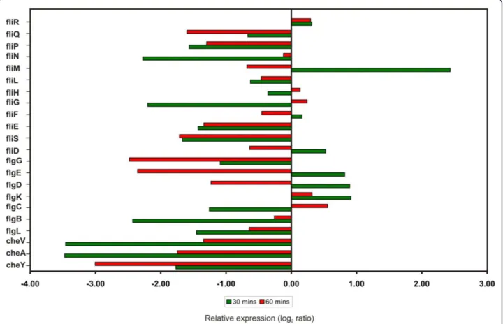
[BC1671] in theB. cereusATCC14579 genome [23,24]. Most of the flagellar and chemotactic genes were down regulated at transcript level upon silver stress are shown in Figure 5. Most of the chemotaxis related genes (CheY, CheA and CheV), flagellar biosynthetic genes (fliP and
fliQ), hook-associated genes (flgL,flgE andfliE), basal body rod genes (flgB andflgG) and motor switch genes (fliG and
fliN)] were down-regulated after silver nitrate treatment. Interestingly, there was no change in the expression level of flagellar motor switch (fliR), a component of type III secretion system, suggesting that it may not have any role in the ionic stress response. In contrast, most of the flagel-lar genes exhibited dramatic changes in their expression within 30 min after silver nitrate treatment. But, few of them were down regulated for continued exposure to sil-ver nitrate for 60 min (Figure 5). The genes encoding fla-gellar components such as basal ring, hook, hook filament junction and cap (fliM,flgK,fliD,flgE,flgD andflgC) were induced inB. cereusduring early stages of silver stress (30 min). The prolonged silver stress leads to the decreased level of expression of genes involved in bio-synthesis of flagellar components. In addition,fliMgene is also involved in the generation of energy from
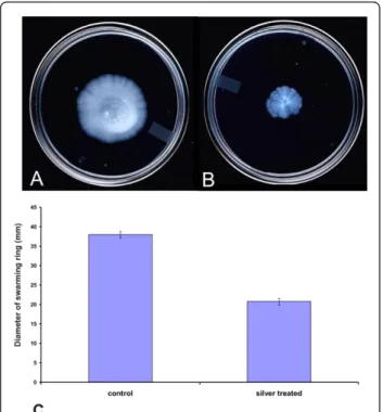
transmembrane electrochemical ion gradients, in the form of Na+/H+antiporter (sodium proton motive force) into mechanical energy [37]. At an early stage of silver stress, the genes involved in ion gradients were induced while the prolonged stress repressed the motive force. Our microar-ray expression profiling study has shown strong evidence for suppression of motility and chemotaxis under silver nitrate stress. This result was supported by the differential expression of chemotaxis and motility related genes inB. cereus, B. subtilis and H. arsenicoxydans strains in response to various stresses [18,23,38]. Further, the che-motactic behaviour of silver stressedB. cereuscells were examined on swarming plates (Figure 6). These results suggested that prolonged exposure of silver nitrate delayed the cell motility.
Identification of sRNAs in Intergenic regions
Bacterial small and un-translated RNAs serve as crucial regulators of cellular physiology to environmental signals. These sRNAs act against the effectors either at transcrip-tion or translatranscrip-tion levels. We used genome based micro-arrays to identify, the expression of candidate sRNAs predicted in the intergenic regions upon silver ionic
stress. An intergenic region is assumed to encode a sRNA and that can bind to its specific cognate probes which are complementary to each other. The distribution of sRNA and the possible transcripts from each and every‘empty’
intergenic region was modelled from the genome ofB.
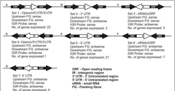
cereusshown in Figure 7 and 8. Possible intergenic tran-scripts as parallel transcriptional outputs from adjacent mRNAs in the same strand are shown in Figure 8A, B &8D. The transcripts expressing from the UTRs of the adjacent genes are shown in Figure 8E &8G. The non-overlapping intact sRNA located within the intergenic region which has diverged orientations with its upstream flanking gene is considered as‘True’sRNA (Figure 8C &8F). The expression pattern of a given transcript was determined in accordance with the computer-assisted algorithm of the Agilent system. In this way, fourteen putative sRNAs collectively from 30 min & 60 min condi-tions were detected according to their diverging orienta-tions with their adjacent flanking genes. Interestingly, the expression of ten putative sRNAs (sRNA1 to sRNA10) were found under early period (30 min) of exposure to
AgNO3stress while other four putative sRNAs (sRNA11
to sRNA14) were found after prolonged (60 min)
expo-sure to AgNO3 stress (Table 1). We have compared the
locations of the fourteen putative sRNAs with the com-putational sRNA predictions by sRNA scanner [39]. This observation has confirmed our initial prediction and detection of fourteen putative silver stress responsive sRNAs proposed from this study. These sRNA candidates are presumed to have regulatory roles under silver ionic stress conditions but needs further validations to under-stand their actual functions. Interestingly, the overexpres-sion of the small RNA binding chaperone protein Hfq [BC1623] was found upon silver ionic stress. It clearly indicates the unknown regulatory cascade of the abun-dant small RNAs and Hfq protein which needs further validation.
Conclusions
Chemical and biological methods are used for the synth-esis of silver nanoparticles with significantly novel struc-tures, improved physicochemical and biological properties. Bacterial species ofBacillus,Pseudomonas
and certain filamentous fungi are widely used for the bio-logical synthesis of silver nanoparticles. Here, we have studied the transcriptomic and phenotypic responses of theB. cereusATCC 14579 during the biosynthesis of sil-ver nanoparticles. The bacteria can activate various cellu-lar and metabolic adaptive mechanisms to reduce the toxicity and precipitate silver as nano-sized particles. Sev-eral microbes are reported to produce silver nanoparti-cles from the aqueous silver nitrate (~1 mM) and several proteins are expected to play vital role in the detoxifica-tion and precipitadetoxifica-tion of silver nanoparticles. The
tran-scriptome analysis ofB. cereusATCC14579 exposed to
Figure 6 Influence of silver nitrate stress on motility of B. cereus. TheB. cereusATCC 14579 was inoculated onto swarming plates without (A) or with exposure to 1 mM silver nitrate (B). After growth at 37°C overnight, plates were observed for swarming capability. (C) The level of motility at 12 h was evaluated as the diameter of the swarming ring in mm. The results presented are the mean value of three independent experiments.
Figure 7Distribution of the putative sRNA-encoding genes along theB. cereusgenome. The origin and terminus of replication are indicated. sRNA genes on the leading and lagging strands are coloured blue and red, respectively.
Ganesh Babuet al.Journal of Nanobiotechnology2011,9:49 http://www.jnanobiotechnology.com/content/9/1/49
silver ionic stress was done using whole-genome DNA microarrays. Approximately 10% of the genes were up-regulated but 20% of the genes were down up-regulated upon silver ionic stress. The SEM along with EDX analy-sis has revealed the accumulation of Ag nanoparticles in the cell-wall. In general, silver stress which has induced the expression of genes involved in the osmoprotection,
transport elements, oxidative stress response and detoxi-fication may have contributed to cross protection. Inter-estingly, silver ionic stress was observed to delay the cell motility. These characteristic phenotypic assessments can contribute to a better understanding of cellular stress adaptation strategies. Finally, fourteen‘putative’
tran-scripts were found to be induced from the ‘empty’
Figure 8Schematic representation of the Intergenic region transcript orientations. Possibility of intergenic transcripts as parallel transcriptional outputs from adjacent mRNAs in the same strand.
Table 1 Summary of RNA transcripts within intergenic regions ofB. cereusATCC14579 regulated in response to silver
nitrate identified by genome based microarray
sRNA id Start End Length (nucleotides)
Flanking gene id Strand* Expression pattern Relative copy numbers Primers
Start End
sRNA1 2840862 2840921 ~60 BC2880/BC2881 <>< ↓↑↓ 54 2840862 2840921
sRNA2 1502573 1502638 ~66 BC1553/BC1554 <>< ↓↑↓ 38 1502576 1502635
sRNA3 5024909 5024970 ~62 BC5122/BC5123 <>< ↓↑↓ 36 5024910 5024969
sRNA4 3372000 3372189 ~190 BC3408/BC3409 <>< ↓↑↓ 181 3372130 3372189
sRNA5 4261246 4261455 ~210 BC4317/BC4318 <>< ↓↑↓ 41 4261395 4261454
sRNA6 4219221 4219336 ~116 BC4271/BC4272 <>< ↓↑↓ 56 4219276 4219335
sRNA7 4040528 4040740 ~213 BC4070/BC4071 <>< ↓↑↓ 169 4040679 4040738
sRNA8 525752 526136 ~385 BC0547/BC0548 ><> ↓↑↓ 142 526073 526132
sRNA9 1106401 1106863 ~463 BC1124/BC1125 ><> ↓↑↓ 44 1106797 1106856
sRNA10 1381925 1382221 ~297 BC1419/BC1420 ><> ↓↑↓ 183 1382156 1382215
sRNA11 5372154 5372598 ~445 BC5447/BC5448 <>< ↓↑↓ 42 5372540 5372599
sRNA12 4790423 4791589 ~1167 BC4864/BC4865 <>< ↓↑↓ 19 4791428 4791487
sRNA13 3802805 3802907 ~103 BC3824/BC3825 <>< ↓↑↓ 16 38028445 3802904
sRNA14 3121534 3121698 ~165 BC3151/BC3152 <>< ↓↑↓ 12 3121639 3121698
intergenic regions upon silver nitrate stress and they are proposed as stress responsive putative sRNAs which could be studied in detail for their role in differential gene expressions.
Materials and methods
Bacterial strain culture conditions and SEM analysis
Gram-positiveBacillus cereusATCC 14579 was used as a model system in this study. The strain was routinely grown in LB broth containing yeast extract (10 g/L), pep-tone (10 g/L) and NaCl (5 g/L) and pH - 8.0) and incu-bated at 37°C with agitation (200 rpm). The overnight cultures were diluted to 1:100 in 100 ml pre-warmed LB broth and incubated at 37°C, with shaking at 200 rpm until the cells were growing exponentially. When an opti-cal density at 600 nm (OD600) of 0.5 - 0.6 was reached, decimal dilutions were prepared using 9 ml of a peptone saline solution and plated on LB agar to determine the viable counts.
To study the effect of silver nitrate exposure, the expo-nentially grown cells were treated with 1 mM silver nitrate and incubated at 37°C. At intervals, aliquots of control and silver treated cultures were diluted and plated on LB agar plates. The plates were incubated for 24 h at 37°C and the viable cells were expressed as log10colony forming units
(CFU). The AgNO3treated cultures and corresponding
controls at two different time points (30 and 60 min) were used for gene expression profiling. For each condition of microarray analysis, biological duplicates were prepared.
The accumulation of silver nanoparticles within the
B. cereus cells after AgNO3 treatment for 60 min was recorded with a JEOL Model JSM - 6390LV and JEOL Model JED - 2300 operating at 1 pA to 1 mA with a 3, 8 and 15 nm resolution (JEOL Model JSM - 6390LV and JEOL Model JED - 2300, Tokyo, Japan). Samples were collected by centrifugation (8000 rpm) for 10 min, dried in oven at 60°C overnight. Approximately, 1 g of finely powdered sample was used for SEM-EDX analysis.
Microarray design
A sense and antisense oligonucleotide microarray slides
complementary to theB. cereusATCC14579 genome was
custom-designed (obtained from Agilent technologies, USA) using the published DNA sequence [NC_004722] [24]. Antisense oligonucleotide sequences targeting the ‘empty’intergenic regions were also designed on the cus-tomized genome array. Each oligonucleotide probe was 60 nucleotides in length and was specifically designed using eArray software from Agilent technologies http://earray. chem.agilent.com/earray. Totally, 15,000 probe sets were designed for Gene Expression profiling ofB. cereususing 8 × 15 k Array AMADID: 23971. It targets the 5234 anno-tated CDS’s, 900‘empty’intergenic regions located on the genome and CDS’s encoded by the 21 plasmids.
Total RNA isolation and cDNA preparation
Bacterial RNA was isolated using the Qiagen RNeasy kit and on-column DNA digestion was carried out according to manufacturer’s instructions (Qiagen, Hilden, Germany), additional DNA removal was done by with DNase I (Ambion, Austin, USA). To perform this, the RNA was precipitated and re-constituted in 85μl of nuclease-free water and then added with 10μl of 10× DNase I buffer and 5μl of (1 U/μl) DNase I. The reaction mixture was incubated at 37°C for 30 min and then chilled on ice. A second RNeasy column purification was performed. The RNA isolation protocol for Gram-positive bacteria was fol-lowed, in which 3 mg/ml of lysozyme was used to degrade the bacterial cell wall. The RNA quality, purity and integ-rity were determined using both NanoDropTM 1000 spec-trophotometer (Thermo Scientific, Wilmington DE, USA) and RNA 6000 Nano Lab Chips with an Agilent 2100 Bioanalyzer (Agilent Technologies, Santa, CA, USA). Stan-dard methods were used for cDNA synthesis, fragmenta-tion and cyanine3 labelling as per the manufacturer’s protocol (Genotypic technologies, Bangalore).
cRNA preparation, microarray hybridization
The synthesized cDNA was transcribed into cRNA using
in vitrotranscription kit (Agilent Technologies, CA, USA) and labelled with cyanine 3 labelled nucleotide according to manufacturer’s protocol and purified with RNeasy Mini columns (Qiagen, Hilden, Germany). The quality of the labelled cRNA sample was verified by the total yield and specificity calculated based on NanoDrop ND-1000 spec-trophotometer (Thermo Scientific, Wilmington DE, USA). Labelled cRNAs with specificity greater than 7 were con-sidered as of high quality and taken for hybridization using thein situhybridization kit plus (Agilent Technolo-gies, Santa, CA, USA). Then, the arrays were incubated at 65°C for 16 h in Agilent’s microarray hybridization cham-bers and the hybridized slides were washed according to the manufacturer’s protocol.
Image processing and Data analysis
Arrays were scanned at 5μm resolution using the Agilent Microarray Scanner G Model G2565BA and images were saved as TIFF format. Data extractions from images were performed with the Feature Extraction software v 10.5.1 and GeneSpring GX version 11.0 software (Agilent Tech-nologies, Santa, CA, USA). Normalization of the data was carried out by GeneSpring GX using the percentile shift normalization which is a global normalization, where the locations of all the spot intensities in an array are adjusted. This normalization takes each column in an experiment independently, and computes thenth percen-tile of the expression values for this array, across all spots (where n has a range from 0-100 and n=75 is the med-ian). It subtracts this value from the expression value of Ganesh Babuet al.Journal of Nanobiotechnology2011,9:49
http://www.jnanobiotechnology.com/content/9/1/49
each entity and normalized to specific samples. After normalization, controls were removed and replicates cor-responding to the same genes within each slide were averaged, provided that there were at least two replicates left after the initial filtering procedure. Otherwise, the gene entry was removed. The average values were com-pared across slides. The model was evaluated on the basis of thatPvalues computed using a false discovery rate correction. Genes were considered regulated at a sta-tistically significant level if they had aPvalue below 0.05 and a ratio of ± 1 cut-off relative to the reference cDNA. Several controls were employed to minimise the technical and biological variations and ensure the quality of the data: 1) each ORFs were present in duplicates in each array 2) array slides were prepared in duplicates for each experiments and 3) two independent RNA batches from each condition were used.
Microarray data submission and accession numbers
The microarray data derived from this study have been deposited in the National Center for Biotechnology Infor-mation (NCBI) Gene Expression Omnibus http://www. ncbi.nlm.nih.gov/geo and are accessible through GEO accession number - [GSE26043].
Motility Assay
Cells from exponentially grown cultures with and
with-out 1 mM AgNO3 treatment for 60 min were recovered
by centrifugation at 10,000 × g at 4°C for 1 min. The sample volume of the silver stressed cells was adjusted to equivalent cell density and inoculated into LB-soft agar (0.4% agar) plates and incubated at 37°C overnight.
Additional material
Additional file 1: Supplemental information to manuscript. Expression patters of genes responding to silver nitrate stress.
Acknowledgements
Authors thank the University Grants Commission (UGC) New Delhi, Government of India for the financial support received through University with Potential for Excellence (UPE) scheme to Madurai Kamaraj University. Authors also thank UGC-Networking Resource Centre in Biological Sciences and Centre for Excellence in Genomic Sciences in School of Biological Sciences for support facilities.
Author details
1Department of Genetics, Centre for Excellence in Genomic Sciences, School
of Biological Sciences, Madurai Kamaraj University, Madurai - 625 021, Tamil Nadu, India.2UGC-Networking Resource Centre in Biological Sciences, School
of Biological Sciences, Madurai Kamaraj University, Madurai - 625 021, Tamil Nadu, India.
Authors’contributions
MMGB has made substantial contributions to conception, design, acquisition, collection, analysis and interpretation of data and wrote the paper. JS carried out the computational analyses of sRNA. PG provided research support and
involved in drafting and revising the manuscript critically for important intellectual content. All authors read and approved the final manuscript.
Competing interests
The authors declare that they have no competing interests.
Received: 26 July 2011 Accepted: 10 November 2011 Published: 10 November 2011
References
1. Mandal D, Bolander ME, Mukhopadhyay D, Sarkar G, Mukerjee P:The use of microorganisms for the formation of metal nanoparticles and their application.Appl Microbiol Biotechnol2006,69:485-492.
2. Reith F, Etschmann B, Grosse C, Moors H, Benotmane MA, Moniseurs P, Grass G, Doonan C, Vogt S, Lai B, Martinez-Criado G, George GN, Nies DH, Mergeay M, Pring A, Southam G, Brugger J:Mechanisms of gold biomineralization in the bacteriumCupriavidus metallidurans.PNAS2009,
106:17757-17762.
3. Shirdam R, Khanafari A, Tabatabaee A:Cadmium, nickel and vanadium accumulation by three strains of marine bacteria.Ira J Biotechnol2006,
4:180-187.
4. Mabbett AN, Lloyd JR, Macaskie LE:Effect of complexing agents on reduction of Cr (VI) byDesulfovibrio vulgarisATCC 29579.Biotechnol Bioeng2002,79:389-397.
5. Cheung K, Ji-Dong G:Chromate reduction byBacillus megateriumTKW3 isolated from marine sediments.World J Microbiol Biotechnol2005,
21:213-219.
6. Strickler DJ:Biomaterials to prevent nosocomial infections: is silver the gold standard?Curr Opin Infect Dis2000,13:389-393.
7. Percival SL, Bowler PG, Russell D:Bacterial resistance to silver in wound care.J Hospital Infection2005,60:1-7.
8. Cohen MS, Stern JM, Vanni AJ, Kelley RS, Baumgart E, Field D, Libertino JA, Summerhayes IC:In vitroanalysis of a nanocrystalline silver-coated surgical mesh.Surg Infec2007,8:397-404.
9. Sondi I, Sondi BS:Silver nanoparticles as antimicrobial agent: a case study onE. colias a model for Gram-negative bacteria.J Colloids Interface Science2004,275:177-182.
10. Mullen MD, Wolf DC, Ferris FG, Beveridge TJ, Flemming CA, Bailey GW:
Bacterial sorption of heavy metals.Appl Environ Microbiol1989,
55:3143-3149.
11. Beveridge TJ, Forsberg CW, Doyle RJ:Major sites of metal binding in Bacillus licheniformiswalls.J Bacteriol1982,150:1438-1448. 12. Kalishwaralal K, Ramkumarpandia SB, Deepak V, Mohammad B,
Sangiliyandi G:Biosynthesis of silver nanocrystals byBacillus licheniformis.
Coll surf B: Biointerface2008,65:153.
13. Ganesh Babu MM, Gunasekaran P:Production and structural
characterization of silver nanoparticles fromBacillus cereusPGN1 isolate.
Coll surf B: Biointerface2009,74:191-194.
14. Beveridge TJ, Murray RGE:Sites of metal deposition in the cell wall of Bacillus subtilis.J Bacteriol1980,141:876-887.
15. Anuradha P, Seema S, Naheed A, Ashok G, Preety S:Synthesis of AgNps By Bacillus cereusbacteria and their antimicrobial potential.J Biomaterial Nanobiotech2011,2:155-161.
16. Wilks JC, Kitko RD, Cleeton SH, Lee GE, Ugwu CS, Jones BD, BonDurant SS, Slonczewski JL:Acid and base stress and transcriptomic responses in Bacillus subtilis.Appl Environ Microbiol2009,75:981-990.
17. Helmann JD, Wu MFW, Gaballa A, Kobel PA, Morshedi MM, Fawcett P, Paddon C:The global transcriptional response ofBacillus subtilisto peroxide stress is coordinated by three transcription factors.J Bacteriol
2003,185:243-253.
18. den Besten HMW, Mols M, Moezelaar R, Zwietering MH, Abee T:
Phenotypic and transcriptomic analyses of mildly and severely salt-stressedBacillus cereusATCC 14579 cells.Appl Environ Microbiol2009,
75:4111-4119.
19. Steil L, Hoffmann T, Budde I, Volker U, Bremer E:Genome-wide transcriptional profiling analysis of adaptation ofBacillus subtilisto high salinity.J Bacteriol2003,185:6358-6370.
20. Mols M, Kranenburg RV, Tempelaars MH, Schaik WV, Moezelaar R, Abee T:
Comparative analysis of transcriptional and physiological responses of Bacillus cereusto organic and inorganic acid shocks.Int J Food Microbiol
21. Moore CM, Gaballa A, Hui M, Ye RW, Helmann JD:Genetic and physiological responses ofBacillus subtilisto metal ion stress.Mol Microbiol2005,57:27-40.
22. Passalacqua KD, Bergman NH, Lee JY, Sherman DH, Hanna PC:The global transcriptional responses ofBacillus anthracisstrain 34F2and asodA1
mutant to paraquat reveal metal ion homeostasis imbalances during endogenous superoxide stress.J Bacteriol2007,189:3996-4013. 23. Kristoffersen SM, Ravnum S, Tourasse NJ, Okstad OA, Kolsto AB, Davies W:
Low concentration of bile salts induce stress responses and reduce motility inBacillus cereusATCC 14570.J Bacteriol2007,189:5302-5313. 24. Ivanova N, Sorokin A, Anderson I, Galleron N, Candelon B, Kapatral V,
Bhattacharyya A, Reznik G, Mikhailova N, Lapidus A, Chu L, Mazur M, Goltsman E, Larsen N, D’Souza M, Walunas T, Grechkin Y, Pusch G, Haselkorn R, Fonstein M, Ehrlich SD, Overbeek R, Kyrpides N:Genome sequence ofBacillus cereusand comparative analysis withBacillus anthracis.Nat2003,423:87-91.
25. Griffiths-Jones S, Moxon S, Marshall M, Khanna A, Eddy SR, Bateman A:
Rfam: annotating non-coding RNAs in complete genomes.Nucl Acid Res
2005,33:D121-D124.
26. Guzman MG, Dille J, Godet S:Synthesis of silver nanoparticles by chemical reduction method and their antibacterial activity.Int J Che Bio Engg2009,2:104-111.
27. Benn TM, Westerhoff P:Nanoparticle silver released into water from commercially available sock fabrics.Env Sci Technol2008,42:4133-4139. 28. Yue H, Eastman PS, Wang BB, Minor J, Doctolero MH, Nuttall RL, Stack R, Becker JW, Montgomery JR, Vainer M, Johnston R:An evaluation of the performance of cDNA microarrays for detecting changes in global mRNA expression.Nucl Acid Res2001,29:e41.
29. Solioz M, Odermatt A:Copper and silver transport by CopB-ATPase in membrain vesicles ofEnterococcus hirae.J Biol Chem1995,270:9217-9221. 30. Prasad J, McJarrow P, Gopal P:Heat and osmotic stress responses of
probioticLactobacillus rhamnosusHN001 (DR20) in relation to viability after drying.Appl Environ Microbiol2003,69:917-925.
31. Periago PM, Van Schaik W, Abee T, Wouters JA:Identification of proteins involved in the heat stress response ofBacillus cereusATCC 14579.Appl Environ Microbiol2002,68:3486-3495.
32. Hu P, Brodie EL, Suzuki Y, McAdams HH, Anderson GL:Whole-genome transcriptional analysis of heavy metal stresses inCaulobacter crescentus.
J Bacteriol2005,187:8437-8449.
33. Kumar SA, Abyaneh MK, Gosavi SW, Kulkarni SK, Pasricha R, Ahmad A, Khan MI:Nitrate reductase-mediated synthesis of silver nanoparticles from AgNO3.Biotechnol Lett2007,29:439-445.
34. Schaik WV, van der Voort M, Molenaar D, Moezelaar R, de Vos WM, Abee T:
Idetification of theσBregulon ofBacillus cereusand conservation ofσB
-regulated genes in low-GC-content Gram-positive bacteria.J Bacteriol
2007,189:4384-4390.
35. Vido K, Spector D, Lagniel G, Lopez S, Toledano MB, Labarre J:A proteome analysis of the cadmium response inSaccharomyces cerevisiae.J Bio Chem2001,276:8469-8474.
36. Bae W, Chen X:Proteomic study for the cellular responses to Cd2+in Schizosaccharomyces pombethrough amino acid-coded mass tagging and liquid chromatography tandem mass spectrometry.Mol cell prot
2004,3:596-607.
37. Trivedi VD, Spudich JL:Photostimulation of a sensory rhodopsin II/HtrII/ Tsr fusion chimera activates CheA-autophosphorylation and CheY-phosphotransferin vitro.Biochemistry2003,42:13887-13892. 38. Arnold JC, Sandrine K, Proux C, Fardeau ML, Dillies MA, Coppee JY,
Ploetze FA, Bertin PN:Temporal transcriptomic response during arsenic stress inHerminiimonas arsenicoxydans.BMC Genomics2010,11:709-718. 39. Sridhar J, Narmada SR, Sabrinathan R, Ou HY, Deng Z, Sekar K, Rafi ZA,
Rajakumar K:sRNAscanner: A computational tool for intergenic small RNA detection in bacterial genomes.Plos one2010,8:e11970.
doi:10.1186/1477-3155-9-49
Cite this article as:Ganesh Babuet al.:Global transcriptome analysis of
Bacillus cereusATCC 14579 in response to silver nitrate stress.Journal of Nanobiotechnology20119:49.
Submit your next manuscript to BioMed Central and take full advantage of:
• Convenient online submission
• Thorough peer review
• No space constraints or color figure charges
• Immediate publication on acceptance
• Inclusion in PubMed, CAS, Scopus and Google Scholar
• Research which is freely available for redistribution
Submit your manuscript at www.biomedcentral.com/submit Ganesh Babuet al.Journal of Nanobiotechnology2011,9:49
http://www.jnanobiotechnology.com/content/9/1/49