Ciliary Dyskinesia
Maimoona Zariwala*, Wanda K. O’Neal*, Peadar G. Noone, Margaret W. Leigh, Michael R. Knowles, and Lawrence E. Ostrowski
Departments of Medicine, Pathology, and Pediatrics, and the Cystic Fibrosis/Pulmonary Research and Treatment Center, University of North Carolina at Chapel Hill, Chapel Hill, North Carolina
Primary ciliary dyskinesia (PCD) is an autosomal recessive dis-ease caused by mutations that affect the proper function of cilia. Recently, deletion of DNA polymerase (Poll) in mice produced a phenotype characteristic of PCD (Kobayashiet al., 2002,Mol. Cell. Biol.22:2769–2776). Because it is unclear how a mutation in a DNA polymerase would result in a specific de-fect in axonemes, the targeting construct was examined fur-ther. Analysis of the genomic region surrounding thePollgene revealed an uncharacterized gene, named Dpcd, that is pre-dicted to be transcribed from the opposite strand relative to
Poll. The deletion of Pollwould also remove the first exon of
Dpcd. Because it is possible that the PCD phenotype observed is due to the absence of either gene, the expression of these genes during ciliogenesis of human airway epithelial cells was examined. Northern analysis demonstrated that DPCD expres-sion increases during ciliated cell differentiation; the expresexpres-sion of POLL decreases. To examine directly whetherDPCDis mu-tated in cases of human PCD, the complete coding sequence ofDPCDwas sequenced from 51 unrelated PCD patients. No disease-causing mutations were confirmed; however, one vari-ant could not be excluded. Therefore,DPCDremains a novel candidate gene for PCD.
Primary ciliary dyskinesia (PCD) is a genotypically hetero-geneous disease, usually inherited as an autosomal recessive trait, caused by mutations that impair the function of cilia and flagella (1–3). The defect in ciliary function in the respi-ratory tract results in impaired or absent mucociliary clear-ance, which is believed to be responsible for the frequent and recurrent episodes of sinusitis and bronchitis that are typical of this disease. The phenotype of the disease is also variable; in severe cases patients develop end-stage
bronchi-(Received in original form September 12, 2003 and in revised form November 14, 2003) * These authors contributed equally to this work, and are considered co-first authors.
Address correspondence to:Lawrence E. Ostrowski, Ph.D., University of North Carolina at Chapel Hill School of Medicine, Cystic Fibrosis/Pulmo-nary Research and Treatment Center, CB# 7248, 6123A Thurston-Bowles Bldg., Chapel Hill, NC 27599-7248. E-mail: [email protected] Abbreviations:deleted in a mouse model of PCD, Dpcd; human bronchial epithelial cells, HBE cells; inner dynein arm, IDA; outer dynein arm, ODA; primary ciliary dyskinesia, PCD; DNA polymerase, Poll.
This article has an online data supplement, which is accessible from this issue’s table of contents online at www.atsjournals.org
Am. J. Respir. Cell Mol. Biol. Vol. 30, pp. 428–434, 2004
Originally Published in Press as DOI: 10.1165/rcmb.2003-0338RC on November 20, 2003 Internet address: www.atsjournals.org
ectasis and require lung transplantation. In addition, the disease affects other organs and processes that require cili-ary or flagellar motion, and patients with PCD may exhibit otitis media, situs inverses totalis, and/or infertility.
The respiratory cilium is a complex structure consisting ofⵑ250 proteins (4, 5). In addition to the highly conserved 9⫹2 arrangement of microtubule doublets that form the basic axonemal structure, cilia contain inner and outer dy-nein arms (IDA, ODA) that provide the force for ciliary beating. Cilia also contain an unknown number of other proteins that may be involved in the assembly, maintenance, and regulation of ciliary function. In theory, a mutation in many of these genes would lead to the same phenotype, that is, impaired mucociliary clearance. However, most patients with PCD exhibit a structural defect in the outer and/or inner dynein arms. In a small percentage of patients with PCD, mutations have been identified in an ODA intermedi-ate chain (DNAI1) (6–9) or an ODA heavy chain (DNAH5) (10, 11). These results confirm that PCD is a genetically heterogeneous disease and indicate that the causative muta-tion in many cases has yet to be identified.
Recently, a deletion of DNA polymerase(Poll) in mice was reported to produce a PCD phenotype that included a lack of inner dynein arms (12). This model exhibited many of the features associated with other models of PCD, includ-ing chronic sinusitis, situs inversus totalis, infertility, and hydrocephalus. Thus,POLLbecame a candidate gene for cases of human PCD. Because it is difficult to reconcile the function of a nuclear DNA polymerase with a specific defect in axonemal structure (missing IDA), we examined the deletion construct in more detail and determined that the expression of another gene was also likely disrupted in the mouse model. This novel gene, which we have namedDpcd
(for deleted in a mouse model of primary ciliary dyskinesia), is predicted to code for a protein of 23 kD of unknown function. In addition, deletion of only the catalytic domain of polin a separate study resulted in mice with a normal phenotype (13). Thus the PCD phenotype observed in the
Pollknockout mice reported by Kobayashi and coworkers (12) is most likely due to the loss of Dpcd.
in vitro.Finally, to examine whether mutations inDPCD
orPOLLcould account for some cases of human PCD, we have sequenced the coding regions of both DPCD and
POLLin a cohort of well-characterized PCD patients. A preliminary report of this work has been presented (14).
Materials and Methods Database Analysis
TheDpcdgene was found in the Celera Discovery System mouse genomic database by searching the region surroundingPoll. Subse-quent analysis identified the same gene in the National Center for Biotechnology (NCBI) databases (http://www.ncbi.nlm.nih.gov). Ensembl (http://www.ensembl.org) was used to visualize the gene relative toPollon the chromosomes of mouse and human.
Analysis of DPCD and POLL Expression
RT-PCR primers were designed to amplify probes for DPCD (458 bp) and POLL (251 bp) based on sequences present in NCBI (XM_058358* and NM_013274). Primers for DPCD were 5⬘ GTTCACTATTTATTCCCAGACGGC 3⬘and 5⬘CACCACAA CCTCCTTTGGCTT 3⬘; primers for POLL were 5⬘ CCTAGG AGGGAAGAGGGAGA 3⬘and 5⬘TGACTTCACCAGCTGA GCAC 3⬘. The coding region of DPCD (687 nt) was amplified using primers 5⬘ TGCTTAGCAGGGGAAAGATG 3⬘ and 5⬘ GCCAAGCCTTGAAGTCTCAC 3⬘, cloned into TopoTA (In-Vitrogen, Carlsbad, CA) and sequenced (deposited in GenBank as accession number AY532267). Human multiple tissue northern blots were purchased from Clontech (Palo Alto, CA) and probed for DPCD expression according to the manufacturer’s instructions. For analysis of expression during ciliogenesis, human bronchial epithelial cells (obtained under protocols approved by the Univer-sity of North Carolina Institutional Review Board) were cultured using conditions described in detail elsewhere (15, 16). Total RNA was harvested from cells grown on plastic for 5 d, cells grown on collagen-coated Millicell-CM culture inserts (Millipore Corp., Bedford, MA) submerged in media for 3 d, and cells cultured on inserts at an air–liquid interface and harvested on Days 7, 29, and 40 of culture. RNA was isolated using RNeasy reagents (Qiagen Inc., Valencia, CA), fractionated on an agarose/formaldehyde gel as previously described (17), transferred to Hybond-N⫹ mem-brane (Amersham Biosciences, Piscataway, NJ), and hybridized with radioactively labeled probes in Rapid-hyb buffer (Amersham Biosciences) according to the manufacturer.
Clinical Evaluation and Ciliary Dynein Arm Evaluation of Patients with PCD
This study was approved by the committee for the protection of the rights of human subjects at the University of North Carolina (UNC)-Chapel Hill. The majority of the PCD families (n⫽44) were seen and evaluated at General Clinical Research Center at UNC, whereas 7 families were evaluated at other institutions. Clinical evaluations, ciliary ultrastructural analysis, and genomic DNA isolations were performed as described earlier (6). Patients were categorized as outer dynein arm defects (ODA⫺), inner dyn-ein arm defects (IDA⫺), both dynein arm defects (Both⫺), and normal dynein arm (other). A cohort of 68 patients belonging to 51 independent families was selected. There was known
consan-* This sequence record has been removed from the NCBI database (note added in proof).
guinity in three families. Ages ranged from 10 mo to 73 yr, and there were 30 males and 38 females. Of the 68 PCD patients, 28 (41%) presented with situs inversus and 1 patient presented with situs ambiguous. We selected one patient with PCD from each of the 51 families for the DPCD gene analysis, and 15 patients with PCD from 15 PCD families for thePOLLgene analysis, focusing on patients with a defect in the IDA.
Mutation Profiling
DNA was extracted from either fresh blood or buccal brushings using standard procedures. All the coding exons ofDPCD(exons 1–6) andPOLL(exons 2–9) and the intron/exon boundries were amplified. All the primer sets were located in the flanking intronic sequences and are available upon request. Amplifications were performed using reagents and AmpliTaq polymerase from Perkin Elmer (Foster City, CA). The initial denaturation was 94⬚C for 5 min, followed by 35 cycles (94⬚C for 30 s, 60⬚C for 30 s, 72⬚C for 45 s) and final extension for 10 min at 72⬚C. Direct DNA sequencing of PCR products was performed on an ABI310 or ABI3100 auto-mated DNA sequencer (PE Biosystem, Foster City, CA), using Prism BigDye primer Cycle Sequencing Ready Reaction kit (PE Biosystem). Intragenic polymorphisms ofDPCDwere used for exclusion mapping. To estimate the frequencies of variants in the general population, 100–110 chromosomes from non-PCD individ-uals were analyzed by sequencing.
Results
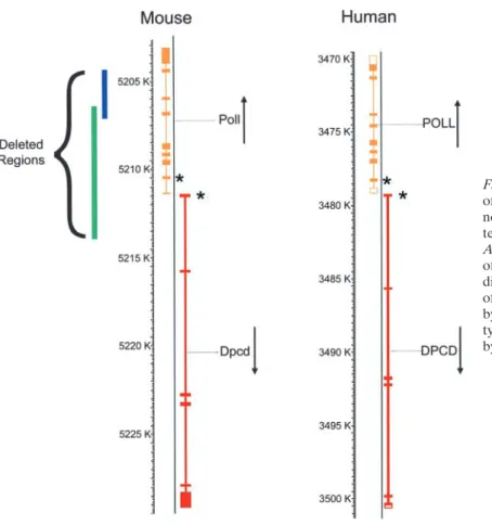
Analysis of mouse chromosome 19 in the region of thePoll
gene revealed an uncharacterized gene (mCG127478 in the Celera mouse genomic database) predicted to be tran-scribed from the opposite strand relative toPoll. The pre-dicted first exon of this novel gene, namedDpcd, lies only 75 bases from exon 1 ofPoll, in the same region encom-passed by the deletion ofPollreported to produce a PCD phenotype (12). The 6.4-kb deletion is predicted to remove the first exon ofDpcd, including the predicted first ATG, and therefore would likely disrupt the function of both
Dpcd and Poll (Figure 1). Thus the phenotype observed could be due to the loss of either gene. For comparison, the deletion reported by Bertocci and colleagues (13) is also shown. This deletion, which replaced three exons of polwith a neo gene, did not produce a PCD phenotype, suggesting the disruption ofDpcdis likely responsible for the PCD phenotype.
Figure 1. Diagram showing the chromosomal location of the DNA polymerase lambda gene (Poll) and the novel geneDpcdon mouse chromosome 19. The syn-tenic region of human chromosome 10 is also shown.
Asterisks indicate the start codons. Targeted deletion of the region indicated by the green line is predicted to disrupt both thePollandDpcdgenes in a mouse model of PCD (12). Targeted deletion of the region indicated by the blue line produced animals with a normal pheno-type (13), suggesting that the PCD phenopheno-type is caused by disruption of the novel gene,Dpcd.
heart. Other tissues examined showed no detectable expres-sion, even after prolonged exposure (14 d). The expression of POLL is also highest in testis, with lower levels of expression reported in ovary, skeletal muscle, pancreas, and heart (18– 20). Thus the expression pattern of DPCD and POLL follow a similar pattern when analyzing RNA extracted from total tissues. However, the absence of a DPCD signal in lung and other tissues may be due to the low percentage of
Figure 2. Expression of DPCD in human tissues. Northern blots containing polyadenylated RNA from the indicated human tissues were probed for DPCD expression. DPCD message was most abundant in testis, with low levels of expression observed in pan-creas, skeletal muscle, and heart. Exposure time for the figure shown was 14 d. Molecular size markers are indicated.
ciliated cells in these total tissue samples. To determine if DPCD was expressed in airway epithelial cells, RT-PCR was used to amplify a full-length cDNA from cultured HBE cells. The sequence of the cDNA was identical to that in the database, verifying the expression of the predicted transcript (not shown). To evaluate more specifically whether DPCD and/or POLL are expressed in ciliated airway epithelial cells and whether their expression correlated with ciliogenesis, HBE cells were grown at an air–liquid interface (15, 16). RNA was isolated from parallel cultures at different stages of differentiation and analyzed by Northern blotting. At early time points in these cultures, the HBE cells are unated and resemble basal cells. With time, the cells differenti-ate to form a heavily cilidifferenti-ated epithelium similar to that ob-servedin vivo. The expression of ciliated cell–specific genes has previously been shown to correlate with the development of ciliated cells in this model system (e.g., Figure 5 in Ref. 21). The results demonstrate that DPCD is expressed in airway cultures and that it becomes more abundant during ciliated cell differentiation, consistent with a potential role in cilia structure or function (Figure 3,top panel). Reprobing of the blot with a POLL-specific probe showed a low level of expression early in culture, which appeared to decrease as differentiation occurred (Figure 3,middle panel).
To examine whether mutations inDPCDcould account for some cases of human PCD, the coding region was se-quenced from a group of 68 patients with well-characterized PCD. Initially, we sequenced the coding region of DPCD
Figure 3. Expression of DPCD and POLL RNA during ciliogenesis of HBE cells. RNA was isolated from cultures of HBE cells cultured on plastic for 5 d or at an air–liquid inter-face for the times indicated. North-ern analysis was performed using probes specific for the novel gene DPCD (top panel) or DNA polymer-ase lambda (POLL) (middle panel). The ethidium bromide stained gel showing the amount of RNA loaded is shown (bottom panel). The expres-sion of DPCD increases at later time points when ciliated cell differentia-tion is occurring, whereas the expres-sion of POLL appears to decrease during differentiation.
of sequence analysis, six sequence variants were detected (Table 1).
To determine if any of these variants could be patho-genic, a population study was performed in individuals with-out PCD (Table 2). Four of these variants were found to be present in individuals without PCD, with varying fre-quencies. Five non-PCD individuals were homozygous for 467T⬎C, demonstrating that this variant is clearly not pathogenic. IVS2⫹59A⬎G and 399C⬎T (N133N) variants were not detected in a homozygous state in the control population; however, given their high minor allele frequen-cies (30% and 22%, respectively), they are most likely poly-morphisms. Also, the 399C⬎T and IVS2⫹59A⬎G variants were found to be in a homozygous state in the unaffected sibling of PCD1 (Figure E1 in the online supplement). The unaffected father of PCD151 was also homozygous for the 399C⬎T variant. IVS2–22_40 del had a minor allele fre-quency of only 1% in the normal population, but this variant was not detected in an affected sibling of PCD163 (Figure E1 in the online supplement). Therefore, this variant is most likely a rare polymorphism. IVS5–48_50 del was not found in the 106 alleles analyzed from the disease-free indi-viduals. However, it also is probably a rare polymorphism because it was detected in one allele from an affected sibling of PCD354, who was wild-type at this locus (Figure E1 in the online supplement). The 168T⬎G (S56R) variant was not detected in 100 alleles analyzed from the control population. The role of this variant is less certain because it was detected in a heterozygous state in only one affected individual (PCD222), who was wild-type for all the other coding nucle-otides and intron–exon junctions in this gene. It is possible that a mutation in the noncoding region of the other allele ofDPCDmay be responsible for PCD in this individual. Further studies will be required to test this hypothesis.
The discovery of intragenic polymorphisms was useful for carrying out exclusion mapping analysis to exclude the
DPCDgene from linkage to PCD, assuming an autosomal recessive mode of inheritance. For PCD1, affected and un-affected siblings shared the same genotype at the polymor-phic loci in intron 2 (IVS2⫹59A⬎G) and in exon 4 (399C⬎T), and both the parents were heterozygous. Thus
DPCD was excluded from linkage to PCD in this family. PCD373 was borne of consanguineous union and was het-erozygous for IVS2⫹59A⬎G; therefore, linkage was also excluded in this family (Figure E1 in the online supple-ment). For PCD Patients 10, 16, 34, 108, 163, and 354, the affected and unaffected siblings were discordant for either one or more polymorphic loci, hence these families were also excluded. Given the proximity of thePOLLgene to
DPCDgene, all the above-mentioned families are probably excluded forPOLLgene as well.
Because the ultrastructural analysis of thePollknockout mouse revealed an absence of IDA (12), we sequenced the coding region of the POLLgene in a patient from all 13 PCD families with solely an IDA defect, and in two patients with PCD with normal dynein arms. During the course of sequence analysis, eight sequence variants were detected (Table E1 in the online supplement); all of these have been identified as intragenic polymorphisms (Table E2 in the online supplement).
Discussion
Although a small number of mutations have recently been identified as causes of PCD, in the majority of cases the underlying mutation is still unknown. Recently, a deletion of DNA polymerase was reported to produce a PCD phenotype in a mouse model (12). The COOH terminal region of DNA polymerase(pol) shows 33% sequence identity with DNA polymerase, and recombinant pol displays DNA polymerase activity (19, 20). In addition, a pol -GFP fusion protein has been shown to localize predominantly to the nucleus (20). Because it is difficult to reconcile the function of a nuclear DNA polymerase with a defect in axonemal structure (missing IDA), we examined the deletion construct in more detail and determined that another gene was also likely disrupted in the mouse model. Other reports have also documented the disruption of more than one gene in mouse models (22, 23). Therefore, the results from any genetically modified animal should be in-terpreted with caution, especially if the phenotype observed does not agree with the known function of the targeted gene. Interestingly, an independent deletion ofPolltargeting the catalytic domain produced mice with a normal phenotype (13). This deletion would not be predicted to disrupt the coding sequence of the novel gene. Thus the PCD pheno-type is most likely due to disruption of the novel gene.
The novel gene, which we have namedDpcd, is predicted to code for a protein of 23 kD of unknown function. Searches of databases as well as protein analysis programs have not revealed any significant homology to known pro-teins or any conserved functional domains to date. The predicted protein contains several potential phosphoryla-tion sites and potential amidaphosphoryla-tion and glycosylaphosphoryla-tion sites, but further studies of the location and function of this novel protein are clearly required.
TABLE 1
A summary ofDPCDsequence analysis in patients with PCD
IVS2⫹59 IVS2– (S56R) (N133N) (L156S) IVS5–48_50 PCD No. Ultrastructure A⬎G 22_40del 168T⬎G 399C⬎T 467T⬎C delGAG
PCD 1 IDA⫺ GG WT/WT TT TT CC WT/WT
*PCD 10 IDA⫺ AA WT/WT TT CC TC WT/WT
*PCD 16 Both⫺ AG WT/WT TT CT TC WT/WT
PCD 26 Unknown AG WT/WT TT CC TT WT/WT
*PCD 34 Unknown AA WT/WT TT CC TC WT/WT
PCD 62 Both⫺ AA WT/WT TT CC TT WT/WT
PCD 66 IDA⫺ AA WT/WT TT CC TT WT/d
†PCD 71 Both⫺ AA WT/WT TT CC TT WT/WT
PCD 78 Other AA WT/WT TT CC TT WT/WT
PCD 79 IDA⫺ AG WT/WT TT CC TC WT/WT
*PCD 108 Other AG WT/WT TT CC TT WT/WT
PCD 116 IDA⫺ AA WT/WT TT CC TT WT/WT
PCD 140 Unknown AA WT/WT TT CC TT WT/WT
PCD 151 IDA⫺ AG WT/WT TT CT TC WT/WT
PCD 157 Other GG WT/WT TT TT CC WT/WT
PCD 158 Both⫺ AG WT/WT TT CT TC WT/WT
*PCD 163 Other AA WT/d TT CC TT WT/WT
PCD 182 Both⫺ AG WT/WT TT CC TT WT/WT
PCD 190 IDA⫺ AG WT/WT TT CT TC WT/WT
PCD 204 Both⫺ AG WT/WT TT CT TC WT/WT
PCD 221 Both⫺ AA WT/WT TT CC TT WT/WT
PCD 222 Both⫺ AA WT/WT TG CC TT WT/WT
PCD 224 Both⫺ AA WT/WT TT CC TT WT/WT
PCD 227 Both⫺ AA WT/WT TT CC TC WT/WT
PCD 254 IDA⫺ AG WT/WT TT CT TC WT/d
PCD 255 IDA⫺ AA WT/WT TT CC TT WT/d
PCD 256 Unknown AG WT/WT TT CT TC WT/WT
PCD 261 IDA⫺ GG WT/WT TT CT TC WT/WT
PCD 262 Both⫺ AA WT/WT TT CC TT WT/d
PCD 264 Other AA WT/WT TT CC TT WT/d
PCD 274 Unknown GG WT/WT TT CT TC WT/WT
PCD 290 Both⫺ AG WT/WT TT CC TT WT/WT
†PCD 299 Both⫺ AA WT/WT TT CC TT WT/WT
PCD 306 Other GG WT/WT TT TT CC WT/WT
PCD 333 ODA⫺ AA WT/WT TT CC TT WT/WT
PCD 340 Both⫺ GG WT/WT TT CC TT WT/WT
PCD 345 Other AG WT/WT TT CT TC WT/WT
*PCD 354 Other AG WT/WT TT CT TC WT/WT
PCD 365 Other AG WT/WT TT CT CC WT/WT
PCD 367 Other AG WT/WT TT CC TC WT/WT
PCD 372 Both⫺ AA WT/WT TT CC TT WT/WT
*†PCD 373 Both⫺ AG WT/WT TT CC TT WT/WT
PCD 383 Other AA WT/WT TT CC TT WT/d
PCD 388 IDA⫺ AA WT/WT TT CC TC WT/WT
PCD 429 IDA⫺ AA WT/WT TT CC TT WT/WT
PCD 460 Unknown AG WT/WT TT CT TC WT/WT
PCD 467 Both⫺ GG WT/WT TT TT CC WT/WT
PCD 481 Both⫺ AA WT/WT TT CC TT WT/WT
PCD 483 Both⫺ AA WT/WT TT CC TT WT/WT
PCD 515 IDA⫺ AG WT/WT TT CT TC WT/WT
PCD 526 Both⫺ AA WT/WT TT CC TT WT/WT
Definition of abbreviations:d, deletion polymorphism; IDA⫺, inner dynein arm defect; ODA⫺, outer dynein arm defect; Both⫺, inner⫹outer dynein arm defect; Other, dynein arms normal; WT, wild-type.
Sequence analysis of theDPCDgene. DNA was isolated from a group of PCD patients using standard procedures and the coding region of theDPCDgene, including intron-exon boundaries, was sequenced.
TABLE 2
Population frequency ofDPCDvariants in PCD and non-PCD individuals
Subject Frequency of Frequency of
Polymorphisms Population Analyzed Wild-type Heterozygous Homozygous Allele 1 Allele 2
IVS2⫹59A⬎G Control 51 20 31 0 0.696 0.304
[Intron 2] PCD 51 25 19 7 0.676 0.323
IVS2–22_40del Control 50 49 1 0 0.990 0.010
[Intron 2] PCD 51 50 1 0 0.990 0.010
168T⬎G (S56R) Control 50 50 0 0 1 0
[Exon3] PCD 51 50 1 0 0.990 0.010
399C⬎T (N133N) Control 52 29 23 0 0.780 0.220
[Exon4] PCD 51 33 14 4 0.784 0.216
467T⬎C (L156S) Control 55 22 28 5 0.655 0.345
[Exon5] PCD 51 27 19 5 0.716 0.284
IVS5–48_50del Control 53 53 0 0 1 0
GAG (Intron 5) PCD 51 45 6 0 0.941 0.059
Single nucleotide polymorphism consortium: http://snp.cshl.org/.399C⬎T (N133N): rs7874, 1,488 chromosomes analyzed. C allele: 0.944. T allele: 0.056. 467T⬎C (L156S): rs7006, 308 chromosomes analyzed. T allele: 0.828; C allele: 0.172.
are expressed at low levels in a wide variety of other tissues, based on this and previous studies and EST data (18–20). However, in cultures of normal HBE cells undergoing ciliogenesis, DPCD increases in a pattern consistent with other cilia-specific genes (21, 24), whereas the expression of POLL remains constant or decreases. These data suggest that DPCD plays a role in the formation or function of ciliated cells.
Because the above data suggest that DPCD may be responsible for some cases of PCD, we examined a group of PCD patients for mutations inDPCD. Thirteen of these patients had a defect in the IDA, which is the same ciliary abnormality observed in the mouse model, and 20 patients had defects in both the IDA and ODA. Six sequence vari-ants were identified, but none were confirmed as disease causing. Although several PCD patients were homozygous for the IVS2⫹59A⬎G and 399C⬎T variants, based on ex-clusion analysis, these are most likely polymorphisms and not disease causing. In addition, RT-PCR was performed on three patients (PCD 1, 157, and 158) and yielded a product of normal size with no mutations detected (not shown).
Although no causative mutations were positively identi-fied inDPCD, one variant, 168T⬎G, which results in a serine to arginine change, cannot be eliminated without further studies. It is possible that in this patient a mutation in the otherDPCDallele may have occurred outside the region sequenced. For example, a mutation in the promoter region may prevent expression of the wild-type allele, creating a
DPCDnull phenotype.
The fact that no disease causing mutations were identi-fied in DPCD is not unexpected. PCD is known to be a heterogeneous disease, and only small numbers of samples are available for analysis. Further, mutations that cause PCD in mouse models may be different than those responsi-ble for human disease. It is interesting to note that hydro-cephalus is a common feature of many animal models of PCD, but is uncommon in the human disease. It is possible that mutations in genes likeDPCDmay not be compatible with life, and so may not be found in the PCD population.
A subset of the patients with PCD, including thirteen with only an IDA defect, were also examined for possible mutations in thePOLLgene. No mutations were detected in the coding region or splice junctions. A recently reported study also failed to identify mutations in the POLLgene in patients with PCD (25). This is perhaps not surprising, as it appears unlikely that the PCD phenotype observed by Kobayashi and coworkers (12) was caused by the disruption of thePollgene.
In conclusion, our results strongly suggest that the dele-tion ofDpcdmay be responsible for the PCD like phenotype in the mouse model previously reported. This conclusion is supported by the expression pattern of Dpcd, the absence of a known function of a DNA polymerase in the structure/ function of cilia, and by the lack of a PCD phenotype in mice in which the catalytic region ofPollwas specifically deleted. Our results also show that mutations in the coding region and splice junctions ofDPCDdo not account for a large percentage of PCD cases, although it is possible that isolated cases may still be found. Further studies are needed to identify the role of DPCD in cilia structure and function.
Acknowledgments:The authors thank Drs. J. Carson, A. Sannuti, M. Hazucha, and S. Minnix for their help in evaluation of the patients with PCD in this study, and Drs. S. Bell, R.U. De Iongh, and L.M. Morgan for providing additional PCD samples. They also thank L. Bauer, B. Brighton, and K. Burns for excellent technical support, L. Brown for help with the figures, and the patients with PCD for their willingness to participate in this study. This work was supported in part by NIH grant HL63103 (L.E.O.), 5-K23-HL04225 (P.G.N.), RR00046–43 (M.R.K.), Cystic Fibrosis Foundation grant R026-CR02 (W.K.O.), and UNC grant RR00046.
References
1. Meeks, M., and A. Bush. 2000. Primary ciliary dyskinesia (PCD).Pediatr. Pulmonol.29:307–316.
2. Schidlow, D. V. 1994. Primary ciliary dyskinesia (the immotile cilia syn-drome).Ann. Allergy73:457–468.
3. Leigh, M. W. 1998. Primary ciliary dyskinesia.InV. Chernick and T. F. Boat, editors. Disorders of the Respiratory Tract of Children, 6th ed. W. B. Saunders, Philadelphia. 819–825.
of flagellar proteins from wild-type and paralyzed mutants of Chlamydo-monas reinhardtii. Proc. Natl. Acad. Sci. USA74:1600–1604.
6. Zariwala, M., P. G. Noone, A. Sannuti, S. Minnix, Z. Zhou, M. W. Leigh, M. Hazucha, J. L. Carson, and M. R. Knowles. 2001. Germline mutations in an intermediate chain dynein cause primary ciliary dyskinesia.Am. J. Respir. Cell Mol. Biol.25:577–583.
7. Pennarun, G., E. Escudier, C. Chapelin, A. M. Bridoux, V. Cacheux, G. Roger, A. Clement, M. Goossens, S. Amselem, and B. Duriez. 1999. Loss-of-function mutations in a human gene related toChlamydomonas reinhardtiidynein IC78 result in primary ciliary dyskinesia.Am. J. Hum. Genet.65:1508–1519.
8. Noone, P. G., M. Zariwala, A. Sannuti, S. Minnix, M. W. Leigh, J. Carson, and M. R. Knowles. 2002. Mutations in DNAI1 (IC78) cause primary ciliary dyskinesia.Chest121:97S.
9. Guichard, C., M. C. Harricane, J. J. Lafitte, P. Godard, M. Zaegel, V. Tack, G. Lalau, and P. Bouvagnet. 2001. Axonemal dynein intermediate-chain gene (DNAI1) mutations result in situs inversus and primary ciliary dyski-nesia (Kartagener syndrome).Am. J. Hum. Genet.68:1030–1035. 10. Ibanez-Tallon, I., S. Gorokhova, and N. Heintz. 2002. Loss of function of
axonemal dynein Mdnah5 causes primary ciliary dyskinesia and hydro-cephalus.Hum. Mol. Genet.11:715–721.
11. Olbrich, H., K. Haffner, A. Kispert, A. Volkel, A. Volz, G. Sasmaz, R. Reinhardt, S. Hennig, H. Lehrach, N. Konietzko, M. Zariwala, P. G. Noone, M. Knowles, H. M. Mitchison, M. Meeks, E. M. Chung, F. Hilde-brandt, R. Sudbrak, and H. Omran. 2002. Mutations in DNAH5 cause primary ciliary dyskinesia and randomization of left-right asymmetry.
Nat. Genet.30:143–144.
12. Kobayashi, Y., M. Watanabe, Y. Okada, H. Sawa, H. Takai, M. Nakanishi, Y. Kawase, H. Suzuki, K. Nagashima, K. Ikeda, and N. Motoyama. 2002. Hydrocephalus, situs inversus, chronic sinusitis, and male infertility in DNA polymerase lambda-deficient mice: possible implication for the pathogenesis of immotile cilia syndrome.Mol. Cell. Biol.22:2769–2776. 13. Bertocci, B., A. De Smet, E. Flatter, A. Dahan, J. C. Bories, C. Landreau, J. C. Weill, and C. A. Reynaud. 2002. Cutting edge: DNA polymerases mu and lambda are dispensable for Ig gene hypermutation.J. Immunol.
168:3702–3706.
14. O’Neal, W. K., M. Zariwala, P. G. Noone, M. R. Knowles, and L. E. Ostrow-ski. 2003. Investigation of the possible role of two novel genes in primary ciliary dyskinesia characterized by a deficiency of inner dynein arms.Am. J. Respir. Crit. Care Med.167:A398. (Abstr.)
15. Gray, T. E., K. Guzman, C. W. Davis, L. H. Abdullah, and P. Nettesheim.
1996. Mucociliary differentiation of serially passaged normal human tra-cheobronchial epithelial cells.Am. J. Respir. Cell Mol. Biol.14:104–112. 16. Bernacki, S. H., A. L. Nelson, L. Abdullah, J. K. Sheehan, A. Harris, C. William Davis, and S. H. Randell. 1999. Mucin gene expression during differentiation of human airway epithelia in vitro. Muc4 and muc5b are strongly induced.Am. J. Respir. Cell Mol. Biol.20:595–604.
17. Ostrowski, L. E., K. Andrews, P. Potdar, H. Matsuura, A. Jetten, and P. Nettesheim. 1999. Cloning and characterization of KPL2, a novel gene induced during ciliogenesis of tracheal epithelial cells.Am. J. Respir. Cell Mol. Biol.20:675–683.
18. Aoufouchi, S., E. Flatter, A. Dahan, A. Faili, B. Bertocci, S. Storck, F. Delbos, L. Cocea, N. Gupta, J. C. Weill, and C. A. Reynaud. 2000. Two novel human and mouse DNA polymerases of the polX family.Nucleic Acids Res.28:3684–3693.
19. Garcia-Diaz, M., O. Dominguez, L. A. Lopez-Fernandez, L. T. de Lera, M. L. Saniger, J. F. Ruiz, M. Parraga, M. J. Garcia-Ortiz, T. Kirchhoff, J. del Mazo, A. Bernad, and L. Blanco. 2000. DNA polymerase lambda (Pol lambda), a novel eukaryotic DNA polymerase with a potential role in meiosis.J. Mol. Biol.301:851–867.
20. Nagasawa, K., K. Kitamura, A. Yasui, Y. Nimura, K. Ikeda, M. Hirai, A. Matsukage, and M. Nakanishi. 2000. Identification and characterization of human DNA polymerase beta 2, a DNA polymerase beta-related enzyme.J. Biol. Chem.275:31233–31238.
21. Zhang, Y. J., W. K. O’Neal, S. H. Randell, K. Blackburn, M. B. Moyer, R. C. Boucher, and L. E. Ostrowski. 2002. Identification of dynein heavy chain 7 as an inner arm component of human cilia that is synthesized but not assembled in a case of primary ciliary dyskinesia.J. Biol. Chem.
277:17906–17915.
22. Ohno, H., S. Goto, S. Taki, T. Shirasawa, H. Nakano, S. Miyatake, T. Aoe, Y. Ishida, H. Maeda, T. Shirai. 1994. Targeted disruption of the CD3 eta locus causes high lethality in mice: modulation of Oct-1 transcription on the opposite strand.EMBO J.13:1157–1165.
23. Sharpless, N. E., N. Bardeesy, K. H. Lee, D. Carrasco, D. H. Castrillon, A. J. Aguirre, E. A. Wu, J. W. Horner, and R. A. DePinho. 2001. Loss of p16Ink4a with retention of p19Arf predisposes mice to tumorigenesis.
Nature413:86–91.
24. Andrews, K. L., P. Nettesheim, D. J. Asai, and L. E. Ostrowski. 1996. Identification of seven rat axonemal dynein heavy chain genes: expression during ciliated cell differentiation.Mol. Biol. Cell7:71–79.