Regulatory T Cells Reduce Acute Lung Injury
Fibroproliferation by Decreasing Fibrocyte Recruitment
Brian T. Garibaldi1, Franco R. D’Alessio1, Jason R. Mock1, D. Clark Files1,2, Eric Chau1, Yoshiki Eto1, M. Bradley Drummond1, Neil R. Aggarwal1, Venkataramana Sidhaye1, and Landon S. King1
1Division of Pulmonary and Critical Care Medicine, Johns Hopkins Asthma and Allergy Center, Johns Hopkins University School of Medicine, Baltimore, Maryland; and2Pulmonary, Critical Care, Allergy and Immunology Division, Department of Internal Medicine, Wake Forest University School of Medicine, Winston-Salem, North Carolina
Acute lung injury (ALI) causes significant morbidity and mortality. Fibroproliferation in ALI results in worse outcomes, but the mech-anisms governing fibroproliferation remain poorly understood. Regulatory T cells (Tregs) are important in lung injury resolution. Their role in fibroproliferation is unknown. We sought to identify the role of Tregs in ALI fibroproliferation, using a murine model of lung injury. Wild-type (WT) and lymphocyte-deficientRag-12/2mice re-ceived intratracheal LPS. Fibroproliferation was characterized by his-tology and the measurement of lung collagen. Lung fibrocytes were measured by flow cytometry. To dissect the role of Tregs in fibropro-liferation, Rag-12/2 mice received CD41CD251 (Tregs) or CD41 CD252Tcells (non-Tregs) at the time of LPS injury. To define the role of the chemokine (C-X-C motif) ligand 12 (CXCL12)–CXCR4 pathway in ALI fibroproliferation,Rag-12/2mice were treated with the CXCR4 antagonist AMD3100 to block fibrocyte recruitment. WT andRag-12/2mice demonstrated significant collagen deposi-tion on Day 3 after LPS. WT mice exhibited the clearance of collagen, butRag-12/2mice developed persistent fibrosis. This fibrosis was mediated by the sustained epithelial expression of CXCL12 (or stromal cell-derived factor 1 [SDF-1]) that led to increased fibrocyte recruitment. The adoptive transfer of Tregs resolved fibroprolifera-tion by decreasing CXCL12 expression and subsequent fibrocyte re-cruitment. Blockade of the CXCL12–CXCR4 axis with AMD3100 also decreased lung fibrocytes and fibroproliferation. These results indi-cate a central role for Tregs in the resolution of ALI fibroproliferation by reducing fibrocyte recruitment along the CXCL12–CXCR4 axis. A dissection of the role of Tregs in ALI fibroproliferation may inform the design of new therapeutic tools for patients with ALI.
Keywords:acute lung injury; fibroproliferative ARDS; fibrocytes; regu-latory T cells; lung injury resolution
Acute lung injury (ALI) and acute respiratory distress syn-drome (ARDS) affect 190,000 individuals in the United States each year, accounting for 75,000 deaths (1). The only treatment that improves outcomes involves a lung-protective strategy in
patients on mechanical ventilation (2). Mortality from ALI/ARDS remains as high as 44% (3).
ALI/ARDS is divided into an exudative phase marked by edema fluid, hyaline membrane formation, and neutrophilic in-filtration, followed in some patients by a fibroproliferative phase (4). Fibroproliferation is part of the normal repair response, and is characterized by the intra-alveolar accumulation of fibroblasts and collagen deposition. If this process is ineffective or continues unabated, patients may develop fibrosis (5). Longer durations of ARDS correspond to increased lung collagen and fibrosis, and portend worse outcomes (6). Fibroproliferative changes on bi-opsy and computed tomography predict mortality (7, 8). The determinants of prolonged fibroproliferation and factors that govern its resolution remain poorly understood.
The fibroblast is a key cell in fibroproliferation (9). The source of activated fibroblasts (myofibroblasts) in ALI remains unclear. Three cell populations likely contribute: (1) resident lung fibroblasts (10), (2) epithelial cells that transform into myo-fibroblasts via epithelial-to-mesenchymal transition (11), and (3) bone marrow progenitors called fibrocytes. Fibrocytes are identified by their expression of CD45, collagen-1 (Col-1), and CXCR4 (12–14). In a model of bleomycin fibrosis, fibrocytes accounted for 20% of myofibroblasts (10, 15). Fibrocytes are contained in the bronchoalveolar lavage (BAL) and blood of patients with idiopathic pulmonary fibrosis (IPF), and may pre-dict exacerbations (14, 16). Recently, fibrocytes were found in the BAL of patients with ARDS, and were associated with increased mortality (17, 18). The mechanisms governing fibro-cyte recruitment and activation in ALI remain unknown.
Persistent inflammation is a hallmark of fibroproliferative ARDS (4). Our group has highlighted a central role for regula-tory T cells (Tregs, i.e., CD41CD251Foxp31 T cells) in the resolution of lung inflammation (19). The role of Tregs in the fibroproliferative response to ALI has not been described, to the best of our knowledge.
Using a well-established model of lung injury involving intratra-cheal LPS (20), we show that lymphocyte-deficientRag-12/2mice demonstrate persistent fibroproliferation, characterized by sus-tained fibrocyte recruitment to the lung. The adoptive transfer of Tregs, but not other CD41T cells, intoRag-12/2mice reduces
(Received in original form May 31, 2012 and in final form August 30, 2012) This work was supported by National Institutes of Health grant NRSA F32HL104908– 02 (B.T.G.), National Institutes of Health grant K99HL103973 (F.D.R.), National Institutes of Health grant R01HL089346 (L.S.K.), and the Johns Hopkins Bayview Scholars Program (L.S.K.).
B.T.G., F.R.D., J.R.M., V.S., and L.S.K. conceived and designed experiments. B.T.G., F.R.D., D.C.F., J.R.M., E.C., Y.E., and N.R.A. performed experiments and analysis. M.B.D. provided assistance with statistical analysis. B.T.G. and L.S.K. wrote the man-uscript and provided creative input.
Correspondence and requests for reprints should be addressed to Brian T. Garibaldi, M.D., Division of Pulmonary and Critical Care Medicine, Johns Hopkins Asthma and Allergy Center, Johns Hopkins University School of Medicine, 5501 Hopkins Bayview Circle, 4A-64, Baltimore, MD 21224. E-mail: [email protected]
This article has an online supplement, which is accessible from this issue’s table of contents at www.atsjournals.org
Am J Respir Cell Mol Biol Vol 48, Iss. 1, pp 35–43, Jan 2013 Copyrightª2013 by the American Thoracic Society
Originally Published in Press as DOI: 10.1165/rcmb.2012-0198OC on September 20, 2012 Internet address: www.atsjournals.org
CLINICAL RELEVANCE
fibrocyte number and resolves fibroproliferation. This fibrocyte reduction is caused by a Treg-mediated decrease in lung CXCL12, a chemokine that induces fibrocyte recruitment. Epithelial cells are an important source of CXCL12, suggesting that Tregs reduce CXCL12 concentrations by interacting with the epithelium. The blockade of CXCR4, the receptor for CXCL12, decreases fibro-cyte numbers and fibroproliferation inRag-12/2mice, providing further evidence for the importance of the CXCL12–CXCR4 axis in ALI fibroproliferation. Our results indicate that Tregs play a critical role in the fibroproliferative response to ALI, and that targeting the CXCL12–CXCR4 pathway may provide new insights into the treatment of fibroproliferative ARDS.
Some of these results were previously reported in the form of an abstract (21).
MATERIALS AND METHODS
Mice
C57BL/6 wild-type (WT) and Rag-12/2 mice (aged 6–8 wk) were
purchased from Jackson Laboratories (Bar Harbor, ME).
Green-fluorescent protein (GFP)-labeled, Foxp31 mice were the gift of
Alexander Rudensky (Sloan-Kettering Institute, New York, NY). Protocols were approved by the Johns Hopkins Animal Care and Use Committee (Baltimore, MD).
Preparation of Mice
Mice were anesthetized with intraperitoneal ketamine and acetylproma-zine (150 and 13.5 mg/kg, respectively) before tracheal exposure.
Escherichia coli LPS (3.75 mg/g, O55:B5 L2880; Sigma-Aldrich, St. Louis, MO) or sterile water was instilled intratracheally via 20-gauge catheter. Animals were killed by vena cava exsanguination (19).
CXCR4 Blockade
Rag-12/2mice underwent daily intraperitoneal injections of AMD3100
(200mg/250ml PBS; Sigma-Aldrich) or PBS (250ml).
Isolation and Adoptive Transfer of Tregs
CD41CD251 and CD41CD252 T cells were isolated from
GFP-Foxp31 spleens using magnetic beads (Miltenyi, Cambridge, MA)
(19). Single-cell suspensions (13106cells in 100ml PBS) were
trans-ferred within 60 minutes after LPS by retro-orbital injection. Treg purity was measured at 85–90% by flow cytometry.
Analysis of BAL Fluid
Two aliquots of 0.7 ml PBS were instilled into the right lung. Cell counts were determined by hemocytometer. Protein was measured using the method of Lowry (19).
Lung Histology and Immunohistochemistry
Left lungs were inflated to 25 cm H2O with 1% low-melting agarose
(Invitrogen, Grand Island, NY) (19) for evaluation by Masson’s tri-chrome. Sections were prepared for immunohistochemistry with anti–SDF-1 AB (1/50, clone 79018; R&D Systems, Minneapolis, MN), followed by a mouse-on-mouse immunodetection technique (Vector Laboratories, Burlingame, CA), peroxidase-conjugated secondary antibody, and development with 3,3-diaminobenzidine.
Lung and BAL Collagen Content
Left lungs were frozen in liquid nitrogen. Lungs were homogenized in 1 ml of Complete Lysis Buffer (Roche, Indianapolis, IN), using an Ultra-Turrax tissue homogenizer (Janke and Kunkel, Wilmington, NC). Col-lagen was measured by Sircol Assay (BioColor, Carrickfergus, UK), according to the manufacturer’s instructions.
Lung CXCL12 Concentrations
CXCL12 concentrations were measured in lung homogenates, using an ELISA duoset (R&D Systems).
Preparation of Lung Homogenates for Flow Cytometry
Right lungs were excised, minced, and incubated at 378C in RPMI
containing 2.4 mg/ml collagenase I and 20mg/ml DNase (Invitrogen),
and then mashed through a 70-mm nylon strainer (BD Biosciences,
San Jose, CA). For epithelial cell isolation, lungs were instilled with 1.2 ml Dispase (BD Biosciences), with 3 U/ml elastase (Worthington, Lakewood, NJ) at removal. Red blood cells were removed using ACK lysis buffer (Invitrogen), and cells were resuspended in FACS buffer.
Flow Cytometry
Cells were prepared for FACS analysis (19). Surface stains included Alexa-700 conjugated anti-CD4 (Ebioscience, San Diego, CA), allophycocyanin-conjugated (APC) anti-CXCR4 (BD Biosciences), APC-Cy7–conjugated anti-CD326, and biotinylated anti-CD45 and CD31 with V450-conjugated streptavidin secondary (Biolegend, San Diego, CA). Intracellular stains included biotinylated rabbit anti–Col-I
antibody (Rockland Chemicals, Gilbertsville, PA) with a PE–Texas red–conjugated streptavidin secondary (BD Biosciences) and APC-conjugated anti-CXCL12 (R&D Systems).
Statistical Analysis
Mortality was analyzed byx2test. Markers of injury were compared
using the Student t test or Mann-Whitney rank sum test. Multiple
groups were compared using one-way ANOVA or one-way ANOVA
on ranks. Pairwise comparisons were performed using the Studentttest
with the Bonferroni correction or the Dunn test. Lung CXCL12 was compared with fibrocyte numbers, using linear regression analysis on log-transformed data. Statistical analysis was performed using Sigma-plot 11.0 (Systat Software, Inc., Chicago, IL).
Further details on adoptive transfers, FACS, the Sircol assay, ELISA, and immunohistochemistry are provided in the online supplement.
RESULTS
Resolution of LPS-Induced Lung Injury Was Impaired in Lymphocyte-Deficient Mice
After the administration of intratracheal LPS, Rag-12/2 mice demonstrated increased mortality when compared with WT mice (Figure 1A). Both WT andRag-12/2mice lost body weight, but WT mice returned to near-baseline body weight by Day 7, whereasRag-12/2mice demonstrated persistently decreased body weight (Figure 1B). Both WT andRag-12/2mice exhibited sim-ilar degrees of lung injury on Day 3, as indicated by BAL cell count (Figure 1C) and protein (Figure 1D). Both BAL cell counts and protein returned to near baseline values by Day 7 in WT mice, whereas these remained elevated inRag-12/2mice (Figures 1C and 1D).
Figure 2. Adoptive transfer of Tregs reduced fibroproliferation inRag-12/2mice after LPS in-jury. (A) Lung sections were stained with Mas-son’s trichrome to highlight collagen deposition on Day 3 and Day 7 after intratracheal LPS or intratracheal water in WT andRag-12/2mice (n> 3 in each group; images are of representative examples,3200 magnification). (B) Lung sections were stained with Masson’s trichrome on Day 7 after intratracheal LPS or intratracheal water in
Rag-12/2 mice, Rag-12/2 mice 1 Tregs, and
Rag-12/2mice1non-Tregs (images are of repre-sentative examples,3200 magnification, n>3 in each group). (C) Lung collagen was measured by Sircol assay in all groups on Day 3 and Day 7 after intratracheal LPS (*P¼0.009 and#P¼0.047, n> 4, all groups). (D) BAL collagen was measured by Sircol assay in all groups on Day 3, Day 7, and Day 10 after intratracheal LPS (*P¼0.002,yP¼
0.003,zP¼0.005, and#P¼ 0.005, n>3, all groups). (E) Lung collagen was positively corre-lated with the natural log of BAL collagen (r¼
of Lung Injury
After magnetic bead isolation, Tregs (CD41CD251cells; 90% Foxp31) or non-Tregs (CD41CD252 cells) were adoptively transferred into intratracheal LPS–exposed Rag-12/2 mice. The adoptive transfer of Tregs but not non-Tregs into Rag-12/2mice 1 hour after intratracheal LPS restored the normal resolution of lung injury, as evidenced by significantly im-proved mortality, increased body weight, and decreased markers of lung injury (Figure 1). Adoptive transfer was con-firmed using flow cytometry to identify Tregs (CD41Foxp31 cells) or non-Tregs (CD41Foxp32 cells) in the BAL, lungs, and spleens of Rag-12/2 mice (see Figure E1 in the online supplement). These results confirm the critical role of Tregs in the normal resolution of LPS-induced lung injury, and es-tablish the pattern of responses against which all other param-eters are compared.
LPS-Induced Fibroproliferation Resolved in the Presence of Tregs
We assessed fibroproliferation by histology as well as by measur-ing collagen concentrations in the BAL and lungs of WT mice,
Rag-12/2mice, andRag-12/2mice that received Tregs or non-Treg CD41cells. Collagen deposition and interstitial thickening were evident in both WT andRag-12/2mice on Day 3 after LPS
injury. However, histologic changes resolved in WT mice by Day 7, whereasRag-12/2mice demonstrated persistent interstitial thickening and collagen deposition (Figure 2A). The adop-tive transfer (AT) of Tregs but not non-Tregs intoRag-12/2 mice led to the histopathologic resolution of fibroproliferation (Figure 2B). Lung collagen concentrations (using the Sircol assay) increased in both WT and Rag-12/2 mice on Day 3, but remained persistently elevated inRag-12/2mice on Day 7 after LPS injury (Figure 2C). The AT of Tregs intoRag-12/2
mice reduced lung collagen concentrations to baseline by Day 7. Lung collagen remained significantly elevated in mice receiving non-Tregs, compared with both WT mice and mice that re-ceived Tregs. We also examined BAL fluid to see if collagen concentrations were altered in the alveolar compartment. WT andRag-12/2mice exhibited increased collagen in BAL fluid on Day 3 after LPS injury (Figure 2D). BAL collagen concentra-tions returned to baseline in WT mice, but remained elevated in
Rag-12/2mice up to Day 10 after LPS injury, compared with WT mice. The AT of Tregs, but not non-Tregs, intoRag-12/2
mice returned BAL collagen concentrations to baseline. BAL collagen was positively correlated with lung collagen, suggesting that fibroproliferation as measured in the alveolar compartment reflects overall lung fibroproliferation (r¼0.73,P, 0.001 by Pearson product moment correlation; Figure 2E). These data strongly support a vital role for Tregs in the resolution of fibro-proliferation after LPS injury.
Tregs Reduced Lung and BAL Fibrocyte Number after ALI
Fibrocytes are circulating progenitor cells that can be recruited to sites of injury to become collagen-producing myofibroblasts. Fibrocytes can be identified according to the expression of the surface markers CD45 and CXCR4, and the intracellular expres-sion of collagen-1 (Col-I) (12, 13) (Figure E2A). Using flow cytometry, we measured fibrocyte numbers in the lungs (Figure 3A) and BAL (Figure 3B) of WT andRag-12/2mice after LPS injury. Despite similar increases in both lung and BAL fibro-cytes on Day 3 after LPS injury, fibrofibro-cytes were persistently elevated inRag-12/2mice compared with WT mice on Day 7. The AT of Tregs, but not non-Tregs, intoRag-12/2mice returned
both lung and BAL fibrocytes to WT concentrations. These results suggest that one of the mechanisms by which Tregs resolve fibro-proliferation after LPS injury occurs through a reduction in lung fibrocyte number.
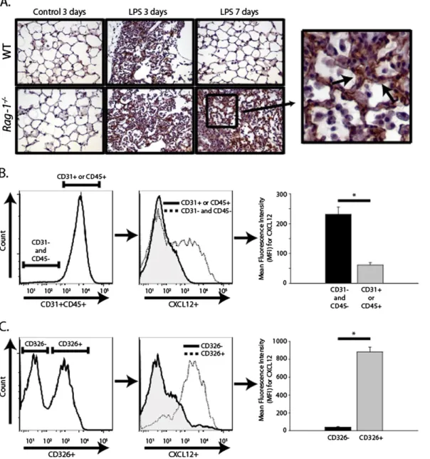
Epithelial-Derived CXCL12 Led to Fibrocyte Recruitment after LPS Injury
CXCL12 (or stromal cell-derived factor 1 [SDF-1]) is the most potent ligand for the CXCR4 receptor (expressed on fibrocytes), and is an important stimulus for fibrocyte recruitment to the lung after injury (12). We used immunohistochemistry to stain for CXCL12 in lung sections from WT andRag-12/2mice that had received either intratracheal water or LPS. CXCL12 was in-creased in both WT and Rag-12/2 mice on Day 3 after lung
injury. On Day 7, CXCL12 returned to near baseline in WT mice, but Rag-12/2 mice demonstrated persistently elevated CXCL12 expression. At higher magnifications, epithelial cells appeared to comprise the predominant cell type that stained positive for CXCL12 after LPS injury (Figure 4A).
To confirm that epithelial cells were the major source of CXCL12 expression after LPS injury, we used multicolor flow cytometry to characterize intracellular CXCL12 expression in var-ious cell populations in both WT andRag-12/2 mice on Day 7 after LPS injury. We used biotinylated CD31 and CD45 antibod-ies with a V450-conjugated streptavidin to distinguish endothelial cells (CD311) and leukocytes (CD451) from other cell populations.
CD31(2)CD45(2) cells exhibited a fourfold higher level of ex-pression of CXCL12 by mean fluorescence intensity, suggesting that leukocytes and endothelial cells were not important sources of elevated lung CXCL12 expression after LPS injury (Figure 4B). Further examination of the CD31(2)CD45(2) cells revealed that the subset of cells that stained positive for the pan-epithelial marker, CD326, demonstrated a 22-fold higher level of CXCL12 expression compared with CD326-cells (Figure 4C). CD326, or epithelial cell adhesion molecule (EpCAM), was recently used to characterize human lung epithelial cell subsets (22). This pro-vides strong evidence that epithelial cells were the primary source of the increased CXCL12 expression after LPS injury.
Tregs Reduced Lung CXCL12 Concentrations after LPS Injury
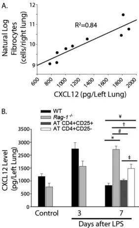
To better quantify CXCL12 expression and to determine the role of Tregs in the regulation of CXCL12 after LPS injury, we mea-sured lung CXCL12 concentrations by ELISA in WT mice, Rag-12/2mice, andRag-12/2mice that received Tregs and non-Treg
CD41 lymphocytes. CXCL12 concentrations predicted lung
fibrocyte numbers according to linear regression analysis (r¼
0.91, R2 ¼ 0.84, P , 0.001; Figure 5A), consistent with the
established role of CXCL12 as a chemokine for fibrocyte recruitment.
CXCL12 concentrations were significantly increased in both
Rag-12/2and WT mice on Day 3 after LPS injury (Figure 5B). CXCL12 concentrations returned to baseline by Day 7 in WT mice, but remained significantly elevated inRag-12/2mice. The AT of Tregs into Rag-12/2 mice reduced CXCL12 concentra-tions to baseline. The AT of non-Tregs intoRag-12/2mice led
to a slight reduction in CXCL12 concentrations, but CXCL12 remained significantly elevated compared with the Rag-12/2
control (92% higher), WT Day 7 LPS (80% higher), and AT Treg groups (45% higher). These data suggest that Tregs re-duced the epithelial expression of CXCL12 after LPS injury, leading to decreased fibrocyte recruitment and the resolution of fibroproliferation.
Blockade of the CXCR4 Receptor with AMD3100 Decreased Fibroproliferation by Reducing Fibrocyte Recruitment to the Lung
To better understand the role of the CXCL12–CXCR4 axis in ALI fibroproliferation, we used the chemical AMD3100 to block the CXCR4 receptor. AMD3100 is a nonpeptide
antagonist of CXCR4 that inhibits the binding and function of CXCL12 with high affinity and specificity (23, 24). Rag-12/2
mice received intratracheal LPS on Day 0, followed by daily
intraperitoneal injections of either AMD3100 (200 mg in
250ml) or sterile PBS (250ml), starting on Day 0. Mortality was no different between Rag-12/2 mice that received AMD3100 and Rag-12/2 mice that received PBS (25% and 27%, respec-tively). Peak weight loss was measured at nearly 25% of initial body weight in both groups, and was similar to the peak weight loss observed in WT andRag-12/2mice that received LPS alone (Figure 6A). BAL protein and cell counts were not significantly different between groups (Figure E4). InRag-12/2mice, CXCR4 receptor blockade with AMD3100 decreased lung fibrocytes (Figure 6B), histologic fibrosis (Figure 6C), and lung collagen (Figure 6D) on Day 7 after intratracheal LPS. Lung CXCL12 concentrations were no different betweenRag-12/2 mice that received AMD3100 or PBS (Figure 6E), indicating that the difference in fibrocyte numbers between groups was attribut-able to the CXCR4 receptor blockade rather than a decrease in CXCL12 expression. WT animals receiving intratracheal LPS and daily injections of AMD3100 did not exhibit reduced inflammation on Day 4 after lung injury, as measured by BAL protein (Figure E4A) and cell count (Figure E4B), demonstrating
sponse to LPS. These results provide further evidence that fibrocytes recruited along the CXCL12–CXCR4 axis play an important role in fibroproliferation after LPS injury, because blocking CXCR4 reduces fibrocyte numbers, collagen deposi-tion, and fibrosis inRag-12/2mice.
DISCUSSION
Fibroproliferation is part of the normal repair response to ALI (5). This repair begins early, as evidenced by increased markers of collagen turnover in the BAL fluid of patients with ARDS within the first days of diagnosis (25, 26). However, excessive fibroproliferation is associated with worse outcomes, because patients develop decreased lung compliance and increased dead space, and require prolonged mechanical ventilation (4, 6, 8). Despite its importance to patient outcomes, the factors that underlie fibroproliferation in ALI remain poorly understood.
Tregs down-regulate lung inflammation in a number of contexts (27–30). Because the fibroproliferative phase of ARDS is charac-terized by persistent interstitial inflammation (4), Tregs might be expected to reduce fibroproliferation, perhaps by dampening the inflammation that leads to myofibroblast accumulation and col-lagen deposition (31). Patients with IPF and colcol-lagen vascular disease–associated pulmonary fibrosis demonstrate a reduced number and function of Tregs in BAL fluid and peripheral blood, compared with controls, suggesting that Tregs may play a role in fibroproliferation (27). However, conflicting data in the literature have emerged regarding Tregs in animal models of pulmonary fibrosis (31–34), suggesting that the role of Tregs in regulating fibroproliferation may be both context-specific and site-specific. To our knowledge, the role of Tregs in the fibro-proliferative response to ALI has not been described.
Our data support a novel role for Tregs in accelerating the res-olution of fibroproliferation by decreasing fibrocyte recruitment to the lung after injury. Fibrocytes are CD451Col-11CXCR41 bone marrow–derived cells that migrate to sites of injury to become collagen-producing myofibroblasts (12, 13). Fibrocytes play an important role in bleomycin models of pulmonary fibro-sis (10, 15). They are present in the BAL fluid and blood of patients with IPF, and increased fibrocyte numbers predict exacerbations (14, 16). Fibrocytes are also found in the BAL fluid of patients with ARDS, and are associated with increased mortality (17, 18).
CXCL12 is constitutively expressed in a number of tissues, and binds to the CXCR4 receptor on fibrocytes to promote migration (12, 14, 35–37). The significant association between lung CXCL12 concentrations and fibrocyte numbers in our model supports pre-vious studies that demonstrated the importance of CXCL12 in fibrocyte recruitment (12, 13). CXCL12 secretion is induced in rat Type II epithelial cells after scratch injury, implicating the epithelium as an important source of CXCL12 (38). In patients with IPF, epithelial cells expressed CXCL12 at higher concentra-tions than in control samples (14). Epithelial CXCL12 increases in both murine and human lungs after ALI (39). Our data indicate that CD3261 epithelial cells are the most abundant source of CXCL12 expression after LPS-induced ALI.
Our study shows that Tregs facilitate the resolution of ALI fibroproliferation by decreasing lung CXCL12 concentrations, leading to a reduction in lung fibrocytes and a decrease in col-lagen deposition and fibrosis. Tregs might alter lung CXCL12 concentrations in a number of ways. Because epithelial cells are the most abundant source of CXCL12 after LPS injury, Tregs likely modulate epithelial CXCL12 expression. Treg–epithelial interactions were previously described. For example, crosstalk between Tregs and the intestinal or retinal epithelium is an
important determinant of both immune tolerance and Treg pro-liferation (40–42). Type II alveolar epithelial cells (AECs) can present antigen to naive Tcells through the Class II major his-tocompatibility complex (MHC), and induce the proliferation of Tregs, in part through a transforming growth factor (TGF)– b–dependent mechanism (43). In the context of our model, Tregs might down-regulate AEC CXCL12 expression in a con-tact-dependent manner. For example, the engagement of Class II MHC receptors on epithelial cells by Tregs might lead to a change in CXCL12 expression. Furthermore, a contact-independent interaction might occur between Tregs and AECs, perhaps through the release of cytokines that decrease AEC CXCL12 production. Tregs may also exert their effects on AECs through a third cell type. For example, our group has shown that Tregs decrease the macrophage production of TNF-ain a contact-dependent manner (19). An alteration in macrophage phenotype might then lead to a decrease in AEC CXCL12 production. Future experiments will seek to identify the specific interactions between Tregs and epithelial cells that govern overall CXCL12 expression and subsequent fibrocyte recruitment in the context of ALI.
The reduction in lung collagen concentrations and fibrocytes in
Rag-12/2 mice after CXCR4 receptor blockade with AMD3100
provides further evidence that the CXCL12–CXCR4 axis is crit-ical in fibrocyte-mediated ALI fibroproliferation, and is consistent with previous studies that demonstrated a reduction in lung fibro-sis in bleomycin models after CXCR4 or CXCL12 blockade (12, 24). The lack of a significant difference in BAL cell count, protein, and lung CXCL12 concentrations between AMD3100-treated
Rag-12/2mice and PBS control mice, as well as the similar peak weight loss in each group, indicate that the difference in fibropro-liferation did not result from reduced inflammation after LPS in-jury, and was instead attributable to the decrease in fibrocyte number. The uncoupling of inflammation and fibroproliferation in this context further supports the notion that the reduction in fibroproliferation seen in our Treg AT experiments was not solely the result of a Treg-dependent decrease in inflammation, but was in part attributable to a reduction in fibrocyte recruitment. Given the potential difficulties of using Tregs as therapy for human au-toimmune and inflammatory diseases (44), the pharmacologic ma-nipulation of downstream pathways such as the CXCL12–CXCR4 axis may provide attractive targets for ALI treatment in the future. Factors in addition to fibrocyte recruitment along the CXCL12– CXCR4 axis likely modulate fibrocyte numbers and subsequent fibroproliferation in the context of ALI. For example, Tregs could influence both fibrocyte proliferation and survival. Moreover, fibro-cyte number alone clearly does not explain the differences in lung collagen concentrations seen between our experimental groups, as evidenced by the finding that lung fibrocytes were lower in WT mice on Day 7 after LPS compared with water control mice, yet lung collagen concentrations were slightly higher in the Day 7 WT mice. The collagen-producing capacity of fibrocytes likely depends in part on their surrounding environment. In addition, our flow cytometry markers may not have identified fibrocytes as they tran-sitioned to a myofibroblast phenotype. In culture, fibrocytes lose the expression of CD34 and CD45 as they transition to a collagen-producing cell (45), making them potentially difficult to track once they transition to a myofibroblast phenotypein vivo. Future
eration, survival, and collagen-producing capacity in the context of ALI fibroproliferation, and to refine the tracking of fibrocytes after they transition to a myofibroblast phenotype.
Our study contains other important limitations. Although the pu-rity of our bead-sorted Tregs used in adoptive transfers was consis-tently greater than 85%, some CD42CD251cells were included in our ATs. Although these represented on average less than 7.5% of overall transferred cells, they may have contributed to the reduction in fibroproliferation. In addition to Tregs, CD25 can be expressed in CD81 T cells, activated B cells, and (at lower concentrations) monocytes. Almost all of the CD42CD251 cells demonstrated a low forward and side scatter by flow cytometry, suggesting they were likely lymphocytes (data not shown). Our group has shown that the AT of splenocytes from CD4-depleted spleens intoRag-12/2 mice does not resolve LPS-induced lung injury. Furthermore, the AT of CD81 lymphocytes into Rag-12/2 mice dose not result in the resolution of lung injury (19). This suggests that CD42CD251cells do not play a role in reducing fibroproliferation in our model. Treg depletion with the CD25-specific antibody PC61 impairs the resolu-tion of lung injury, further emphasizing the critical role of Tregs in our model of lung injury (19).
The small but significant decrease in CXCL12 concentrations after the AT of non-Tregs intoRag-12/2mice might suggest that CD41 lymphocytes other than Tregs play a role in reducing fibroproliferation. However, this small reduction in CXCL12 did not result in decreased biochemical or histologic markers of fibroproliferation. The decrease in CXCL12 may also have been the result of a small number of Tregs present in the non-Treg ATs, either from bead separation impurities or from the induction of naive CD41lymphocytes into Tregs (46). Although we did not find significant numbers of Tregs in the lungs of Rag-12/2mice that received non-Tregs on Day 7 after AT, the num-ber of Tregs required to reduce CXCL12 concentrations and the kinetics of this response remain unknown.
Although our study highlighted an important role for fibro-cytes in ALI fibroproliferation, it did not address the potential contributions of other cell types to the activated myofibroblast pool. Resident fibroblasts were shown to be an important source of collagen deposition and fibrosis in both animal and human models. Recently an interest has emerged in the concept of epithelial-to-mesenchymal transition, whereby epithelial cells may either transform into collagen-producing myofibroblasts or secrete profibrotic growth factors and cytokines that can lead to fibroproliferation (10, 47). Future experiments will seek to define the role of these cell types in our injury model.
In conclusion, we report on a unique role for Tregs in the res-olution of ALI fibroproliferation through the control of fibrocyte recruitment to the lung after LPS injury. A better understanding of the role of Tregs in the fibroproliferative response will poten-tially uncover pathways that might lead to new diagnostic and therapeutic options in patients with ALI.
Author disclosuresare available with the text of this article at www.atsjournals.org.
Acknowledgments: The authors thank James Watkins and Andre Robinson at the
Johns Hopkins Pulmonary Histology Core, Allen Myers at the Johns Hopkins Bay-view Immunohistochemistry Core, and Joe Chrest and Mark Soloski at the Johns Hopkins Bayview Flow Cytometry Core.
References
1. Rubenfeld GD, Caldwell E, Peabody E, Weaver J, Martin DP, Neff M, Stern EJ, Hudson LD. Incidence and outcomes of acute lung injury.N Engl J Med2005;353:1685–1693.
2. The Acute Respiratory Distress Syndrome Network. Ventilation with lower tidal volumes as compared with traditional tidal volumes for acute lung injury and the acute respiratory distress syndrome.N Engl J Med2000;342:1301–1308.
Scales DC, Stather DR, Li A, Jones A,et al. Has mortality from acute respiratory distress syndrome decreased over time? A systematic re-view.Am J Respir Crit Care Med2009;179:220–227.
4. Ware LB, Matthay MA. The acute respiratory distress syndrome.N Engl J Med2000;342:1334–1349.
5. Meduri GU, Chinn AJ, Leeper KV, Wunderink RG, Tolley E, Winer-Muram HT, Khare V, Eltorky M. Corticosteroid rescue treatment of progressive fibroproliferation in late ARDS: patterns of response and predictors of outcome.Chest1994;105:1516–1527.
6. Zapol WM, Trelstad RL, Coffey JW, Tsai I, Salvador RA. Pulmonary fibrosis in severe acute respiratory failure.Am Rev Respir Dis1979; 119:547–554.
7. Martin C, Papazian L, Payan M, Saux P, Gouin F. Pulmonary fibrosis correlates with outcome in adult respiratory distress syndrome.Chest1995;107:196–200. 8. Ichikado K, Suga M, Muranaka H, Gushima Y, Miyakawa H, Tsubamoto M, Johkoh T, Hirata N, Yoshinaga T, Kinoshita Y,et al. Prediction of prognosis for acute respiratory distress syndrome with thin-section CT: validation in 44 cases.Radiology2006;238:321–329. 9. McAnulty RJ, Campa JS, Cambrey AD, Laurent GJ. The effect of
transforming growth factor [beta] on rates of procollagen synthesis and degradationin vitro.Biochim Biophys Acta1991;1091:231–235. 10. Tanjore H, Xu XC, Polosukhin VV, Degryse AL, Li B, Han W, Sherrill
TP, Plieth D, Neilson EG, Blackwell TS, et al. Contribution of epithelial-derived fibroblasts to bleomycin-induced lung fibrosis.Am J Respir Crit Care Med2009;180:657–665.
11. Kim KK, Wei Y, Szekeres C, Kugler MC, Wolters PJ, Hill ML, Frank JA, Brumwell AN, Wheeler SE, Kreidberg JA,et al. Epithelial cell alpha3-beta1 integrin links beta-catenin and SMAD signaling to promote myofi-broblast formation and pulmonary fibrosis.J Clin Invest2009;119:213–224. 12. Phillips RJ, Burdick MD, Hong K, Lutz MA, Murray LA, Xue YY, Belperio JA, Keane MP, Strieter RM. Circulating fibrocytes traffic to the lungs in response to CXCL12 and mediate fibrosis.J Clin Invest2004;114:319–321. 13. Strieter RM, Keeley EC, Hughes MA, Burdick MD, Mehrad B. The role of circulating mesenchymal progenitor cells (fibrocytes) in the path-ogenesis of pulmonary fibrosis.J Leukoc Biol2009;86:1111–1118. 14. Andersson-Sjoland A, de Alba CG, Nihlberg K, Becerril C, Ramirez R,
Pardo A, Westergren-Thorsson G, Selman M. Fibrocytes are a po-tential source of lung fibroblasts in idiopathic pulmonary fibrosis.Int J Biochem2008;40:2129–2140.
15. Hashimoto N, Jin H, Liu T, Chensue SW, Phan SH. Bone marrow-derived progenitor cells in pulmonary fibrosis.J Clin Invest2004;113:243–252. 16. Moeller A, Gilpin SE, Ask K, Cox G, Cook D, Gauldie J, Margetts PJ,
Farkas L, Dobranowski J, Boylan C,et al. Circulating fibrocytes are an indicator of poor prognosis in idiopathic pulmonary fibrosis.Am J Respir Crit Care Med2009;179:588–594.
17. Quesnel C, Piednoir P, Nardelli L, Garnier M, Lec¸on V, Lasocki S, Bouadma L, Elbim C, MentréF, Dehoux M. Alveolar fibrocyte per-centage is an independent predictor of poor outcome in patients with acute lung injury.Crit Care Med2012;40:21–28.
18. Quesnel C, Nardelli L, Piednoir P, Lecon V, Marchal-Somme J, Lasocki S, Bouadma L, Philip I, Soler P, Crestani B,et al. Alveolar fibroblasts in acute lung injury: biological behaviour and clinical relevance.Eur Respir J2010;35:1312–1321.
19. D’Alessio FR, Tsushima K, Aggarwal NR, West EE, Willett MH, Britos MF, Pipeling MR, Brower RG, Tuder RM, McDyer JF,et al. CD41CD251Foxp31Tregs resolve experimental lung injury in mice and are present in humans with acute lung injury.J Clin Invest2009;119:2898– 2913.
20. Matute-Bello G, Downey G, Moore BB, Groshong SD, Matthay MA, Slutsky AS, Kuebler WM, on behalf of the Acute Lung Injury in Animals Study Group. An official American Thoracic Society work-shop report: features and measurements of experimental acute lung injury in animals.Am J Respir Cell Mol Biol2011;44:725–738. 21. Garibaldi B, D’Alessio F, Files DC, Chau E, Sidhaye V, Fraig M, King
LS. Regulatory T-lymphocytes play a key role in the fibroproliferative response to acute lung injury [abstract].Am J Respir Crit Care Med
2010;181:A3772.
23. Rosenkilde MM, Gerlach LO, Jakobsen JS, Skerlj RT, Bridger GJ, Schwartz TW. Molecular mechanism of AMD3100 antagonism in the CXCR4 receptor.J Biol Chem2004;279:3033–3041.
24. Song JS, Kang CM, Kang HH, Yoon HK, Kim YK, Kim KH, Moon HS, Park SH. Inhibitory effect of CXC chemokine receptor 4 antagonist AMD3100 on bleomycin induced murine pulmonary fibrosis. Exp Mol Med2010;42:465–476.
25. Armstrong L, Thickett DR, Mansell JP, Ionescu M, Hoyle E, Billinghurst R, Poole R, Millar AB. Changes in collagen turnover in early acute respiratory distress syndrome.Am J Respir Crit Care Med1999;160: 1910–1915.
26. Marshall RP, Bellingan G, Webb S, Puddicombe A, Goldsack N, McAnulty RJ, Laurent GJ. Fibroproliferation occurs early in the acute respiratory distress syndrome and impacts on outcome.Am J Respir Crit Care Med2000;162:1783–1788.
27. Kotsianidis I, Nakou E, Bouchliou I, Tzouvelekis A, Spanoudakis E, Steiropoulos P, Sotiriou I, Aidinis V, Margaritis D, Tsatalas C,et al. Global impairment of CD41CD251FOXP31 regulatory T cells in idiopathic pulmonary fibrosis.Am J Respir Crit Care Med2009;179: 1121–1130.
28. Hartl D, Koller B, Mehlhorn AT, Reinhardt D, Nicolai T, Schendel DJ, Griese M, Krauss-Etschmann S. Quantitative and functional impair-ment of pulmonary CD41CD25hiregulatory T cells in pediatric asthma.
J Allergy Clin Immunol2007;119:1258–1266.
29. Tosiek MJ, Gruber AD, Bader SR, Mauel S, Hoymann HG, Prettin S, Tschernig T, Buer J, Gereke M, Bruder D. CD41CD251Foxp31 reg-ulatory T cells are dispensable for controlling CD81T cell–mediated lung inflammation.J Immunol2011;186:6106–6118.
30. Schreiber TH, Wolf D, Tsai MS, Chirinos J, Deyev VV, Gonzalez L, Malek TR, Levy RB, Podack ER Therapeutic Treg expansion in mice by TNFRSF25 prevents allergic lung inflammation.J Clin Invest2010; 120:3629–3640.
31. Wilson MS, Wynn TA. Pulmonary fibrosis: pathogenesis, etiology and regulation.Mucosal Immunol2009;2:103–121.
32. Trujillo G, Hartigan A, Hogaboam C. T regulatory cells and attenuated bleomycin-induced fibrosis in lungs of CCR72/2mice.Fibrogenesis Tissue Repair2010;3:18.
33. Boveda-Ruiz D, Alessandro-Gabazza CN, Toda M, Takagi T, Naito M, Matsushima Y, Matsumoto T, Kobayashi T, Gil-Bernabe P, Chelakkot-Govindalayathil AL,et al. Differential role of regulatory T cells in early and late stages of pulmonary fibrosis.Immunobiology(In press) 34. Lo Re S, Lecocq M, Uwambayinema F, Yakoub Y, Delos M, Demoulin
JB, Lucas S, Sparwasser T, Renauld JC, Lison D, et al.
PDGF-producing CD41 Foxp31 regulatory T lymphocytes promote lung fibrosis. Am J Respir Crit Care Med 2011;184:1270–1281.
35. Santiago B, Calonge E, Rey MJD, Gutierrez-Canas I, Izquierdo E, Usategui A, Galindo M, Alcami J, Pablos JL. CXCL12 gene expression is upregulated by hypoxia and growth arrest but not by inflammatory cytokines in rheumatoid synovial fibroblasts.Cytokine2011;53:184–190. 36. Karin N. The multiple faces of CXCL12 (SDF-1) in the regulation of immunity during health and disease.J Leukoc Biol2010;88:463–473. 37. Reilkoff RA, Bucala R, Herzog EL. Fibrocytes: emerging effector cells
in chronic inflammation.Nat Rev Immunol2011;11:427–435. 38. Ghosh MC, Makena PS, Gorantla VK, Sinclair SE, Waters CM. CXCR4
regulates migration of lung alveolar epithelial cells through activation of Rac1 and matrix metalloproteinase–2 (MMP-2). Am J Physiol Lung Cell Mol Physiol2012;302:L846–L856.
39. Petty JM, Sueblinvong V, Lenox CC, Jones CC, Cosgrove GP, Cool CD, Rai PR, Brown KK, Weiss DJ, Poynter ME,et al. Pulmonary stromal– derived factor–1 expression and effect on neutrophil recruitment during acute lung injury.J Immunol2007;178:8148–8157.
40. Chen X, Song CH, Feng BS, Li TL, Li P, Zheng PY, Chen XM, Xing Z, Yang PC. Intestinal epithelial cell–derived integrin AB6 plays an important role in the induction of regulatory T cells and inhibits an antigen-specific Th2 response.J Leukoc Biol2011;90:751–759. 41. Beswick EJ, Pinchuk IV, Earley RB, Schmitt DA, Reyes VE. Role of
gastric epithelial cell–derived transforming growth factor beta in reduced CD41T cell proliferation and development of regulatory T cells during
Helicobacter pyloriinfection.Infect Immun2011;79:2737–2745. 42. Kawazoe Y, Sugita S, Keino H, Yamada Y, Imai A, Horie S, Mochizuki
M. Retinoic acid from retinal pigment epithelium induces T regula-tory cells.Exp Eye Res2012;94:32–40.
43. Gereke M, Jung S, Buer J, Bruder D. Alveolar Type II epithelial cells present antigen to CD41T Cells and induce Foxp31regulatory T cells.Am J Respir Crit Care Med2009;179:344–355.
44. Riley JL, June CH, Blazar BR, Human T. Regulatory cell therapy: take a billion or so and call me in the morning [abstract].Immunity2009; 30:656–665.
45. Mori L, Bellini A, Stacey MA, Schmidt M, Mattoli S. Fibrocytes con-tribute to the myofibroblast population in wounded skin and originate from the bone marrow.Exp Cell Res2005;304:81–90.
46. Josefowicz SZ, Rudensky A. Control of regulatory T cell lineage com-mitment and maintenance.Immunity2009;30:616–625.