Original Research Article
Role of stromal CD10 expression in classifying phyllodes tumors
Hemavathi Manoharan
1*, Kavitha Manoharan
1, Padmavathi Rajagopalan
2INTRODUCTION
Phyllodes tumor(PTs) is a rare fibroepithelial neoplasm containing both epithelial and mesenchymal component that occur in middle aged women.1 Though it contain both components, neoplastic component is formed by stroma which determines the behaviour of the tumor.2
Phyllodes tumors is subclassified into benign, borderline and malignant phyllodes tumors based on Stromal
cellularity and overgrowth, tumor margin, Cellular atypia and number of mitosis per 10 high power field (HPF). Grading of tumor is important as it determines the biological behaviour of the tumor with recurrence rate of 8 to 65% depending on grade and distant metastasis of tumor encountered in up to 22% of malignant tumors.3,4
There is interobserver variation in grading phyllodes especially in intermediate variant as tumors were more atypical than the benign but does not fulfil the criteria of malignancy. Hence, there is a need for a marker for 1Department of Pathology, Government Medical College and ESI hospital, Coimbatore, Tamilnadu, India
2Department of Pathology, Madras Medical College, Chennai, Tamilnadu, India
Received: 17 June 2019
Revised: 11 July 2019
Accepted: 08 August 2019
*Correspondence:
Dr. Hemavathi Manoharan, E-mail: [email protected]
Copyright: © the author(s), publisher and licensee Medip Academy. This is an open-access article distributed under the terms of the Creative Commons Attribution Non-Commercial License, which permits unrestricted non-commercial use, distribution, and reproduction in any medium, provided the original work is properly cited.
ABSTRACT
Background: Phyllodes tumor is a rare fibroepithelial tumor of the breast comprising less than 1% of all primary breast tumor. Phyllodes tumors are classified into benign, borderline and malignant based on histological criteria. Grading of phyllodes tumor is important as it determines the biological behaviour of the tumor. The aim of the present study was to identify the incidence, pathological features of benign, borderline and malignant phyllodes tumors and to compare the CD10 expression in benign, borderline and malignant phyllodes, in order to highlight its diagnostic significance.
Methods: This is a retrospective study conducted in Department of Pathology, Madras Medical College, Chennai for a period of 3 years. The clinical and pathological findings of phyllodes tumors were retrieved from the surgical pathology records. Totally 50 case were selected randomly (38 benign, 6 borderline and 6 malignant) and their representative formalin fixed paraffin embedded tissue samples were subjected to immunohistochemistry for CD10 expression.
Results: In the 38 cases of benign phyllodes tumors, only three cases (7.9%) were CD10 positive. Three out of six cases (50%) of borderline phyllodes tumors showed CD10 positivity, whereas five out of six cases (83.3%) of malignant phyllodes tumor showed CD10 positivity.
Conclusions: CD10 expression correlated well with grade of phyllodes tumors, which is of statistical significance and therefore it can be used in the determination of tumor grade and this may pave way for development of targeted therapies.
Keywords: CD10, Immunohistochemistry, Benign, Borderline, Malignant phyllodes tumor
proper grading and evaluating its clinical behaviour for proper treatment. CD10 known as common acute lymphoblastic leukemia antigen is a zinc metalloproteinase which is normally expressed in myoepithelial cells of breast. Its expression in stromal cells increases with increasing malignant grade.5
In this study CD10 expression in the stromal cells of benign, borderline and malignant phyllodes which are already histologically classified is studied and its role in grading of tumor is evaluated.
METHODS
This is a retrospective study conducted in Department of Pathology, Government Medical College, Chennai for a period of 3 years . The clinical and histopathological findings of phyllodes tumors were retrieved from the surgical pathology records. Of 83 cases of phyllodes tumor retrieved, hematoxylin and eosin (H and E) stained slides were reviewed again and grading was done based on stromal cellularity, stromal overgrowth, cellular atypia, mitosis and marginal status.
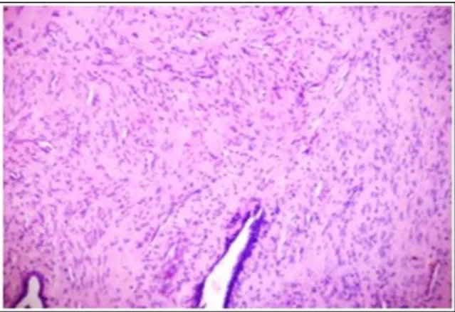
Based on the above criteria, cases with well circumscribed border, no extensive stromal overgrowth, Mitosis ≤2 per 10HPF and absence of nuclear pleomorphism were classified as benign phyllodes tumor (Figure 1). Malignant phyllodes were diagnosed when it showed features of infiltrating border, extensive stromal overgrowth, marked nuclear pleomorphism and mitotic figure ≥5 HPF (Figure 2). When histopathological features were more atypical than benign but not fulfilling the criteria of malignancy were classified as borderline phyllodes tumor (Figure 3).
Figure 1: Benign phyllodes tumor-monotonous
spindle cell with bland nuclei and no mitosis (H and E) 100x.
Out of 83 cases, 50 cases comprising 38 benign tumors, 6 borderline tumors and 6 malignant phyllodes tumor based on pathological features were randomly selected and their representative formalin fixed paraffin embedded tissue samples were subjected to immunohistochemistry for CD10 expression.
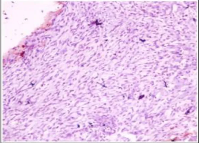
Figure 2: Borderline phyllodes tumor-Increased stromal cellularity and stromal overgrowth with
nuclear atypia (H and E) 100x.
Figure 3: Malignant Tumor-marked stromal overgrowth, marked nuclear atypia and increased
mitosis (H and E) 100x.
Immunohistochemistry
Immunohistochemical analysis of marker CD10 were done in paraffin embedded tissue samples using Super-sensitive polymer HRP system based on non-biotin polymeric technology. Four micron thick sections from formalin fixed paraffin embedded tissue samples were transferred onto gelatin coated slides. Heat induced antigen retrieval was done. The antigen was bound with mouse monoclonal antibody (Pathnsitu) against CD10 antigen and then detected by the addition of secondary antibody conjugated with horse radish peroxidase-polymer and diaminobenzidine substrate. Slides were air dried and mounted with DPX. Every section was examined under Olympus microscope for CD10 immunostaining of stromal tumor cells. Positive control- Myoepithelial cells of breast (Internal control).
as 0, +1,+2,+3 which corresponds to no staining, weak, moderate and strong. The tumor was considered positive for CD10 if 20% or more of the stromal cells exhibit +2 or +3 expression.6
Statistics
The statistical analysis was performed using statistical package for social science software version SPSS 17.0. The p value was considered significant if below 0.05.
RESULTS
The study included 50 cases of phyllodes tumor ( 38 benign, 6 borderline and 6 malignant) classified based on Stromal cellularity and overgrowth, tumor margin, Cellular atypia and number of mitosis per 10 high power field (HPF). The patient age ranged from 17 years upto 60 years and tumor sizes ranged from 3cm to 19cm in maximal diameter.
In the 38 cases of benign phyllodes tumors, the patients’ ages ranged from 17 years up to 57 years (mean 39.7) and the tumor sizes ranged from 3 cm up to 15 cm in maximal diameters (mean 5.6). In the six cases of borderline phyllodes tumors, the patient’s ages ranged from 35 years up to 60 years (mean 47.8) and the tumor sizes ranged from 5 cm up to 14 cm in maximal diameters (mean 8.5).
As for the remaining six cases of malignant phyllodes tumors the patients’ ages ranged from 51 year up to 60 years (mean 54) and the tumors sizes ranged from 7 cm up to 19 cm in maximal diameters (mean 10.8). These results showed highly significant correlation between the patients’ ages (P=0.001) and the tumor sizes (P=0.031) on one hand and the tumor grade on the other hand. These results were summarized in Tables 1 and 2, respectively.
Table 1: Relationship between patients age and tumor grade.
Tumor grade
Youngest
age Oldest age Mean
Benign 17 57 39.7 Borderline 35 60 47.8 Malignant 51 60 54 *p value <0.001
Table 2: Relationship between tumor size and tumor grade.
Size Smallest size
(cm)
Largest size (cm)
Mean (cm)
Benign 3 15 5.6 Borderline 5 14 8.5 Malignant 7 19 10.8
*p value<0.031
It was found that highly significant correlation exist between CD10 expression and the tumor grade (P=0.0001).
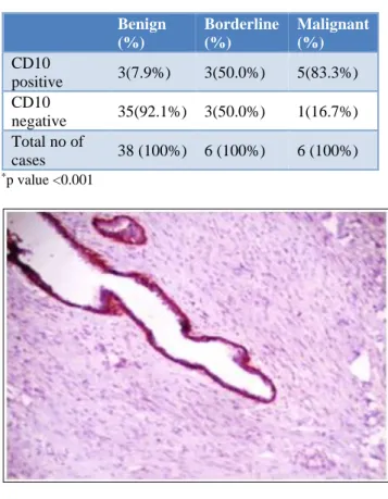
Table 3: CD10 positivity/negativity in different subtypes.
Benign (%)
Borderline (%)
Malignant (%)
CD10
positive 3(7.9%) 3(50.0%) 5(83.3%) CD10
negative 35(92.1%) 3(50.0%) 1(16.7%) Total no of
cases 38 (100%) 6 (100%) 6 (100%) *p value <0.001
Figure 4: Benign phyllodes tumor -CD10 positive only in myoepithelial cells (arrow). Negative CD10 staining
in tumor cells 100x.
Figure 5: Borderline phyllodes tumor - showing strong CD10 immunostaining(3+) in >20% of stromal
tumor 100x.
the borderline phyllodes tumors showed positive CD10 reactivity (Figure 5) where all three (50%) were strong CD10 positive in >20% of cells, remaining 3 cases (50%) were negative for CD10 (Figure 6). As for cases of malignant phyllodes tumors, the majority of cases showed positive CD10 staining constituting five cases (83.3%) (Figure 7), distributed as one (16.7%) moderately stained case and four (66.7%) strongly stained cases in >20% of cells, while the remaining one (16.7%) was CD10 negative. These results were summarized in Table 3.
Figure 6: Borderline phyllodes tumor- Negative for CD10 immunostaining in tumor cells 100x.
Figure 7: Malignant phyllodes tumor showing strong CD10 immunostaining(3+) in most tumor cells (>20%
stromal cells) 400x. DISCUSSION
Phyllodes tumor is a rare fibroepithelial tumor accounting for <1% of all primary breast tumors and constitute 2.5% of fibroepithelial tumors. Annual incidence is estimated to be 2.1 per million women in a population based study conducted in USA.7 Age group most commonly affected are between 35 and 55 years with peak incidence of 45 years. Behaviour of Phyllodes tumor varies from completely benign to highly malignant tumor.8 Thus phyllodes tumor has been sub classified into benign and
malignant with some category not fitting into both will come under borderline category. Though benign tumors are indolent, it sometimes has aggressive growth with local recurrence but do not metastasize whereas it is common in malignant phylodes tumor where adjuvant therapies are needed. Thus grading of tumor has important prognostic significance.
CD10 is a metalloproteinase which degrades bioactive peptides suggesting its expression in tumor stroma is associated with aggressiveness of tumor and making it a important prognostic marker.9 In this study expression of CD10 in stromal cells correlates with phyllodes tumor grading as evidenced by 7.9% of benign, 50% borderline and 83.3% malignant show CD10 immunoreactivity there by helping us to classify into benign, borderline and malignant phyllodes.
Similar conclusion was also obtained in some studies which shows CD10 immunoreactivity in 16.7%, 60% and 80% of benign, borderline and malignant tumors with p value of 0.0001 whereas study CD10 immunoreactivity in 5.9%, 31.4% and 50% of benign, borderline and malignant tumors with p value <0.001.5,10 According to CD10 positivity in benign, borderline and malignant were 43.8%,60% and 82.4% respectively with p value of 0.02 showing significant correlation.6
CONCLUSION
CD10 expression is more in borderline and malignant tumors when compared to benign tumors. In this study CD10 expression is significantly associated with increasing grade of tumor. This concludes the role of CD10 expression in classifying the tumor. A large sample study in future may show the significance of CD10 as a prognostic marker and might pave way for developing targeted therapy.
ACKNOWLEDGEMENTS
Authors would like to thank our colleagues, laboratory technician and our family members for their support and co-operation during the study period.
Funding: No funding sources Conflict of interest: None declared
Ethical approval: The study was approved by the Institutional Ethics Committee
REFERENCES
1. Palmer ML, De Risi DC, Pelikan A, Patel J, Nemoto J, Rosner D, et al. Treatment options and recurrence potential for cystosarcoma phyllodes. Surg Gynecol Obstet. 1990;170(3):193-6.
2. Parker SJ, Harries SA. Phyllodes tumours. Postgrad Med J. 2001;77(909):428-35.
phyllodes tumors. Breast Cancer Res Treat. 1999; 57(3):291-5.
4. Moffat CJ, Pinder SE, Dixon AR, Elston CW, Blamey RW, Ellis IO. Phyllodes tumours of the breast: a clinicopathological review of thirty‐two cases. Histopathol. 1995 Sep;27(3):205-18.
5. Tse GM, Tsang AK, Putti TC, Scolyer RA, Lui PC, Law BK, et al. Stromal CD10 expression in mammary fibroadenomas and phyllodes tumors. J Clin Pathol. 2005;58(2):185-9.
6. Masri MA,Darwazeh G, Sawalhi S, Mughrabi A, Sughayer M, Al-Shatti M, et al. Phyllodes tumor of the breast: Role of CD10 in predicting metastasis. Ann surg Oncol. 2012 Apr 1;19(4):1181-4.
7. Bernstein L, Deapen D, Koss RK. The descriptive epidemiology of malignant cystosarcoma phyllodes tumors of the breast. Cancer. 1993;71(10):3020-4. 8. Margaret IL, Bhuvaneswari R, Cynthia CP, Michael
TM, Gayle G, Gerard JN, et al. Giant breast tumors:
Surgical management of phyllodes tumors, potential for reconstructive surgery and a review of literature. World J Surg Oncol. 2008;6(1):117.
9. Nikita AM, Malcolm H, Beverley AC, Shahriar D, Gilks CB, David GH. Stromal CD10 expression in invasive breast carcinoma correlates with poor prognosis, estrogen receptor negativity, and high grade. Mod Pathol. 2007;20(1):84-9.
10. Ibrahim WS. comparison of strmal Cd10 expression in benign, borderline and malignant phyllodes tumors among Egyptian female patients. Indian J Pathol Microbiol. 2011;54(4):741-4.