Sanjeev Kumar et al JMSCR Volume 06 Issue 10 October 2018 Page 902
A Randomized Study to Evaluate the Serum Iron and Ferritin Levels in
Relation with Cholelithiasis
Authors
Sanjeev Kumar
1, Ashima Badyal
2*, Vidyut Gupta
31
Physician, Sub-District Hospital Hiranagar, Kathua, J&K, India
2
Lecturer, Department of Biochemistry, GMC Jammu, J&K, India
3
PG, Department of Surgery, GMC Jammu, J&K, India *Corresponding Author
Ashima Badyal
Email: [email protected]
Abstract
Gallstone disease is a common clinical entity affecting the adult population of both genders. It has been studied that iron deficiency results in altered mobility of gall bladder, leading to increased cholesterol crystal formation in the gall bladder bile.
The study was conducted in the department of biochemistry & department of surgery/medicine, GMC Jammu. 100 Patients within the age group of 15 – 70 years, suffering from gall stone confirmed by ultra-sonography, were included as cases. There were more females in this group (79%). The age group of 30-39 showed the maximum prevalence of gallstone disease. In females, the serum ferritin was low in 38.0% cases and 23.1% controls respectively. Further, this study also revealed that low iron and low ferritin, combined for 18% of cases while Iron normal and ferritin normal, combined, for 50% of cases, which was significant (p<0.05). The study further found that patients who had gallstones in the age group of 30-39 had low serum iron 33% and low serum ferritin 35%, which was significant for females of age group: 30-39 years. Iron deficiency has been shown to alter the activity of several hepatic enzymes, leading to increases gall bladder’s bile cholesterol saturation and promotion of cholesterol crystal formation, playing a significant role in gallstone pathogenesis. This gender related difference showing more prevalence of cholelithiasis in females could be linked to pregnancy and female sex hormones along with iron deficiency. A considerable no of people falling in age group of 30-49 years, particularly females, show low total body iron as well as, increased risk of cholelithiasis, because of low body stores of iron. Patients above 30 years age should be screened for serum iron and serum ferritin to diagnose their progression towards severe iron deficiency and as an early marker for gallstone disease.
Keywords: Gallstone Disease, cholesterol saturation, cholelithiasis.
Introduction
The old axiom that a typical gall stone sufferer is a fat, fertile, female of fifty, is only partially true, as the disease is found in women soon after their first delivery and also in underweight and thin people.[1]
Gallstone disease is a common clinical entity affecting the adult population of both sexes. The earliest known gallstones date back to the 21st Egyptian dynasty discovered in the mummy of a priestess of Amenen (1085-945 BC).[2]
www.jmscr.igmpublication.org Impact Factor (SJIF): 6.379
Index Copernicus Value: 79.54 ISSN (e)-2347-176x ISSN (p) 2455-0450
Sanjeev Kumar et al JMSCR Volume 06 Issue 10 October 2018 Page 903
Cholelithiasis is a common abdominal disorder resulting in increasing hospital admissions. About 10-12% of adults develop gallstones.[3] The prevalence of common bile duct stones in patients with gallstones varies from 8 to 16%.[4] Pure cholesterol stones are uncommon and account for less than 10% of all stones. Whether pure or of mix nature, the common primary event in the formation of cholesterol stone is super saturation of bile with cholesterol which is almost always is caused by cholesterol hyper secretion rather than reduced secretion of phospholipids or bile salts. Pigment stones contain less than 20% cholesterol and are dark because of presence of calcium bilirubinate. Black pigment stones are usually small, brittle, black and sometimes spiculated. They are formed by super saturation of calcium bilrubinate, carbonate and phosphate and occur most often secondary to hemolytic disorder such as hereditary spherocytosis and sickle cell anaemia. Like cholesterol stones they are almost always found in gall bladder. Brown pigment stones are usually less than 1 cm in diameter, brownish-yellow and soft, often mushy. They may form either in gallbladder or in bile ducts, usually secondary to bacterial infection caused by bile stasis. Precipitated calcium bilirubinate and bacterial cell bodies compose the major part of the stone.[5]
While, iron deficiency was found to be new and interesting aetiological factor in the formation of gall stones. Gallstones may produce several hepatic enzymes, leading to increased gall symptoms, or
may remain asymptomatic. Over half the
asymptomatic cases are usually detected by abdominal ultrasound. Today the incidence of gall stone disease has increased considerably with the invention of ultra-sonography.[6]
Iron acts as acoenzyme for nitric oxide synthetase which synthesize nitric oxide synthase and that is important for the maintenance of basal gall bladder tone and normal relaxation. It was found that the iron deficiency resulted in altered motility of gall bladder and sphincter of oddi, leading to biliary stasis and thus increased cholesterol crystal formation in the gall bladder bile. Serum iron, total
iron binding capacity and transferring saturation are not good indicators of iron status in individuals. In infection free situation, serum ferritin is an ideal indicator for diagnosis of iron deficiency and response to iron therapy in a community. If the prevalence of iron deficiency in a population must be described with a single number, serum ferritin
should be used and complemented with
haemoglobin in all programmed evaluations.[7] The serum iron concentration is found least in patients with pigment stones hence enforcing the role of iron in gall stone formation. Nutritional anaemia is a major public health problem in India and is primarily due to iron deficiency Haemoglobin concentration alone cannot be used to diagnose iron
deficiency. However, the concentration of
haemoglobin should be measured, even though not all anaemia is caused by iron deficiency. The prevalence of anaemia is an important health indicator and when it is used with other measurements of iron status, the haemoglobin concentration can provide information about the severity of iron deficiency.[8] Serum ferritin will act as more specific indicator for iron deficiency anaemia.
If we can predict which factors contribute to the development of gall stone disease, then its prevention could be affected by modifying these factors. This study was planned to study the correlation between serum iron and serum ferritin in patients suffering from gallstone disease.
Material and Methods
Sanjeev Kumar et al JMSCR Volume 06 Issue 10 October 2018 Page 904
Inclusion criteria
For control group: Patients in surgery ward within the age group of 15 – 70 years, not suffering from gall stone disease, confirmed by ultra-sonography but without a previous history of biliary track surgery.
For case group: All patients with Cholelithiasis confirmed by ultrasonography within the age group of 15 - 70 years.
Exclusion criteria
Patients taking iron for anaemia and/or patients with previous case of biliary tract surgery.
Procedure
In this study, a detailed history was taken from all the patients suffering from with and without gall stone disease. Routine investigations such as ultrasound of abdomen were done in all the patients. Venous blood sample of 4 ml was taken in red vacutainer for evaluation of serum iron and serum ferritin level. Serum ferritin level was assessed by chemiluminescence.[9] Serum iron was estimated by the ferrozine kit method.[10]
Results
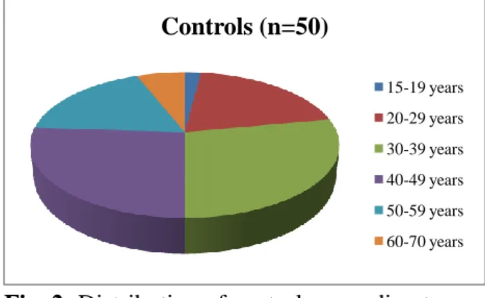
The population studied was divided into two groups. The first group consisted of healthy controls, 50 in number, with age group: 15 to 70 years, admitted in surgery ward but without gallstone disease. The second group consisted of 100 patients (cases) showing gallstone on ultrasonography, admitted in surgery ward, fulfilling inclusion criteria. There were more females in cases group 79 (79%) than control group 26 (52%) and males in case were (21) 21% and 24 (48%) in control. The age distribution of patients in second group having gall stone was: 2 (2%) in age group of 15-19 years, 15(15%) in the age group of 20-29, 27 (27%) in the age group of 30-39, 25 (25%) in the age group of 40-49, 20 (20%) in the age group 50-59, and 11 (11%) in age group of 60-70 years as shown in figure 1. There was low prevalence of gallstones in younger population, as compared to the older ones, and highest prevalence of gallstones in 30-39 years and 40-49 years of age.
Fig. 1: Distribution of patients according to age (Mean±SD= 43.0±13.2)
Fig. 2: Distribution of controls according to age (Mean±SD= 40.8±11.6)
Table 1: Distribution of Serum Iron
Males: Case Group (n= 21)
Control Group (n=24)
P Value
Serum iron level (µg/dl)
Low 8 (38.1%) 5 (20.8%) Normal 13 (61.9%) 19 (79.2%)
Above
Normal 0 (0%) 0 (0%)
Mean±SD 67.1± 22.4 64.8± 20.2 0.280
Females Case Group (n= 79)
Control Group (n=26)
P Value
Serum iron level (µg/dl)
Low 31 (39.2%) 8 (30.8%) Normal 48 (60.8%) 18 (69.2%)
Above
Normal 0 (0%) 0 (0%)
Mean±SD 54.4± 19.5 62.5± 24.3 0.077 P value>0.05 (not significant)
In this study in males, the normal reference value was supplied with the kit, for male normal value was 60-160 µg/dl. Serum iron levels were low in 38.1% of cases and 20.8% of controls. Serum iron was normal in 61.9% of cases and in 79.2% of controls. The mean serum iron among cases stood at 67.1±22.4 µg/dl and that of controls: 64.8± 20.2. P value was >0.05 which was not significant. On the other hand the normal reference value for females was estimated at 45-145 µg/dl. Here serum iron was low in 39.2% of cases and 30.8% of controls. Serum
Cases (n=100)
15-19 years 20-29 years 30-39 years 40-49 years
50-59 years 60-70 years
Controls (n=50)
15-19 years 20-29 years
Sanjeev Kumar et al JMSCR Volume 06 Issue 10 October 2018 Page 905
iron was normal in 60.8% of cases and 69.2% of controls. The mean serum iron was found to be 54.4± 19.5in cases and 62.5± 24.3in controls. P value was >0.05 which was also not significant however. (Table 1)
Table 2: Serum ferritin distribution
Males Case Group (n=
21)
Control Group (n=24)
P Value
Serum ferritin level (ng/ml)
Low 9 (42.9%) 6 (25.0%) Normal 12 (57.1%) 14 (58.3%)
Above
Normal 0 (0%0 4 (16.7%)
Mean±SD 39.6± 19.7 65.4± 33.8 <0.05
Females Case Group (n=
79)
Control Group (n=26)
P Value
Serum ferritin level (ng/ml)
Low 30 (38.0%) 6 (23.1%) Normal 49 (62.0%) 16 (61.5%)
Above
Normal 0 (0%) 4 (15.4%)
Mean±SD 27.9± 11.5 45.9± 14.3 <0.05 P value<0.05 (significant)
For serum ferritin levels, the normal reference range for males 23-336 ng/ml was considered and similarly for females: 11-306 ng/ml. In males, the serum ferritin was low in 42.9% of cases and 25.0% of controls. The figures in females were at 38.0% and 23.1% respectively. Serum ferritin was normal for 57.1% of cases and 58.3% of controls in males. The same stood at 62.0% and 61.5% in females. However it was similarly placed at 16.7% and 15.4% for above normals, in controls, in males and females respectively. P value was in each case significant, ie<0.05. (Table 2)
Table 3: Distribution of low serum iron and low serum ferritin within cases (n=100)
Parameter Low Iron Normal Iron
Low ferritin 18 (18%) 21 (21%)
Normal ferritin 11 (11%) 50 (50%)
Total 29 (27%) 71 (73%) P value<0.05 (significant)
Further, this study also revealed that low iron and low ferritin, combined for 18% of cases while Iron normal and ferritin normal, combined, for 50% of cases, which was a significant result (p<0.05). (Table 3) Besides this, the study further found that patients who had gallstones in the age group of 15-19 years had low serum iron in 2% and low serum
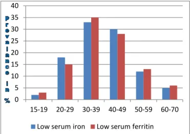
ferritin in 3%, those were in the age group of 20-29 had low serum iron in 17 % and low serum ferritin in 15%, in the age group of 30-39 had low serum iron 33% and low serum ferritin 35%; in the age group of 40-49 the respective values were 30% and 28% respectively, as shown in figure 3. The p value is <0.05, indicating a significant prevalence of low serum iron and low serum ferritin in case of females of age group: 30-39 years.
Fig. 3: Prevalence of low Serum iron and low serum ferritin in various age groups
Discussion
Iron deficiency has been shown to alter the activity of several hepatic enzymes, leading to increases gall bladder’s bile cholejsterol saturation and promotion of cholesterol crystal formation. Iron acts as a coenzyme for nitric oxide synthetase (NOS), and that is important for the maintenance of basal gall bladder tone and normal relaxation.[11] It was found that iron deficiency resulted in altered mobility of gall bladder and sphincter of Oddi, leading to biliary stasis and thus increases cholesterol crystal formation in the gall bladder bile.[12] Hence, iron seems to play a significant role in gallstone pathogenesis. Therefore, regulation of serum iron by ferritin needs to be studied; as ferritin is the most specific marker for iron levels in the body.
Body ferritin levels, in contrast to haemoglobin, are not affected by altitude of a place or smoking behaviour of a person.[13] Therefore, ferritin can more closely reflect relationship with iron deficiency and can be its more specific indicator,
0 5 10 15 20 25 30 35 40
15-19 20-29 30-39 40-49 50-59 60-70
Sanjeev Kumar et al JMSCR Volume 06 Issue 10 October 2018 Page 906
thus enabling to assess the relation between gall bladder stone and iron deficiency anaemia.
While, in the current study, gallstones were more common in females between 30-39 years, at 83%, but there was low prevalence of gallstone in otherwise younger population. This gender related
difference showing more prevalence of
cholelithiasis in females could be linked to pregnancy and female sex hormones along with iron deficiency. In a study conducted in 2012 by Prasad et al,[8] there were 62% female patients with gallstone disease who had serum iron levels below the normal value (59-158g/dl). There were 38% females in the healthy volunteer group whose serum iron levels were below normal. There were only 12% female patients with gallstones whose serum iron levels were normal, which were 38% females in healthy control group. The results corresponded to our present study.
The study conducted by Halgaonkar et al[14] in 2015 and also by P K Misra et al in 2014, reached similar conclusions.
A considerable no of people falling in age group of 30-49 years and particularly females, most commonly involved with low serum ferritin, showing low total body iron as well as, indicating the increased risk of cholelithiasis in this age group because of low body stores of iron. It is because of the very reason that every patient with gallstones and having age higher than 30 years should be screened for serum iron, while serum ferritin be used as marker of iron store in the body. This way low serum iron status could be diagnosed at early stage and their further progression towards severe iron deficiency can be checked.
References
1. S. Sahu, R. Jain, A. Prakash, D.Bahl, P.Sachan; Correlation Of Gallstone Disease
With Iron-Deficiency Anaemia: A
Prospective Study; Volume 14, 2; The Internet Journal of Surgery; 1-4.
2. G. R. Verma, A. K. Pandey, S. M. Bose, R.
Prasad: Study of serum calcium and trace elements in chronic cholelithiasis. Aust NZ J
Surg. 2002;72:596–599. doi:
10.1046/j.1445-2197.2002.02485.
3. A. K. Diehl: Epidemiology and natural history of gall stone disease. Gastroenterol Clin North Am. 1991;20:1-19.
4. W. Kratzer, R. A. Mason, V. Kachele: Prevalence of gallstones in sonographic surveys worldwide. J Clin Ultrasound. 1999;27;1-7.
5. K. Conlon: The gall bladder and bile ducts. In: Williams Ns, Bulstrode C, O’Connel PR, Editors. Bailey & Love’s short practice of surgery. 26 ed.CRC Press; 2013;67:1097-117.
6. J. J. Roslyn, R. L. Conter, E. Julian, M. Z. Abedin: The role of dietary iron in pigment gallstone formation. Surgery. 1987;102:327– 333
7. S. M. Johnston, K. P. Murray, S. A. Martin, K. Fox-Talbot, P. A. Lipsett, K. D. Lillemoe, et al: Iron deficiency enhances cholesterol gallstone formation. Surgery. 1997;122:354– 361. doi: 10.1016/S0039-6060(97)90027-1 8. P. C. Prasad, S. Gupta, N. Kaushik: To study
serum iron levels in patients of gall bladder stone disease and to compare with healthy individuals. Indian J Surg. 2015;77(1):19-22. 9. D. White, D. Kramer, G. Johnson, F. Dick
and H. Hamilton: A.m.J. Clin. Path 72; 346; 1986.
10. L Stookey, Analytical Chemistry, Vol. 42, 7, June 1970; 779.
11. D. A. Swartz-Basile, M. I. Goldblatt, C. Blaser, P. A. Decker, S. A. Ahrendt, S. K.
Sarna: Iron deficiency diminishes
gallbladder neuronal nitric oxide synthase. J Surg Res. 2000;90:26-31.
12. M. I. Goldblatt, D. A. Swartz-Basile, S. H. Choi, P. Rafiee, A.Nakeeb, S. K. Sarna, et al: Iron deficiency transiently suppresses biliary neuronal nitric oxide synthase. J Surg Res. 2001;98:123-8.
Sanjeev Kumar et al JMSCR Volume 06 Issue 10 October 2018 Page 907
Nutrition Information System. Geneva, World Health Organization, 2011