MUSCLE FIBER-TYPE SWITCH IN
ACYL-COA SYNTHETASE-1 KNOCKOUT MICE
Lawrence Bacudio
A senior honor’s thesis in conjunction with the Department of Nutrition in the University of North Carolina Chapel Hill Gillings School of Global Public Health as part of the
Bachelor of Science in Public Health (Nutrition) program.
Chapel Hill 2018
Approved by:
_______________________ Rosalind A. Coleman
ABSTRACT
Lawrence Bacudio:
Muscle fiber-type switch in acyl-CoA synthetase-1 knockout mice
(under direction of Rosalind A. Coleman)
The product of the Acsl1 gene activates fatty acids for β-oxidation in aerobic respiration.
Without ACSL1, type I fibers, which primarily depend on fatty acids, may not be able to produce
energy. In this study, we have shown that muscle-specific ACSL1 is important in skeletal muscle
fiber type and structure. Mice with muscle-specific Acsl1 deletions developed centronuclear
myopathies, accompanied by an increase in creatine kinase activity and myofiber conversion
from glycolytic to oxidative. To investigate this myofiber conversion, molecular activators of
PGC1α, such as SIRT1 and AMPK, were measured but we found that their role is not
significant. Our findings reveal an essential role of ACSL1 in myofiber identity, skeletal muscle
ACKNOWLEDGEMENTS
Firstly, I would like to express my sincere gratitude to my advisor Dr. Rosalind A.
Coleman for continually supporting me and my research. Her guidance helped me in my
research and writing of this thesis. I could not have asked for a more encouraging advisor for
my honors thesis.
I would also like to thank my thesis committee: Dr. Rosalind A. Coleman and Dr. Eric L.
Klett, for their insightful remarks and support, as well as intriguing questions that made me all
the more curious about my research.
I would especially like to thank my mentor, Liyang Zhao, for guiding me in every step of
the way, whether it be experimental methods or data analysis. Through working together, I
believe I have grown tremendously as a researcher. I could not imagine a nicer and more
helpful research mentor.
I would also like to thank everyone in Coleman Lab for the support and insightful
questions that they asked during my lab presentations. Without them, I would have not been
able to complete my project.
Last but not the least, I would like to thank my family for the spiritual support in my
TABLE OF CONTENTS
LIST OF FIGURES……….…...vi
LIST OF ABBREVIATIONS……….……….……vii
CHAPTER 1: INTRODUCTION………..…..1
1.1 ACSL1………..….1
1.2 Muscle fiber types………1
1.3 The PGC1α pathway……….………...4
1.4 Obesity and Diabetes………..……...5
CHAPTER 2: STUDY AIMS AND HYPOTHESIS……….…..7
CHAPTER 3: MATERIALS AND METHODS……….….8
3.1 Materials………...8
3.2 Methods……….…8
3.2.1 Generation of Acsl1M-/- and collection of gastrocnemius samples………..…...8
3.2.2 mRNA expression of MHC isoforms……….….….8
3.2.3 Quantification of type I muscle fibers……….…9
3.2.4 Measuring glycogen content……….…..9
3.2.5 Measuring creatine kinase activity………..……….10
3.2.6 Western blot analysis………...……..10
3.2.7 Fluorescence microscopy……….…….10
3.2.9 Statistical analysis………..11
CHAPTER 4: RESULTS………..…….…...12
4.1 mRNA expression of MHC isoforms and fiber typing via SDH staining………...……...12
4.2 Glycogen content, creatine kinase activity, Sirt1 and AMPK protein expressions………....…..14
4.3 Muscle fiber typing via immunofluorescence and subcellular localization of nuclei……….……….16
CHAPTER 5: DISCUSSION………..….….18
5.1 Muscle fiber type switch……….……..18
5.2 Molecular mechanism of fiber type switch………....19
5.3 ACSL1 knockout and muscle fiber damage………..…19
5.4 Limitations………...………...20
5.5 Future directions……….….……..21
LIST OF FIGURES
Figure 1. Transcriptional effects of PGC1-α………..….4
Figure 2. mRNA expression of MHC isoforms and fiber typing via SDH staining………...13
Figure 3. Glycogen content, creatine kinase activity,
Sirt1 and AMPK protein expressions………...………15
Figure 4. Muscle fiber typing via immunofluorescence and
LIST OF ABBREVIATIONS
ACSL acyl-CoA synthetase
AcslM-/- skeletal muscle-specific ACSL1 knockout
AMPK AMP-activated protein kinase
ATP adenosine triphosphate
BSA bovine serum albumin
cAMP cyclic adenosine monophosphate
CNM centronuclear myopathy
DAG diacylglycerol
EDTA ethylenediaminetetraacetic acid
FA fatty acid
GLUT4 glucose transporter type 4
IRS insulin receptor substrate
LCFA long-chain fatty acids
MHC myosin heavy chain
mTOR mammalian target of rapamycin
NGS normal goat serum
PBS phosphate-buffered saline
PDH pyruvate dehydrogenase
PFA paraformaldehyde
PGC1α peroxisome proliferator-activated receptor gamma coactivator 1-alpha
PI3K phosphatidylinositol 3-kinase
PIP2 phosphatidylinositol 4,5-bisphosphate
PKC protein kinase C
PPARα peroxisome proliferator-activated receptor alpha
qPCR real-time polymerase chain reaction
RT room temperature
SDH succinate dehydrogenase
SDS-PAGE sodium dodecyl sulfate-polyacrylamide gel electrophoresis
TAG triacylglycerols
TBS-T tris-buffered saline with Tween
TSC2 tuberous sclerosis complex 2
T2DM type 2 diabetes mellitus
CHAPTER 1: INTRODUCTION
1.1 ACSL1
Biological enzymes govern which specific fuel substrates would be used by skeletal
muscles. Long-chain acyl-CoA synthetases (ACSLs) activate long-chain fatty acids (LCFAs)
either for β-oxidation or for biosynthesis of cholesterols, triacylglycerols (TAGs), or
phospholipids1. Once activated, LCFAs are converted to acyl-CoAs, molecules known to
participate in a wide-range of anabolic and catabolic reactions. A separate gene encodes each
of the five isoforms (ACSLs 1, 3-6) of the ACSL family of enzymes, whereby each isoform
affects how fatty acids (FAs) are to be metabolically used2. In particular, the ACSL1 isoform
specifically targets LCFAs for β-oxidation especially in highly oxidative tissues, but it may also
target them for lipid biosynthesis in other tissues3. Although the ACSL isoforms have been
shown to participate in FA metabolism, their role in skeletal muscle remains largely unknown.
Recent reports indicate, however, that skeletal muscle-specific Acsl1 knockout (Acsl1M-/-) have
significantly decreased FA oxidation, greater insulin sensitivity, and greater glucose use
compared to control mice4. Similarly, hearts with temporarily induced Acsl1 knockout had
greater glucose use, more mitochondria, and reduced activated AMP-activated protein kinase
(AMPK) compared to control mice9.
1.2 Muscle fiber types
Skeletal muscle type I and type II are the main types of skeletal muscle fibers that are
vital for support and locomotion. Type I fibers maintain posture and support; thus they are
predominant in posterior muscles such as the soleus, erector spinae, and quadratus
lumborum19. Type I fibers contain high concentrations of myoglobin, and they are densely
bright red color16. The slow contraction of these fibers is due to the slow calcium release and
reabsorption of the sarcoplasmic reticuli and slow adenosine triphosphate (ATP) hydrolysis17.
Type I fibers primarily depend on aerobic respiration to produce energy; thus limiting lactic acid
production22. Saladin reports that type I fibers are thinner, more oxidative, and more
mitochondria-rich than type II fibers. A greater number of type I fibers is linked to higher
ATPmax and VO2max, which means a greater metabolic capacity compared to a greater type II
fiber content, but the reason for this correlation is still unknown15.
Type II fast twitch muscle fibers contract forcefully and quickly; accordingly, they are
predominant in forelimb muscles such as the gastrocnemius and biceps brachii14. Calcium
release and reabsorption by the sarcoplasmic reticulum and ATP hydrolysis are relatively fast in
these fibers17. In human skeletal muscle, scientists have subdivided type II fibers into 7 muscle
fiber types20. However, in rat skeletal muscle, histochemical staining only recognizes three
subdivisions: type IIa, type IIb, and type IIx. Type IIb fibers are fast glycolytic, quickly producing
ATP22 but oxidative fibers produce more ATP. Type IIb fibers heavily rely on anaerobic
respiration and fermentation, thus maximizing lactic acid production and minimizing fatigue
resistance. Saladin reports that type IIb fibers have lower myoglobin content, fewer
mitochondria, and fewer capillaries than type I fibers. This low myoglobin content gives type IIb
fibers their pale color. On the other hand, type IIa fibers are oxidative glycolytic, heavily relying
on glycolysis and aerobic respiration to produce ATP19. Similar to type I, type IIa fibers have
abundant mitochondria and great fatigue resistance15. Scientists have found that in cat
gastrocnemius, succinate dehydrogenase (SDH) staining is stronger in type IIa but weaker in
type IIb fibers11. Type IIx fiber, an intermediate between type IIa and IIb fiber, was identified in
rodent skeletal muscle18.
Since myosin is a motor protein involved in muscle contraction, muscle fiber types are
each muscle fiber type expresses only a single MHC isoform12. Accordingly, Smerdu et al. found
that genes encoding rodent MHC IIa, IIb, and IIx differ from one another. Humans have similar
MHC composition but histochemical analysis do not detect the expression of MHC IIx.
Therefore, type IIx fibers do not exist in human skeletal muscles21 and mice models of skeletal
muscle fiber type composition may not be as representative to humans.
Because FA use as fuel becomes limited without ACSL1, which activates FAs for β
-oxidation for aerobic respiration, we hypothesized that muscle fibers would switch from type I to
type II in Acsl1M-/-. To examine fiber type switching, the mRNA expression of different MHC
isoforms in mouse gastrocnemius was quantified using real-time polymerase chain reaction
(qPCR). If type I fibers do indeed switch to type II fibers in Acsl1M-/-gastrocnemius, MHC I
expression should be lower than in wildtype gastrocnemius. Consequently, type II fibers should
increase in both type II-predominant and type I-predominant muscles. To investigate the
presence of myofiber conversion in Acsl1M-/-, we used immunohistochemical analysis using a
stain for SDH since type I fibers stain more intensely than type II fibers. Another distinction is
relative diameter of the fiber, since the circumference of type I fibers is smaller than type II
fibers17. Although fiber type switching may occur, the signaling pathway for the switch is
unknown. For example, oxidative fibers type I and type IIa switch to glycolytic fibers type IIb and
type IIx in skeletal muscle-specific peroxisome proliferator-activated receptor gamma
coactivator 1-alpha (PGC1α) knockout mice13. Handschin et al. found that compared to control
mice, these knockout mice had lower fatigue resistance and greater fiber damage after
exercising on a treadmill for 3 days. Frequency of performing endurance exercises and the
number of type I /IIa fibers were positively correlated, and this fiber-type switch increased insulin
sensitivity15. Conversely, lack of physical activity correlated with fewer type I and type IIa fibers
than control mice15. Compared to control mice, peroxisome proliferator-activated receptor alpha
overexpression had a lower proportion of type I fibers; thus, abnormal PPARα expression
induces fiber type switching.
1.3 The PGC1α pathway
Figure 1. Transcriptional effects of PGC1α. Activated AMPK phosphorylates PGC1α and then SIRT1 deacetylates PGC1α. PGC1α then binds to the promoter region of PGC1α to increase the mRNA synthesis related to
mitochondrial biogenesis. With increased mitochondria, the capacity for aerobic respiration, and thus ATP production,
is increased. Since a greater number of mitochondria is attributed to MHC I, PGC1α is thought to also increase MHC I
and therefore increase type I muscle fibers.
AMPK
+ AMP
- ATP
SIRT1
PGC1α
– AC
P –
PGC1α
– AC
P –
PGC1α
PGC1α promoter
Mitochondrial
biogenesis
MHC I
phosphorylation
PGC1α is a transcriptional coactivator that has a key role in mitochondrial biogenesis
and muscle energy metabolism28. With increased mitochondrial biogenesis, increase in type 1
fibers is a strong possibility when PGC1α is hyperactivated. Like other transcriptional
coactivators, many proteins are involved in activating PGC1α. For example, AMPK must first
phosphorylate PGC1α before it gets deacetylated by SIRT1. Increased AMPK activation is
moderated by its dephosphorylation, typically regulated by high AMP/ATP ratio, which results
from exercise, starvation, or resveratrol29. SIRT1 functions as a deacetylase for both proteins
and histones, which increases gene transcription usually as a result of calorie restriction29. The
interactions between AMPK and SIRT1 also regulate other target molecules besides PGC1α.
1.4 Obesity and Diabetes
During normal insulin signaling, insulin binding to its receptor in the extracellular matrix
activates the receptor’s tyrosine kinases, which autophosphorylate their own specific tyrosine
residues. Intracellularly, insulin receptor substrate 1 (IRS1) binds to the receptor, which then
activates it by phosphorylating tyrosine residues. Activated IRS1 then binds to and activates
phosphatidylinositol 3-kinase (PI3K). When activated, PI3K phosphorylates phosphatidylinositol
4,5-bisphosphate (PIP2) to form phosphatidylinositol-3,4,5-triphosphate (PIP3). PIP3
phosphorylates and activates AKT, a kinase known to activate transcription factors, glycogen
synthesis, protein synthesis, and glucose transporter type 4 (GLUT4) mobilization. Furthermore,
AKT inhibits tuberous sclerosis complex 2 (TSC2), a protein that blocks the mammalian target
of rapamycin (mTOR). mTOR is a pro-growth kinase that inhibits autophagy.
Obese individuals typically have high serum lipids that contribute to TAG increase in
non-adipocytes. As second messengers, excess intracellular lipid metabolites may increase
serine phosphorylation of IRS-1, thereby preventing activation through tyrosine
that phosphorylate IRS-1 at serine and threonine residues6, prohibiting the signaling cascade
that would otherwise occur with insulin signaling. Since AKT could not produce a downstream
signal to mobilize GLUT4, glucose uptake by muscles and adipocytes in limited, resulting in high
serum glucose, which is a diagnostic marker for diabetes and insulin resistance. In addition, due
to hepatic insulin resistance, gluconeogenesis is continuously activated since there is limited
cleavage of cyclic AMP (cAMP) to AMP by phosphodiesterase, an enzyme activated by insulin
signaling. When subjects with Type II diabetes were treated with acipimox, a lipolytic inhibitor
which should reduce lipid metabolites such as intramyocellular long-chain acyl-CoAs, their
insulin sensitivity increased8. As such, high serum lipids, as seen in obesity, are associated with
insulin resistance10.
The study of muscle fiber type switching is important because muscle fiber types
determine which substrate will be used for fuel. The manipulation of fiber type switching to
increase FA use may allow patients to increase insulin sensitivity and thus decrease the effects
of type 2 diabetes mellitus (T2DM) but more research need to be performed in order to explore
this idea. For example, increasing the frequency of performing endurance exercises increases
CHAPTER 2: STUDY AIMS AND HYPOTHESIS
Our body is largely composed of skeletal muscles, which are important for structure,
movement, and temperature homeostasis. In our study, we looked at whether muscle-specific
Acsl1 knockout could induce myofiber conversion. ACSL1 activates LCFAs for β-oxidation in
aerobic respiration. We previously found that Acsl1M-/- have significantly greater mitochondrial
biogenesis than control mice in gastrocnemius. Since abundance of mitochondria distinguishes
a fiber type, it may be possible that ACSL1 has a role in myofiber conversion. We hypothesize
that Acsl1 deletion modulates an oxidative-to-glycolytic fiber type switch since activation of
LCFAs for β-oxidation would be limited. To test our hypothesis, we have the following aims:
AIM1. Determine what MHC isoform predominates inAcsl1M-/-
Since lack of ACSL1 limits β-oxidation, the use of fatty acids for fuel would decrease. As such,
we hypothesize that there would a switch towards the glycolytic fiber type. Thus, we would
expect an increase in MHC IIb in Acsl1M-/-.
AIM2. Determine what molecular mechanisms generated increased mitochondrial biogenesis inAcsl1M-/-
PGC1α is a transcription factor involved in mitochondrial biogenesis and it is activated by SIRT1
and AMPK. Thus, we hypothesized that there is increased expression of SIRT1 and AMPK in
Acsl1M-/-.
AIM3. Determine if Acsl1M-/- has myopathies
It has been shown that myofiber conversion results in muscle fiber damage. Thus, we
hypothesize that Acsl1M-/- have a centronuclear myopathy phenotype and increased kinase
CHAPTER 3: MATERIALS AND METHODS
3.1 Materials
15-week-old female mice (Acsl1M-/- and Acsl1loxp/loxp) were obtained from Liyang Zhao at
Coleman Lab (NC). RNeasy Plus Mini Kit was obtained from Qiagen (MD). Universal SYBR
Green Supermix and iScript cDNA Synthesis Kitwere obtained from Bio-Rad (CA). Creatine
kinase kit was obtained from Stanbio (TX). Colorimetric glucose kit was obtained from
Sigma-Aldrich (MO). Anti-Sirt1, anti-pAMPK, and anti-AMPK antibodies were obtained from Cell
Signaling Technology (MA). Anti-GAPDH was obtained from Abcam (MA). Anti-MHC IIb,
anti-MHC I, and anti-anti-MHC IIa antibodies were obtained from Developmental Studies Hybridoma
Bank (IA). Secondary antibodies were obtained from Invitrogen (CA). Wheat germ agglutinin
(WGA) was obtained from Fisher Scientific (NH).
3.2 Methods
3.2.1 Generation of Acsl1M-/- and collection of gastrocnemius samples
Controlled by the human skeletal actin (HSA) promoter, Cre recombinase-expressing
mice were mated with floxed mice (Acsl1loxp/loxp). The resulting littermates (Acsl1loxp/loxp-HSACre)
were mated with Acsl1loxp/loxp to generate Acsl1M-/- and the control Acsl1loxp/loxp. Protocols were
approved by the University of North Carolina Institutional Animal Care and Use Committee
(IACUC). Mice were anesthetized using tribromoethanol and white gastrocnemius was
surgically separated from red gastrocnemius. Tissues were flash frozen in liquid nitrogen and
manually pulverized using a mortar and pestle.
3.2.2 mRNA expression of MHC isoforms
Between 25-30 mg of the pulverized red and white gastrocnemius were homogenized.
20 µL RNAse-free water was synthesized via reverse transcription by iScript cDNA Synthesis
Kit. Using Bio-Rad CFX Connect Real-time, qPCR was performed to determine the gene
expression of MHC I, MHC IIa, MHC IIb, MHC IIx, and a housekeeping gene (18S) in each of
the cDNA samples. For the RT-PCR, CFX_3StepAmp+Melt protocol was used and Universal
SYBR Green Supermix was used as a marker. The result was quantified by relative
quantification method. Briefly, the Cq values of the myosin heavy chains were normalized to the
Cq values of the housekeeping gene, and the average gene fold change in Acsl1M-/- mice was
normalized by the gene fold change in Acsl1loxp/loxp.
3.2.3 Quantification of type I muscle fibers
Microscopy was performed on muscle tissue from Acsl1M-/- and Acsl1loxp/loxp mice. the
gastrocnemius was cut transversely and collected. SDH staining was performed to detect
mitochondria. The slides were digitally scanned by the UNC Translational Pathology Lab and
images were analyzed using ImageJ. the scanned images were converted to grayscale and the
color threshold was manually adjusted to select for the cells that stained most intensely. A
threshold was adjusted such that all the cells were selected in order to quantify the total area of
the specimen. Type I fibers stain more intensely than type II fibers. For each genotype, the
percentage of area covered by each fiber type was analyzed through a double-blinded study.
3.2.4 Measuring glycogen content
Acsl1M-/- and Acsl1loxp/loxp mice were either fed or fasted for 17 h overnight. Tissue
collection is outlined under 3.2.1 under Methods section. From each tissue, 20 mg of sample
was used. Samples were then homogenized in 1M HCl using a blade-type homogenizer. To
determine background glucose, 100 µL of homogenate was transferred on 1M NaOH.
Remaining homogenates were heated at 95°C for 90 min. After cooling to room temperature
(RT), samples were neutralized by adding 1M NaOH and spun at 14,000 g for 10 min. The
fasted mice. Total volume for each well was kept at 20 µL using 1:1 NaOH:HCl. Using a glucose
kit, 300 µL of working reagent was added in each well. Samples were incubated at 37°C for 5
min. Color intensity, which is proportional to the glycogen concentration, was measured at 490
nm.
3.2.5 Measuring creatine kinase activity
Blood was collected from mice, added to 0.5M ethylenediaminetetraacetic acid (EDTA),
and spun at 10,000 rpm for 2 min at 4°C. Supernatants were collected and frozen in liquid
nitrogen until used. Plasma samples were thawed and diluted 1:4 in phosphate-buffered saline
(PBS) then 10 µL of samples were pipetted onto wells. From a creatine kinase kit, a working
reagent was set at 37°C for 4 min and 200 µL were added to each well. Using the Cytation 3
machine in the Hursting Lab (NC), measurements were collected after 2, 3 and 4 min at 37°C.
3.2.6 Western blot analysis
White fiber gastrocnemius were homogenized and protein concentration in each sample
was determined using the protein assay. 50 µg of protein was loaded onto 12% sodium dodecyl
sulfate-polyacrylamide gel electrophoresis (SDS-PAGE) gels and was separated at 120V for 90
min. Separated proteins were transferred to nitrocellulose at 100V for 1 h and subjected to
western blot analysis as follows: first, 5% non-fat dry milk was used to block non-specific
binding to the membrane at 23°C for 30 min. The membranes were then incubated overnight at
4°C with primary antibodies (Sirt1 1:1000, pAMPK 1:1000, AMPK 1:1000, GAPDH 1:30,000)
diluted in 5% bovine serum albumin (BSA) in tris-buffered saline with Tween (TBS-T). The
membranes were then incubated with secondary antibodies (1:5000) conjugated to horseradish
peroxidase. Proteins were visualized using SuperSignal West Pico Chemiluminescent Substrate
(Thermo Fisher Scientific, MA) and Molecular Imager VersaDoc (BioRad).
3.2.7 Fluorescence microscopy
Core Facility (UNC). For fiber typing, tissues were fixed in 4% paraformaldehyde for 10 min on
ice. Slides were washed in ice-cold PBS for 5 min, then cell membranes were permeabilized
using 0.3% Triton for 15 min on ice. slides were washed in PBS twice for 5 min each. To block
non-specific binding, 5% normal goat serum (NGS) was used. Slides were incubated with 5%
NGS/PBS containing mouse anti-MHC IIb (1:25) for 2 h at RT and then they were incubated
with goat anti-mouse-IgM-FITC (1:500) for 60 min at RT in the dark. Afterwards, slides were
washed in PBS three times for 5 min each. Similarly, slides were fixed in 4% paraformaldehyde
(PFA) and washed in PBS. Then, they were incubated with 5% NGS/PBS containing mouse
anti-MHC I (1:25) for 2 h at RT. Afterwards, slides were incubated with goat anti-mouse
IgG-Rho (1:500) for 60 min at RT in the dark. Slides were washed in PBS three times for 5 min
each. Fluoroshield with DAPI histology mounting medium (Sigma-Aldrich, MO) was used to
mount the coverslips onto the slides. To detect fluorescence, excitation wavelengths of 358 nm,
488 nm, and 594 nm were used via EVOS FL Cell Imaging System at Hursting Lab (NC).
3.2.8 Locating central nuclei
Tissues were fixed in 10% neutral buffered formalin for 10 min at RT and then were
washed three times in PBS. WGA conjugate stock solution was diluted to 5 µg/mL in PBS.
Tissues were incubated with labeling solution for 10 min at RT and then were washed twice in
PBS. Fluoroshield with DAPI histology mounting medium was used to mount the coverslips onto
the slides. To detect fluorescence, excitation wavelengths of 358 nm and 488 nm were used via
EVOS FL Cell Imaging System at Hursting Lab (NC).
3.2.9 Statistical analysis
Student’s t-test was used to compare the two genotypes. For the mRNA expression of
MHC isoforms, Student’s t test was used to compare between the two genotypes for each MHC
CHAPTER 4: RESULTS
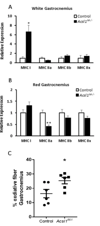
4.1 mRNA expression of MHC isoforms and fiber typing via SDH staining
To investigate fiber type switching at the protein level, we measured mRNA expression
of MHC isoforms via qPCR. In Acsl1M-/- gastrocnemius, mRNA expression of MHC I and MHC
IIa differed from that of control mice (Fig. 2A and B). In white fiber gastrocnemius, Acsl1
M-/-mRNA expression of MHC I was 563% higher compared to control mice (Fig. 2A). Compared
with controls, Acsl1M-/- mRNA expression of MHC IIa was 58% lower in red fiber gastrocnemius
(Fig. 2B). In white and red fiber gastrocnemius, Acsl1M-/- mRNA expression of MHC IIb and MHC
IIx was not significantly different from that of controls (Fig. 2A and B). The percentage of
oxidative fiber area in Acsl1M-/- gastrocnemius were significantly greater than that of control mice
Figure 2. mRNA expression of MHC isoforms and fiber typing via SDH staining. Acsl1 deletion
increased MHC I expression and type I fibers. A: mRNA expression of MHC isoforms in white fibers of gastrocnemius relative to 18S (females; nKO = 5, nWT = 3). B: mRNA expression of MHC isoforms in red fibers of gastrocnemius
relative to 18S (females; nKO = 7, nWT = 3). C: Percentage of type I fiber area in total area of transversely sliced
gastrocnemius stained with succinate dehydrogenase (females; n = 6). D: Histological sections of WT and Acsl1
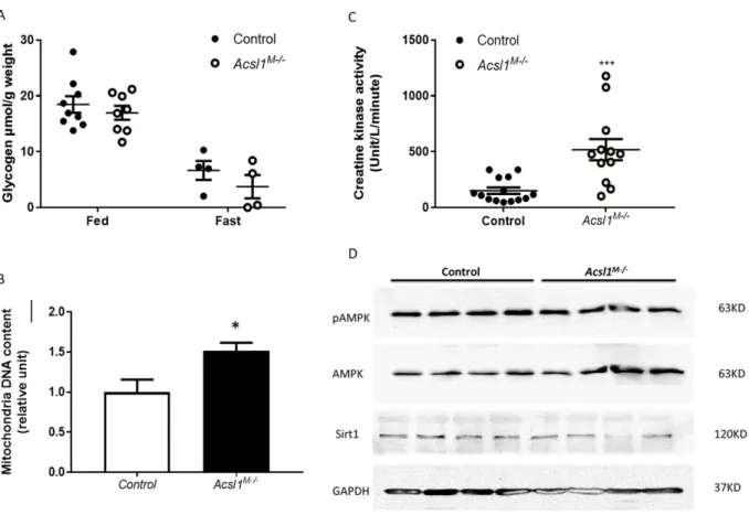
4.2 Glycogen content, creatine kinase activity, Sirt1 and AMPK protein expressions
We investigated difference in glycogen content between control mice and Acsl1M-/- in fed
and 17-h fasted states. No significant difference was observed between control mice and
Acsl1M-/- in either the fed or fasted state (Fig. 3A). Oxidative fibers, such as type I and type IIa,
contain more mitochondria than glycolytic fibers, such as type IIb. By knocking out Acsl1,
mitochondrial content in white gastrocnemius was significantly greater than that of control mice
(Fig. 3B). We also investigated muscle damage by measuring creatine kinase activity. Creatine
kinase activity is a useful serum marker of muscle injury and oxidative stress23, resulting in
increased cellular membrane permeability and therefore, increased movement of
intramyocellular substrates, like creatine kinase, to the extracellular fluid. The creatine kinase
activity was significantly greater in Acsl1M-/- compared to controls (Fig. 3C). Furthermore, we
investigated the proteins involved in PGC1α activation, which is implicated in mitochondrial
biogenesis28. We investigated AMPK and SIRT1, proteins involved in activating the
transcriptional coactivator PGC1α. Via western immunoblotting, no significant differences were
observed in SIRT1, AMPK, and pAMPK protein expression between Acsl1M-/-and control mice
Figure 3. Glycogen content, creatine kinase activity, SIRT1 and AMPK protein expressions. Acsl1
deletion increased creatine kinase activity but did not affect protein expression of SIRT1 and AMPK. A: Glycogen content of fed and 17hr-fasted mice (nfed = 8-9/GT, nfast = 4/GT). B: Mitochondrial DNA content in white fibers of
gastrocnemius (n = 5). C: Creatine kinase activity measured from blood serum (nKO = 12, nWT = 14). D: A western
4.3 Muscle fiber typing via immunofluorescence and subcellular localization of nuclei.
We conducted immunofluorescence to analyze muscle fiber types. In Acsl1M-/-and control
gastrocnemius, anti-MHC I and anti-MHC IIb antibodies were conjugated to a red-emitting
fluorophore and a green-emitting fluorophore, respectively (Fig. 4A). We also further
investigated muscle fiber damage by detecting central nuclei, a marker of myopathy24. Acsl1
M-/-gastrocnemius has more central nuclei compared to that of control mice (Fig. 4B). However,
central nuclei were absent in both Acsl1M-/- and control soleus (Fig. 4B).
Control Acsl
`
Figure 4. Muscle fiber typing via immunofluorescence and subcellular localization of nuclei. Acsl1 plays a role
in subcellular localization of nuclei in gastrocnemius tissue. A: Cross-sections of gastrocnemius were immunostained for MHC I (red), and MHC IIb (green). B: Cross-sections of gastrocnemius and soleus were immunostained for nuclei (blue) sialic acid and N-acetylglucosaminyl residues on outer cell surfaces (green). Arrows indicate central nuclei.
B
ControlAcsl
M-/-So
leu
s
Gas
tro
cne
m
CHAPTER 5: DISCUSSION
5.1 Muscle fiber type switch
Type I muscle fibers have more oxidative enzymes and mitochondria than type II muscle
fibers, which have more glycolytic enzymes. As such, the preferred substrate for type I fibers is
FA whereas for type II fibers is glucose. ACSL1 is specifically required in skeletal muscle
tissues for fatty acid oxidation therefore Acsl1 deletion should result in a more glycolytic
phenotype when FA use becomes limited. Thus, we expected that a switch from type I to type II
muscle fibers would occur in Acsl1M-/-.
Our data suggest that the deletion of Acsl1 in mice results in the increase of type I fibers
and decrease of type IIa fibers. This is a surprising result because deletion of Acsl1, which is
required in skeletal muscle fatty acid oxidation, should have decreased the need for
mitochondria since FAs cannot enter the mitochondria for oxidative phosphorylation. An
increase in the number of type I fibers would increase the number of mitochondria, thus
maximizing oxidative phosphorylation. However, β-oxidation reduction from Acsl1 deletion
should decrease the acyl-CoA concentration that would otherwise participate in oxidative
phosphorylation. As such, we did not expect type I fibers to increase because they rely primarily
on aerobic respiration.
Cellular ATP production must still be maximized even though oxidative phosphorylation is
limited. Because FA use is limited within the muscle, the major fuel substrate becomes glucose
and amino acids. However, glycolysis may not be the major pathway for energy production
since this would only provide 2 net ATPs per glucose molecule. As such, most ATP would still
be generated via oxidative phosphorylation in the through NADH produced from pyruvate
decarboxylation and the TCA cycle. To maximize ATP production, oxidative phosphorylation
decreased.
5.2 Molecular mechanism of fiber type switch
Our data suggest that Acsl1 deletion results in increased mitochondrial biogenesis.
PGC1α, which is activated by SIRT1 deacetylation and AMPK phosphorylation, increases
mitochondrial biogenesis28 and MHC I expression13. SIRT1, AMPK and pAMPK expression was
similar in Acsl1M-/- and control gastrocnemius muscles thus the PGC1α activation must also be
similar.
Because AMPK and pAMPK expression were similar, having similar cellular AMP/ATP
between Acsl1M-/- and control gastrocnemius could be a strong possibility. Since increased
AMP/ATP triggers the activation of AMPK and Acsl1M-/- and control gastrocnemius have similar
AMP/ATP, the expression of activated PGC1α should also be similar.
Interestingly, if PGC1α activation was similar in Acsl1M-/- and control gastrocnemius then
mitochondrial biogenesis should also be similar. However, Acsl1M-/- gastrocnemius has more
mitochondria than control and if PGC1α activation is similar, then it must mean that
mitochondrial biogenesis is activated by another pathway. Because we only measured SIRT1,
AMPK, and pAMPK, it may be possible that other proteins are upregulated to increase PGC1α.
5.3 ACSL1 knockout and muscle fiber damage
The increase in creatine kinase activity and the greater number of central nuclei suggest
that muscle fiber damage is greater in Acsl1M-/-than control mice. It is possible that Acsl1M-/- has
more intramuscular FAs than control mice when reduced β-oxidation limits FA use. Thus, FA
accumulation will lead to crowding along the mitochondrial membrane, which will facilitate the
entry of neutral FAs into the mitochondrial matrix30. With increased FAs that are more likely to
that would permeate the mitochondrial membrane25. The mitochondria would then release
oxidative-inducing enzymes, which would permeate the cellular membrane and lead to cell
death. The dead muscle would release various cellular substrates including creatine kinase.
With more impaired mitochondria, oxidative capacity is reduced and therefore may lead to
increased mitochondrial biogenesis as seen in our Acsl1M-/-.
Myocellular damage is often accompanied by myocellular regeneration, which is evident
by the presence of central nuclei26. In Acsl1M-/-, the presence of central nuclei is more apparent
in gastrocnemius than soleus, which contains mostly type I fibers. It is possible that the muscle
fiber type switch from type II to type I could trigger muscle damage. In the absence of miR-133a,
a muscle-specific microRNA, centronuclear myopathy (CNM) phenotype was also evident in
type II fibers but not type I24. Similarly, the knockout of Srpk3, a muscle-specific protein kinase,
also resulted in a CNM phenotype, specifically for type II fibers27. Both miR-133a and SRPK3
are muscle-specific proteins involved in normal myofiber growth and development2424,32 and
deficiency in either protein resulted in skeletal myopathy similar to Acsl1M-/-. Thus SRPK3,
miR-133a, and ACSL1 might act together to regulate muscle structure and nuclear localization.
However, the mechanism through which this occurs remains largely unknown.
5.4 Limitations
A major limitation of this study is that only one isoform of Acsl is knocked out. Thus,
other isoforms of Acsl could play a role in activating FAs for β-oxidation which could then result
in more subdued muscle fiber type switching, myocellular damage, and mitochondrial
biogenesis. Other ACSL isoforms (ACSL 3, ACSL 4, ACSL5, and ACSL 6) have tissue-specific
expression and differ in activity. ACSL3 and ACSL6 are predominantly expressed in skeletal
muscle however ACSL1 have a more predominant role in FA oxidation use, constituting 90% of
Another limitation of this study is the sole use of gastrocnemius in most of our
experiments. Different muscles have different composition of fiber types so it may be possible
that the effects of muscle-specific Acsl1 KO might vary. Acsl1M-/- soleus has increased TAG,
muscle weight to body weight ratio. Acsl1 deletion resulted in increased central nuclei for
soleus, but the percentage of central nuclei is lower than that of gastrocnemius. In soleus, fatty
acid oxidation and ACSL activity are reduced, but they are not as pronounced as in
gastrocnemius.
It might also be possible that Acsl1M-/- require greater PDH activity than control mice to
reach similar energy levels when oxidative phosphorylation is limited due to lack of oxidized FA;
thus, it would be informative to measure PDH activity. While we did measure the proteins
involved in PGC1α activation, measuring PGC1α protein levels could offer a more direct relation
for the increased mitochondrial biogenesis. Phenotypically Acsl1M-/- mice cannot run as far as
control mice when oxidative capacity of FA is limited4. Thus, when glucose and amino acids are
exhausted, SIRT1 and AMPK expression may also differ between Acsl1M-/- and control
gastrocnemius to meet energy requirements by increasing mitochondrial biogenesis through
activation of PGC1α, which is also activated by exercise29. Although SDH provides a rough
estimate of oxidative fibers, it does not distinguish between type IIa and type I because it only
detects Complex II in mitochondria. To more accurately perform fiber type analysis, different
muscle fibers should be labeled with fluorophores of different emission wavelengths.
5.5 Future directions
In this work, we have explored the effects of absent ACSL1 on muscle fiber type
switching. By knocking out Acsl1 in mice gastrocnemius, more MHC I mRNA was present
oxidative fiber phenotype. However, this switch in phenotype results in muscle fiber damage
which was shown through the increased creatine kinase activity and presence of centronuclear
myopathy. It would be expected that Acsl1M-/- has more FAs and other lipid metabolites in
skeletal myocytes. Therefore, AKT signaling would be impaired4 which could result in decreased
signal transduction that would otherwise result from insulin binding. As such, Acsl1 may
REFERENCES
1. Ellis JM, Frahm JL, Li LO, Coleman RA. (2010). Acyl-coenzyme A synthetases in metabolic
control. Current Opinion in Lipidology. 21: 212–217.
2. Li LO, Klett EL, Coleman RA. (2010). Acyl-CoA synthesis, lipid metabolism and lipotoxicity.
Biochimica et Biophysica Acta. 1801: 246–251.
3. Li LO, Ellis JM, Paich HA, Wang S, Gong N, Altshuller G, Thresher RJ, Koves TK, Watkins
SM, Muoio DM, Cline GW, Shulman GI, Coleman RA. (2009). Liver-specific loss of long
chain acyl-CoA synthetase-1 decreases triacylglycerol synthesis and beta-oxidation and
alters phospholipid fatty acid composition. Journal of Biological Chemistry. 284: 27816–
27826.
4. Li LO, Grevengoed TJ, Paul DS, Ilkayeva O, Koves TR, Pascual F, Newgard CB, Muoio DM,
Coleman RA. (2015). Compartmentalized acyl-CoA metabolism in skeletal muscle regulates
systemic glucose homeostasis. Diabetes. 64: 23-35.
5. Samuel VT, Shulman G. (2012). Mechanisms for insulin resistance: Common threads and
missing links. Cell. 148: 852–871.
6. Savage DB, Petersen KF, Shulman GI. (2007). Disordered lipid metabolism and the
pathogenesis of insulin resistance. Physiological Reviews. 87: 507–520.
7. Ragheb R, Shanab GM, Medhat AM, Seoudi DM, Adeli K, Fantus IG. (2009). Free fatty
acid-induced muscle insulin resistance and glucose uptake dysfunction: Evidence for PKC
activation and oxidative stress-activated signaling pathways. Biochemical and Biophysical
8. Bajaj M, Suraamornkul S, Romanelli A, Cline GW, Mandarino LJ, Shulman GI, DeFronzo
RA. (2005). Effect of a sustained reduction in plasma free fatty acid concentration on
intramuscular long-chain fatty Acyl-CoAs and insulin action in type 2 diabetic
patients. Diabetes. 54: 3148–3153.
9. Grevengoed TJ, Cooper DE, Young PA, Ellis JM, Coleman RA. (2015). Loss of long-chain
acyl-CoA synthetase isoform 1 impairs cardiac autophagy and mitochondrial structure
through mechanistic target of rapamycin complex 1 activation. The FASEB Journal. 29(11):
4641-4653.
10. Boden G. (2009). Obesity and Free Fatty Acids (FFA). Endocrinology Metabolism Clinics of
North America. 37(3): 635-ix.
11. Burke RE, Levine DN, Zajac FE. (1971). Mammalian motor units: physiological
histochemical correlation in three types in cat gastrocnemius. Science. 174: 709-712.
12. Fry AC, Housh TJ, Cramer JB, Weir JP, Beck TW, Schilling BK, Miller JD, Nicoll JX. (2016).
Non-invasive assessment of skeletal muscle myosin heavy chain expression in trained and
untrained men. Journal of Strength and Conditioning.
13. Handschin C, Chin S, Li P, Liu F, Maratos-Flier E, LeBrasseur NK, Yan Z, Spiegelman B.
(2007). Skeletal muscle fiber type switching exercise intolerance, and myopathy in PGC-1α
muscle-specific knock-out animals. Journal of Biological Chemistry. 282: 30014-30021.
14. Harridge SDR, Bottinelli R, Canepari M, Pellegrino MA, Reggiani C, Esbjornsson M, Saltin
B. (1996). Whole muscle and single fibre contractile properties and myosin heavy chain
isoforms in humans. Pflϋgers Archive. 432: 913-920.
15. Liu J, Liang X, Gan Z. (2015). Transcriptional regulatory circuits controlling muscle fiber type
switching. Science China Life Sciences. 58: 321.
17. Saladin KS. (2014). Muscle metabolism. Anatomy and Physiology: The Unity of Form and
Function. 7: 421-423.
18. Schiaffino S, Gorza L, Sartore S, Saggin L, Ausoni S, Vianello M, Gundersen K, Lomo T.
(1989). Three myosin heavy chain isoforms in type 2 skeletal muscle fibres. Journal of
Muscle Research and Cell Motility. 10: 197-205.
19. Schiaffino S, Reggiani C. (2011). Fiber types in mammalian skeletal muscles. Physiological
Reviews. 91(4): 1147-1531.
20. Scott W, Stevens J, Binder-MacLeod, SA. (2001). Human skeletal muscle fiber type
classifications. Physical Therapy. 81(11): 1810-1816.
21. Smerdu V, Karsch-Mizrachi I, Campione M, Leinwand L, Schiaffino S. (1994). Type IIx
myosin heavy chain transcripts are expressed in type IIb fibers of human skeletal muscle.
American Journal of Physiology - Cell Physiology. 267: C1723-C1728.
22. Zierat JR, Hawley JA. (2004). Skeletal muscle fiber type: influence on contractile and
metabolic properties. PLOS Biology. 2(10): e348.
23. Brancaccio P, Lippi G, Maffulli N. (2010). Biochemical markers of muscular damage. Clinical
Chemistry and Laboratory Medicine. 48(6).
24. Liu N, Bezprozvannaya S, Shelton JM, Frisard MI, Hulver MW, McMillan RP, Wu Y, Voelker
KA, Grange RW, Richardson JA, Bassel-Duby R, Olson EN. (2011). Mice lacking microRNA
133a develop dynamin 2-dependent centronuclear myopathy. The Journal of Clinical
Investigation. 121(8): 3258-3268.
25. Schrauwen P, Hesselink MKC. (2004). Oxidative capacity, lipotoxicity, and mitochondrial
damage in type 2 diabetes. Diabetes. 53(6): 1412-1417.
26. Pichavant C, Pavlath GK. (2014). Incidence and severity of myofiber branching with
27. Nakagawa O, Arnold M, Nakagawa M, Hamada H, Shelton JM, Kusano H, Harris TM, Childs
G, Campbell KP, Richardson JA, Nishino I, Olson EN. (2005). Genes and Development.
19(17): 2066-2077.
28. Cantó C, Auwerx J. (2009). PGC-1alpha, SIRT1, AMPK, an energy sensing network that
controls energy expenditure. Current Opinion in Lipidology. 20(2): 98-105.
29. Ruderman NB, Xu XJ, Nelson L, Cacicedo JM, Saha AK, Lan F, Ido Y. (2010). AMPK and
SIRT1: a long-standing partnership. American Journal of Physiology-Endocrinology and
Metabolism. 298: E751-E760.
30. Ho JK, Duclos RI Jr, Hamilton JA. (2002). Interactions of acyl carnitines with model
membranes: a (13)C-NMR study. Journal of Lipid Research. 43: 1429-1439.
31. Russell AP, Gastaldi G, Bobbioni-Harsch E, Arboit P, Gobelet C, Deriaz O, Golay A,
Witztum JL, Giacobino JP. (2003). Lipid peroxidation in skeletal muscle of obese as
compared to endurance-trained humans: a case of good vs. bad lipids? FEBS Letters. 551:
104-106.
32. Nakagawa O, Arnold M, Nakagawa M, Hamada H, Shelton JM, Kusano H, Harris TM, Childs
G, Campbell KP, Richardson JA, Nishino I, Olson EN. (2005). Centronuclear myopathy in
mice lacking a novel muscle-specific protein kinase transcriptionally regulated by MEF2.
Genes and Development. 19: 2066-2077.
33. Teodoro BG, Sampaio IH, Bomfirm LHM, Queiroz AL, Silveira LR, Souza AO, Fernandes
AMAP, Eberlin MN, Huang T, Zheng D, Neufer PD, Cortright RN, Alberici LC. (2017).
Long-chain acyl-CoA synthetase 6 regulates lipid synthesis and mitochondrial oxidative capacity