Effects of an extension-constraint functional knee brace on the ankle mechanics of adolescent athletes in a jump-landing task 6 months post-ACL reconstruction
By Sarah Wilkey
Honors Thesis Exercise and Sport Science
University of North Carolina at Chapel Hill April 2014
Approved:
_______________________ Meredith Petschauer, PhD _______________________ Robin Queen, PhD
Abstract
Context: The prevalence of secondary ACL injury is highest among adolescents.1 It is important to understand how both surgery and rehabilitation affect lower extremity biomechanics in this population because poor lower extremity mechanics can contribute to ACL tears.2 Functional knee bracing following reconstructive surgery could potentially reduce the occurrence of secondary tears by correcting harmful mechanics at the knee. Altering knee kinetics and kinematics may also have an impact on the mechanics of other joints in the kinematic chain, particularly the ankle.3 It is important to monitor for these effects because poor ankle mechanics can potentially contribute to further injury at any of the joints along the kinematic chain, including secondary ACL tear.
Hypothesis: It was hypothesized that wearing a functional knee brace would increase vertical ground reaction force, decrease ankle range of motion in the sagittal plane, and increase ankle moments in the sagittal plane.
Study Objective: To determine whether or not wearing a functional knee brace during rehabilitation affects ankle mechanics in patients six months following ACL
reconstruction surgery.
paired-Results: There were no significant differences found between the braced and non-braced conditions for any of the six variables being observed. These variables included peak vertical ground reaction force, peak dorsiflexion angle, peak plantar flexion angle, total ankle range of motion, peak plantar flexion moment, and peak dorsiflexion moment. Conclusions: Extension-constraint functional knee bracing seems to have no effect on the mechanics of the ankle on the surgical side.
Table of Contents
Chapter I: Introduction ... 4
Research Question ... 7
Hypothesis ... 7
Definitions ... 8
Limitations ... 8
Delimitations ... 9
Assumptions ... 10
Variables ... 10
Chapter II: Literature Review ... 11
Introduction ... 11
Biomechanics ... 11
ACL Injury and Rehabilitation ... 13
Knee Mechanics ... 18
Ankle Mechanics ... 19
Relationship Between the Knee and Ankle ... 20
Conclusion ... 21
Chapter III: Methodology ... 23
Subjects ... 23
Data Collection Procedure ... 23
Data Reduction and Analysis ... 25
Chapter IV: Results ... 27
Chapter V: Conclusion ... 30
Chapter I: Introduction
Normal biomechanics throughout the kinematic chain of the lower extremity (hip, knee, and ankle) allow for the most efficient attenuation of force in weight-bearing activities.32 It is important to maintain normal mechanics, which limit the magnitudes of the forces experienced at each joint, in order to prevent injury. When injury does happen at one of these joints, avoidance is likely to occur due to pain and instability. The subsequent change in range of motion (ROM) and decrease in weight bearing at the effected joint will result in altered ROM and increased loads in the other joints along the chain.1,3 Greater demands may also be placed on the contralateral limb for weight-bearing and force absorption. Surgery and proper rehabilitation are typically employed to repair a severely injured joint and reintroduce normal mechanics. Prophylactic devices, such as functional knee braces, are designed to provide stability to the joint and prevent re-injury in recovery and return to sport.1,17-20 It is beneficial to look at the entire kinematic chain to assess benefits as well as risks when evaluating the effectiveness of rehabilitative devices.
If the ACL is not surgically repaired after rupture, the patient will experience instability in the effected joint.1 There will also be a significant decrease in sagittal plane range of motion at the knee. These asymmetries will result in greater demands on the uninjured knee, increasing the risk for contralateral ACL rupture.9 Another potential consequence is altered mechanics of the ipsilateral ankle joint due to avoidance and decreased range of motion at the knee joint.10,33 The quadriceps and gastrocnemius have a neural connection, so a decrease in neuromuscular control at the quadriceps from an ACL rupture could yield weakness in the gastrocnemius.33
Because of the loss of stability and ROM, reconstructive surgery is often required when the ACL is ruptured. In one of the more common procedures, the surgeon will use a graft of the patient’s own semitendinosus (hamstring) tendon to replace the torn ligament. Some issues that patients face following the procedure include muscle weakness, loss of muscle mass, considerably reduced joint ROM, and impaired joint proprioception.1 It is hypothesized that these symptoms are due to a diminished ability to activate motor units, otherwise known as muscle inhibition. Muscle inhibition may result in long-term strength deficits, causing alterations in joint kinetics of the effected joint and throughout the kinematic chain.1
Surgery is often done within a few weeks of injury and athletes are cleared to return to sport 2 to 6 months following the procedure.29 It is important to begin rehabilitation as soon as possible following surgery in order to prevent degradation of the ligamentous, muscular, and articular structures of the knee joint.14 Research has shown that even after a successful rehabilitative program, patients are likely to continue to experience limb asymmetries 6 to 9 months following surgery.16 Some rehabilitation programs have adapted to these findings by emphasizing early weight bearing, knee ROM restoration, and muscle strengthening programs in order to reduce the magnitude of the asymmetries and return the joint to near-normal function in a shorter period of time.16
The Fourcepoint brace, by DonJoy Orthopedics, is a newer design that was developed to limit knee extension in order to prevent stiffening of the ACL and allow for greater absorption of GRFs by increasing the amount of knee flexion.23 The brace uses an incremental resistance hinge that resists knee extension between 40° and 10°. It does this by gradually increasing resistance to a maximum of 3.5 Nm just before 10°, at which point a stop mechanism is employed to prevent further extension.38,39 The outcomes of this device seem ideal, but Dai et. al. found that it may not work as advertised because the reduction in knee ROM was less than the suggested 30°.38 Also, research on its effectiveness appears to be limited to the knee joint.
Further investigation should be done to assess the effects it has on the mechanics of other important joints in the kinematic chain. If the brace negatively alters kinetic and/or kinematic aspects of the ankle, training with the brace could result in lasting mechanical adaptations that may increase the risk of future injury. Specific variables that should be analyzed are: peak VGRF, peak dorsiflexion angle, peak plantar flexion angle, total ankle ROM, and peak dorsiflexion moment, and peak plantar flexion moment.
I. Research Question
a. What effect does an extension-constraint functional knee brace have on peak vertical ground reaction force when performing a jump/landing task at 6 months post-ACL reconstruction?
c. What effect does an extension-constraint functional knee brace have on dorsiflexion and plantar flexion moments when performing a jump/landing task at 6 months post-ACL reconstruction?
II. Hypothesis
a. The functional knee brace will result in a significantly larger peak vertical ground reaction force.
b. The functional knee brace will significantly reduce total ankle range of motion by decreasing peak dorsiflexion and plantar flexion angles.
c. Functional knee bracing will result in a greater dorsiflexion and plantar flexion moments.
II. Definitions
a. Range of motion (ROM): The angle through which a joint has the ability to move.1
b. Dorsiflexion: Rotation of the foot towards the anterior tibia in the sagittal plane.1
c. Plantar flexion: Rotation of the foot away from the tibia in the sagittal plane.
d. Ground reaction force (GRF): The force exerted by the ground on a body when they come in contact with one another.1
e. Valgus force: A force that drives the joint medially with respect to the distal segment. .1
g. Kinematic Chain: A succession of joints that are linked together in the body.2
III. Limitations
a. Information regarding subject compliance with wearing the brace for 4 weeks is limited.
b. Information on whether or not brace was worn during high loading activities during the 4 weeks is limited.
c. The testing order was not randomized.
d. The study only looked at adolescents, limiting the external validity to other age populations.
e. Concomitant meniscal tears in some of the subjects could have affected the results.
f. Ankle data were not compared between limbs.
g. Data does not exist for the pre-injury, lower-extremity mechanics of the subjects.
IV. Delimitations
a. All subjects had undergone ACL reconstruction surgery via a tibial tunnel-independent technique with a hamstring graft.
b. All subjects had completed a physical therapy program under the direction of a licensed physical therapist.
d. Prior to testing, all participants had been released to return to sport by their treating surgeons.
e. All subjects were issued the same design of knee brace and provided a custom fitting by a trained company representative.
f. All subjects were asked to wear the knee brace for a minimum of 4 weeks prior to testing.
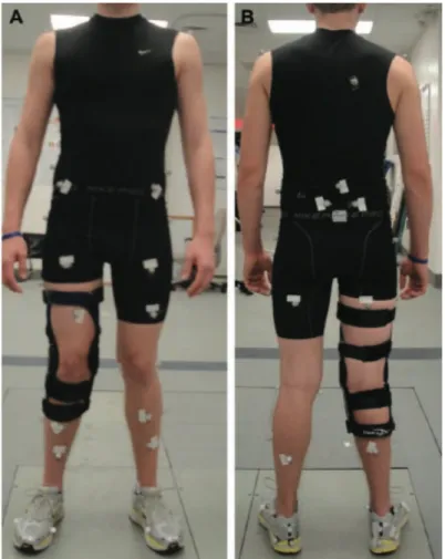
g. All subjects donned similar attire and footwear in the motion analysis. h. Retro-reflective markers were placed on the same landmarks on each
subject for motion capture and analysis.
i. All subjects performed the same jump-landing task in both the braced and un-braced conditions.
j. Appropriate time for rest was provided in-between trials to minimize the effects of fatigue
V. Assumptions
a. It is assumed that subjects complied with wearing the brace 4 weeks prior to the study.
b. It is assumed that the braces were properly fitted to each subject. VI. Variables
a. Independent:
i. Extension-constraint knee brace (DonJoy Orthopaedics LLC, Vista, CA, USA)
b. Dependent:
ii. Peak dorsiflexion angle iii. Peak plantar flexion angle iv. Total ankle ROM
Chapter II: Literature Review
Introduction
Ankle biomechanics have important implications on the mechanics of the knee joint, especially with regard to force transfer.1,3 Along the same lines, altered knee mechanics, specifically reduced range of motion in quick flexion and extension, can result in greater demands on the ankle joint for force absorption.3 Anterior cruciate ligament injuries and reconstructive surgery limit the mobility of the knee.1,6-9,12-16 Prophylactic bracing is often prescribed during rehabilitation to stabilize the knee joint and prevent re-injury in recovery and return to sport.1,17-20 These devices are used to stabilize and protect a post-operative knee joint, but can limit postural control, muscle activation, and athletes’ speed and agility.17-20 The clinical benefits of functional knee bracing following ACL reconstruction surgery are limited and bracing can have significant negative effects on the kinetics and kinematics of the ankle joint in demanding activities.18,19,20
Biomechanics
Biomechanics is the science through which mechanical principles are used to analyze the human body in motion and at rest.1 Biomechanical principles are particularly useful in analyzing specific mechanisms of injury. Understanding these mechanisms of injury can be beneficial in determining risk factors and minimizing them to prevent initial and secondary injury. These principles are often applied when evaluating preventative equipment to determine its potential to reduce or eliminate risk factors.22
Range of Motion
injury. Pain, muscle weakness, and even disunion can cause a joint to behave not as it should. Decreased range of motion at one joint often results in greater demands on the contralateral limb and the other joints of the ipsilateral limb.
Ground Reaction Force
The force exerted by the ground on a body when they come in contact with one another is known as the ground reaction force (GRF).1 Ground reaction forces are divided into vertical force, fore-aft shear, and medial-lateral shear. The concept of this type of force is based off Newton’s third law, which states that for every action there must be an equal and opposite reaction.1 In the case of a landing from a jump-landing task, the action is landing and the reaction is the force (equal in magnitude to the action force) that the ground places back on the body. Magnitudes of these forces can be obtained in a lab setting through the use of force plates.
Valgus Force
The term valgus refers to the outward angulation of the distal segment of a bone or joint when viewed in the frontal plane.1,27,28 A valgus force is one that drives the joint medially with respect to the distal segment. Valgus forces are key risk factors in both contact and non-contact ACL injuries.
Moment
Kinematic Chain
With respect to biomechanics, the term “kinematic chain” refers to a succession of joints that are linked together in the body. These chains can be further classified as “opened” or “closed,” based on the continuity of the structure. Kinematic chain refers only to the intrinsic structure of the chain, not the extrinsic movements associated with treatment of it. The lower extremity, which consists primarily of the ankle, knee, and hip joints, is considered an open kinematic chain due to its discontinuous nature.2
Kinetic Chain
The term “kinetic chain” is primarily used in the context of physical therapy to describe treatment exercises. 2 “Open kinetic chain” refers to exercises where the distal limb is not fixed against resistance. These activities put greater demand on the ligamentous structures of the joint in motion as there is less muscle activation involved. Oppositely, “closed chain kinetic chain” exercises involve some sort of resistance at the distal limb; in these exercises there is greater stability at the joint as a result of increased muscle activation.
ACL Injury and Rehabilitation
Injury Mechanisms
extension it accepts more of the forces applied at the joint.1 A ground reaction force of high magnitude stresses the ACL and is a risk factor for injury. Large GRFs are related to significant knee valgus displacement and moment.27 Valgus forces create large moments at the knee and the ACL naturally resists these during landing.24 This resistance translates to greater strain of the ACL and higher potential for rupture.24 An outside valgus force applied to an extended knee, like in a side-tackle, is most often the cause of injuries in contact cases.1 Knee rotation in combination with a valgus force generates an even greater strain on the ACL, increasing potential for rupture.34 In both contact and non-contact cases, the injury usually occurs at footstrike with the knee approaching full extension.1
Implications
there to be knee stability during cutting and pivoting in an ACL deficient individual, but it is more common for local instability and other debilitating effects to occur in those suffering from ACL ruptures.10 A potential implication of ACL rupture is altered mechanics of the ipsilateral ankle joint.10 This would be the result of higher demands on the ankle joint for force absorption due to avoidance and decreased range of motion at the knee joint. Further investigation is needed to confirm this suggestion.
Reconstructive Surgery
Since a high degree of stability and range of motion is lost after an ACL tear, surgical intervention is often required to improve knee mechanics. Reconstruction of the ligament can be done using either an autograft, the patient’s own tissue, or an allograft, tissue foreign to the patient.11 Grafts are commonly done using the middle portion of the patellar tendon, the semitendinosus, or a combination of the semitendinosus and gracialis.1 Quadriceps atrophy and strength deficits are commonly experienced after the operation. Even with proper rehabilitation, the size and strength of the quadriceps does not return to normal for years following surgery.12 Patients receiving a semitendinosus autograft utilize an adapted landing strategy resulting in decreased activation of the hamstrings muscle and decreased hip extension. This is likely due to the fact that a graft of the semitendinosus reduces hamstring fixation to the bone. A patellar tendon graft does not run this risk and would not affect the hamstrings. The decreased range of motion at the hip, in combination with the limited mobility of the knee, results in increased plantar flexion at the ankle.15 This significant increase in the demands on the ankle is worth investigating for further ramifications.
reduce the magnitude of these asymmetries and return the joint to near-normal function in a shorter period of time.25
Arguments for Bracing
The biomechanical behavior of the knee joint never returns to normal after an ACL graft.14 Though current reconstruction procedures yield normative behavior of the ACL in anterior tibial loading, the integrity of the kinematics of the ACL during rotational and muscle loading is not restored. It takes a long time for the graft to re-vascularize and heal, so many patients use functional knee braces to provide a protective strain shielding effect on the ACL.14 Braces can protect the knee from anterior shear loads and internal moment applied to the knee in non-weight bearing conditions.14 Bracing can also improve performance during certain tasks requiring somatosensory input.18 A new line of braces has been developed that limit knee extension. They were designed to limit the stiffening of the ACL in extension as well as reduce ground reaction forces through increased knee flexion.23 By reducing some of the loads placed on the ACL during activity, like the anterior shear placed on the tibia by the patellar tendon in extension, these extension-constraint braces may be valuable in rehabilitating ACL injuries.23
Arguments Against Bracing
anterior shear loads and internal torques that are applied to the knee in non-weight bearing conditions, however increased magnitudes of these forces decrease the strain-shielding effect.14 Further, bracing improves performance during tasks requiring limited somatosensory input, but this does not carry over to more functionally relevant tasks where performance becomes important.18 Another negative implication of bracing is that it can hinder athletic performance and speed months after ACL reconstructive surgery.19 This may be partially explained by the finding that knee bracing causes delays in muscle recruitment and activation.20 Though Yu and colleagues found an extension-constraining knee brace to decrease knee extension angles by an average of 5° in a jump-landing task, the brace did not significantly affect peak ground reaction forces during landing.23 The potential benefits and/or drawbacks of functional bracing can further be assessed by observing the effect it has on ankle mechanics.
Knee Mechanics
Range of Motion
Sagittal plane rotation of the knee involves flexion and extension. The CDC’s Normal Joint Range of Motion Study summarizes typical range of motion values in adolescent
muscles results in hip extension, knee flexion, and medial tibial rotation. The antagonist muscles to the hamstrings are the quadriceps. They cross the hip and knee joints anteriorly and their contraction causes hip flexion and knee extension. Contraction of the quadriceps is a primary source of knee extensor moment as they generate an anteriorly directed force on the tibial tuberosity via the patellar tendon.26 This moment is related to the force that is transferred to the ACL during walking.26 The medial and lateral condyles of the femur differ slightly in size and shape from the medial and lateral condyles of the tibia. This asymmetry results in what is known as the “screw-home” mechanism, where the tibia rotates laterally on the femur during last few degrees of extension in order to “lock” the knee joint.1 Since there is significant genetic variability in the structure of the tibial plateau, the knee joints of certain individuals may be less stable and more prone to injury than others.1 The gastrocnemius muscle, which aids in knee flexion and plantar flexion, originates at the posterior, distal base of the femur and inserts on the Achilles tendon of the heel.1 The bi-articularity of the muscle allows work to be transferred from the knee extensor to the ankle, resulting in plantar flexion.4 Actions of these bi-articular muscles are highly relied upon for force production as well as ground reaction force absorption in gait and jump/landing mechanics.4
Ankle Mechanics
Movement at the ankle joint primarily involves the sagittal plane. Ankle rotation in this plane is described by the terms dorsiflexion and plantar flexion.
Dorsiflexion is when the top of the foot is brought towards the anterior tibia.1 The primary muscles responsible for dorsiflexion, located anteriorly to the tibia and fibula, are the tibialis anterior, the extensor digitorum longus, and the peroneus tertius.
Plantar Flexion
The movement of the top of the foot away from the tibia is referred to as plantar flexion. The main plantar flexors, situated posteriorly to the tibia and fibula, are the gastrocnemius and the soleus.1 The gastrocnemius is also one of the muscles responsible for knee flexion.
Range of Motion
According to the CDC’s Normal Joint Range of Motion Study, normal ankle dorsiflexion is 17.3 ± 1.7 degrees for adolescent females and 16.3 ± 1.4 degrees for adolescent males. Normal ankle plantar flexion according to this study is 57.3 ± 2.5 degrees for adolescent females and 52.8 ± 2.0 degrees for adolescent males. Injury at any joint along the kinetic chain can threaten normal ROM in the ankle joint.
Relationship Between Knee and Ankle
these joints, not only will the normal range of motion of the affected joint be altered, but the normal range of motion at the other joints in the kinematic chain will also be disturbed.28 Patellofemoral pain and instability result in greater demands on the plantar flexors due to a decreased range of motion at the knee.28 When the plantar flexors are increasingly loaded they become stiff, decreasing dorsiflexion ROM at the ankle joint.28 Decreased dorsiflexion ROM is associated with lower than normal knee flexion and greater GRFs.27,28 Since amplified GRFs are associated with increased valgus displacement in landing tasks, these altered kinetics put the ACL at risk of re-injury.27, 28 Clinical techniques to reduce plantar flexor stiffness and increase dorsiflexion ROM would benefit ACL injury prevention and rehabilitation.
Conclusion
*Disclaimer: Duke University’s Dr. Robin M. Queen and colleagues developed these methods.
Chapter III: Methodology
Subjects
This study looked at thirteen female and seven male adolescent patients (Age: 15.8 ± 1.2 yr; Height: 1.7 ± 0.1 m; Mass: 71.7 ± 16.8 kg; Time following ACL-R: 6.4 ± 0.5 months). All subjects received tibial tunnel-independent ACL reconstruction surgery with a hamstring auto-graft. Of the 20 subjects, 13 had simultaneous meniscus repair or meniscotomy. Patients with a previous history of ACL reconstruction or other lower extremity surgery were excluded from the study. All subjects were high school or collegiate athletes who planned on returning to sports involving cutting and jumping following surgery. After reconstructive surgery, each subject completed a standard rehabilitation program under the supervision of a licensed physical therapist. Prior to involvement in the study, all subjects signed informed consent that had been pre-approved by Duke University Medical Center’s institutional review board.
Data Collection Procedure
procedures required subjects to perform two sets of 5 vertical stop jump tasks. The first set was performed with a functional knee brace on the surgical limb and the second set was performed with no brace. Each subject was provided a custom fit functional knee brace for this study (DonJoy Orthopaedics LLC, Vista, CA, USA). The brace was made to resist knee extension by gradually increasing resistance between 40° and 10° of knee flexion. A maximum resistance of 3.5 Nm is achieved by the brace at 10° of knee
Data Reduction and Analysis
The coordinate data were filtered using a low-pass Butterworth filter at 12 Hz. The ground reaction force data were filtered using a low-pass Butterworth filter at 100Hz. Time series data for the kinematics and kinetics variables were calculated using Visual 3D software (C-Motion, Bethesda, Maryland, USA). Joint angles were calculated as Cardan angles between adjacent local segments and joint moments were calculated through an inverse dynamic approach and transferred into the local segment coordinate system and were expressed as internal moments. Ground reaction forces were normalized to body weight. Joint moments were normalized to body weight and height. All data were analyzed from the first point of contact on the force plates, the initial phase of landing, to when the subject left the plate again at take-off.
Sagittal plane variables at specific events were extracted for analysis due to their importance in ACL loading. These variables also play an important role in ankle loading and range of motion (ROM). Using subroutines developed in Matlab R2010a (MathWorks Inc., Natick, MA, SA), key variables were extracted from the time-series output for analysis: peak vertical ground reaction force (VGRF), peak dorsiflexion angle, peak plantar flexion angle, total ankle ROM, peak dorsiflexion moment, and peak plantar flexion moment.
Chapter IV: Results
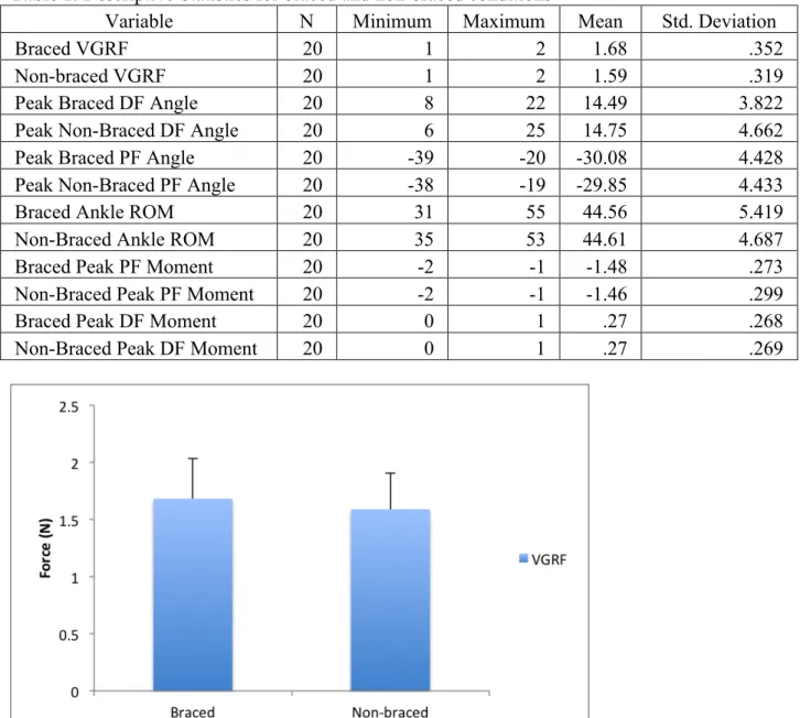
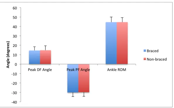
The paired samples t-tests revealed that there were no statistically significant differences between the braced condition and the non-braced condition for any of the six variables: vertical ground reaction force, peak dorsiflexion angle, peak plantar flexion angle, total ankle range of motion, peak plantar flexion moment, and peak dorsiflexion moment.
Table 1. Descriptive Statistics for braced and non-braced conditions
Variable N Minimum Maximum Mean Std. Deviation
Braced VGRF 20 1 2 1.68 .352
Non-braced VGRF 20 1 2 1.59 .319
Peak Braced DF Angle 20 8 22 14.49 3.822
Peak Non-Braced DF Angle 20 6 25 14.75 4.662
Peak Braced PF Angle 20 -39 -20 -30.08 4.428
Peak Non-Braced PF Angle 20 -38 -19 -29.85 4.433
Braced Ankle ROM 20 31 55 44.56 5.419
Non-Braced Ankle ROM 20 35 53 44.61 4.687
Braced Peak PF Moment 20 -2 -1 -1.48 .273
Non-Braced Peak PF Moment 20 -2 -1 -1.46 .299
Braced Peak DF Moment 20 0 1 .27 .268
Chapter V: Discussion
The purpose of this study was to evaluate the effects of knee bracing on ankle mechanics during a jump-landing task in adolescent patients 6 months following ACL reconstruction. When comparing the braced condition to the non-braced condition, it was hypothesized that under the braced condition the involved limb would experience significantly greater vertical ground reaction force and that the ankle would, in the sagittal plane, experience decreased dorsiflexion range of motion, decreased plantar flexion range of motion, and decreased total range of motion as well as increased dorsiflexion and plantar flexion moments. The results failed to support any of these hypotheses. No significant differences were found between the braced and non-braced conditions in vertical ground reaction force, range of motion at the ankle, or moment at the ankle. Some of these results were inconsistent with previous findings, however the related literature is limited.
knee which results in stiffer landing, leading to a larger vertical ground reaction force and increased demands on the ankle joint for force absorption.17,3 The disparity between current and previous findings may be the result of different experimental conditions, like the type of brace used.
Queen and colleagues, who designed and implemented the current study, found that the extension-constraint knee brace allowed for greater range of motion at the knee by increasing the peak knee flexion angle, suggesting fewer demands may be placed on the ankle for force absorption. This assumption was not validated by the results of the current study. When kinetic and/or kinematic alterations are made to a joint along the kinematic chain, the other joints of the limb may adapt to these changes. Since significant mechanical changes were seen at the knee and no mechanical changes were observed at the ankle joint, it is likely that the hip joint, rather than the ankle, responded to these changes. In the future it would be advantageous to observe the movement patterns of the hip joint for any mechanical alterations.
undergoing rehabilitation. The results can also be explained due to the fact that participants were healthy and probably displayed normal movement patterns that did not need correcting.
Based on the results of this study, bracing seems to have no significant effect on ankle dorsiflexion or plantar flexion moments. Joint moments are a good indicator of normal muscle activity and the loads placed on the musculotendinous structures of the joint. Moment is plotted against joint angle to calculate musculotendinous stiffness and to generate a hysteresis loop.37 If dorsiflexion moment had increased in the braced condition while range of motion stayed the same, it could be assumed that the ligaments, tendons, and muscles were under more stress. As previously discussed, this could lead to muscle stiffness and decreased range of motion in the ankle, knee, and hip. Since these values did not change, it may be concluded that bracing would not increase stiffness in the plantar flexors of the surgical limb.
In many cases, it is recommended that patients use functional knee braces during rehabilitation because bracing can protect the knee from anterior shear loads and internal moments.14 Though they were designed to provide protection to the knee, it is possible that these braces could actually put patients at a higher risk of re-injury or secondary injury if joint mechanics along the kinematic chain are altered in a negative way. For example, decreased range of motion at the knee can lead to increased ground reaction forces and greater loading of the other joints within the kinematic chain. Along with this, asymmetries can develop between limbs increasing the risk of contralateral ACL injury.
collections. Because no significant differences were found between the braced and non-braced conditions, it may seem that functional knee bracing has no effect on the mechanical patterns at the ankle. Alternatively, however, these results could mean that the patients had adapted their movement patterns after wearing the extension-constraint knee brace for four weeks, perhaps the result of neuromuscular adaptations to bracing. Queen and colleagues found no differences in knee kinematics between limbs when subjects wore the brace on the surgical side. Even so, kinetic asymmetries were found to persist between the surgical and non-surgical limbs when wearing the brace.
A limitation to this study is that it only looked at the six-month data. Because of this, comparisons could not be made between different stages of rehabilitation. Therefore neither of these conclusions can be affirmed. Ideally, data would be collected multiple times in the months following surgery. This data could be used to assess for the changes in movement patterns that occur throughout the rehabilitation process. The use of a control group would also be useful for determining more specifically whether or not bracing actually causes the ankle to adapt to new movement patterns. If the case were that adaptations had been made in response to bracing, it is also not clear whether the outcomes of these changes would be beneficial or detrimental in terms of risk of another injury.
reduce these irregularities following ACL reconstruction. It would be disadvantageous if knee bracing seemed to increase asymmetries in ankle mechanics.
There are several different functional knee brace designs on the market today and it is important to note that the results from this study are not necessarily applicable to all knee braces. Part of the reason that no significant differences were found in this study may be partially attributed to the fact that the braces were custom fitted to the subjects. Custom fitting likely minimized perturbations to subjects’ movement patterns. The extension-constraint component of this specific brace may also have contributed to the results. In some ways, the use of a single brace design is a limitation to the study. Further research could compare multiple functional knee brace styles for their effects on ankle mechanics.
There were other limitations to this study as well. There is limited information with regards to subject compliance in wearing the brace during the four weeks leading up to data collection, specifically whether or not the brace was worn during high-loading activities. Also, the study only looked at adolescent athletes, limiting the external validity to other populations.
References
1. Hall S. J. Basic Biomechanics. 6th ed. New York, NY: The McGraw Hill Companies, Inc; 2012
2. Di Fabio R. Making jargon from kinetic and kinematic chains. The journal of orthopaedic and sports physical therapy. 1999-03;29:142-
3. Devita P. Effect of landing stiffness on joint kinetics and energetics in the lower extremity. Medicine and science in sports and exercise. 1992-01;24:108-15. 4. van Ingen Schenau G. The unique action of bi-articular muscles in complex
movements. Journal of anatomy. 1987-12;155:1-5.
5. Boden B. Mechanisms of anterior cruciate ligament injury. Orthopedics (Thorofare, N.J.). 2000-06;23:573-8.
6. Cochrane J. Characteristics of anterior cruciate ligament injuries in Australian football. Journal of science and medicine in sport. 2007-04;10:96-104.
7. Gardinier E. Gait and neuromuscular asymmetries after acute anterior cruciate ligament rupture. Medicine and science in sports and exercise. 2012-08;44:1490-6.
8. Berchuck M. Gait adaptations by patients who have a deficient anterior cruciate ligament. Journal of bone and joint surgery. American volume. 1990-07;72:871-7. 9. Swärd P. Risk factors for a contralateral anterior cruciate ligament injury. Knee
10. Stergiou N. The effect of the walking speed on the stability of the anterior cruciate ligament deficient knee. Clinical biomechanics (Bristol). 2004-11;19:957-63.
11. Shybut T. Functional outcomes of anterior cruciate ligament reconstruction with tibialis anterior allograft. Bulletin of the Hospital for Joint Diseases (2013). 2013;71:138-43.
12. Gerber J. Effects of early progressive eccentric exercise on muscle structure after anterior cruciate ligament reconstruction. Journal of bone and joint surgery. American volume. 2007-03;89:559-70.
13. Hasegawa S. Effect of early implementation of electrical muscle stimulation to prevent muscle atrophy and weakness in patients after anterior cruciate ligament reconstruction. Journal of electromyography and kinesiology. 2011-08;21:622-30. 14. Beynnon B. Anterior cruciate ligament injury rehabilitation in athletes.
Biomechanical considerations. Sports medicine (Auckland). 1996-07;22:54-64. 15. Decker M. Landing adaptations after ACL reconstruction. Medicine and science
in sports and exercise. 2002-09;34:1408-13.
16. Xergia S. Asymmetries in functional hop tests, lower extremity kinematics, and isokinetic strength persist 6 to 9 months following anterior cruciate ligament reconstruction. The journal of orthopaedic and sports physical therapy. 2013-03;43:154-62.
18. Birmingham T. Knee bracing after ACL reconstruction: effects on postural control and proprioception. Medicine and science in sports and exercise. 2001-08;33:1253-8.
19. Wu GK. Effects of knee bracing on the functional performance of patients with anterior cruciate ligament reconstruction. Archives of physical medicine and rehabilitation. 2001-02;82:282-5.
20. Smith J. Effects of functional knee bracing on muscle-firing patterns about the chronic anterior cruciate ligament-deficient knee. Archives of physical medicine and rehabilitation. 2003-11;84:1680-6.
21. Etnoyer J. Instruction and jump-landing kinematics in college-aged female athletes over time. Journal of athletic training. 2013-03;48:161-71.
22. Chan K. Orthopaedic sport biomechanics - a new paradigm. Clinical biomechanics (Bristol). 2008;23 Suppl 1:S21-30.
23. Yu B. Immediate effects of a knee brace with a constraint to knee extension on knee kinematics and ground reaction forces in a stop-jump task. The American journal of sports medicine. 2004-07;32:1136-43.
24. Quatman C. Preferential Loading of the ACL Compared With the MCL During Landing: A Novel In Sim Approach Yields the Multiplanar Mechanism of
Dynamic Valgus During ACL Injuries. The American journal of sports medicine. 2013-10-11;null.
reconstruction. The journal of orthopaedic and sports physical therapy. 2013-03;43:154-62.
26. Singer J. The effect of functional knee brace design and hinge misalignment on lower limb joint mechanics. Clinical biomechanics (Bristol). 2008-01;23:52-9. 27. Fong C. Ankle-dorsiflexion range of motion and landing biomechanics. Journal of
athletic training. 2011-01;46:5-10.
28. Macrum E. Effect of limiting ankle-dorsiflexion range of motion on lower extremity kinematics and muscle-activation patterns during a squat. Journal of sport rehabilitation. 2012-05;21:144-50.
29. Biggs A. Rehabilitation for Patients Following ACL Reconstruction: A Knee Symmetry Model. North American journal of sports physical therapy. 2009-02;4:2-12.
30. Macrum E. Effect of limiting ankle-dorsiflexion range of motion on lower extremity kinematics and muscle-activation patterns during a squat. Journal of sport rehabilitation. 2012-05;21:144-50.
31. Dudziński K. The effect of limitation in ankle dorsiflexion on knee joint function. A pilot study. Ortopedia, traumatologia, rehabilitacja. 2013-03;15:159-68.
32. Donatelli R. Abnormal biomechanics of the foot and ankle. The journal of orthopaedic and sports physical therapy. 1987;9:11-6.
33. Thomas A. Lower Extremity Muscle Strength After Anterior Cruciate Ligament Injury and Reconstruction. Journal of athletic training.
34. Shimokochi Y. Mechanisms of noncontact anterior cruciate ligament injury. Journal of athletic training. 2008-07;43:396-408.
35. Kvist J. Rehabilitation following anterior cruciate ligament injury: cur- rent recommendations for sports participation. Sports Med. 2004; 34(4):269-280. 36. Paterno MV, Ford KR, Myer GD, Heyl R, Hewett TE. Limb asymme- tries in
landing and jumping 2 years following anterior cruciate liga- ment reconstruction. Clin J Sport Med. 2007;17(4):258-262.
37. Hansen AH. The human ankle during walking: implications for design of biomimetic ankle prostheses. Journal of biomechanics. 2004-10;37:1467-74. 38. Dai B. Anterior cruciate ligament reconstruction in adolescent patients: limb
asymmetry and functional knee bracing. The American journal of sports medicine. 2012-12;40:2756-63.