Abstract
Purpose—To develop a software tool named Automatic Extra-Axial Cerebrospinal Fluid (Auto EACSF) for automatic computation of brain extra-axial cerebrospinal fluid (EA-CSF) in magnetic resonance image (MRI) scans of infants.
Background—Elevated extra-axial fluid volume is a possible biomarker for Autism Spectrum Disorder (ASD). Auto EACSF, is a tool that aims to automatically calculate the volume of EA-CSF and could be used for early diagnosis of Autism. Early detection of autism could improve the treatment and intervention of the disease and ameliorate outcomes. In addition, the tool could help diagnosis of other neurologic diseases that use EA-CSF as a biomarker.
Methods—Auto EACSF is a user-friendly application that guides setting the parameters for computation via its Graphical User Interface (GUI). The tool generates a Python application to calculate the volume of EA-CSF with the user input, T1-weighted image (T1w). The
application is run via the GUI but it provides the user an advanced use mode via the generated python scripts.
Result—An open-source, interactive tool for automatic computation of EA-CSF volume. The application provides an advanced mode of utilization that allows execution of different steps by themselves via Python and XML scripts.
1. Introduction
symptoms such as having problems with emotional and communication skills, repeating specific actions, and feeling challenged when using words or motions to express their needs (Centers for Disease Control and Prevention).
Early detection of the disease allows early intervention, therefore, better outcomes for patients. Recent research suggests there is a possible biomarker for ASD. The Brain Image Group at UNC-Chapel Hill published “Early Brain Enlargement and Elevated Extra-Axial Fluid in Infants Who Develop Autism Spectrum Disorder.” The study established three hypotheses:
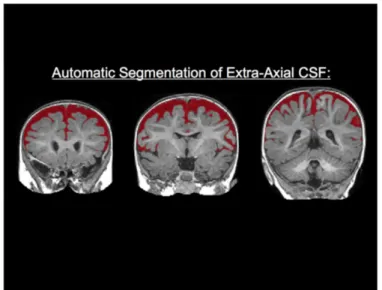
1. High-risk infants later diagnosed with ASD show increased EA-CSF volume at 6 months compared with high-risk and low-risk infants who do not develop ASD.
2. High-risk ASD infants show persistently increased EA-CSF through 24 months. 3. Increased EA-CSF is associated with autism severity as well as early motor deficits.
(Shen, 2)
Figure 1. Automatic Segmentation of EA-CSF; Mark Shen; March 2017
Following the findings in this research, we developed a tool to standardized the calculation of the existing volume of EA-CSF. We intend to further verify and confirm the relation between the amount of EA-CSF and ASD; Auto EACSF would allow early diagnosis of Autism. By accurately measuring EACSF in infants at high risk of ASD, early intervention and treatment can be provided to the patients. Additionally, the tool could help diagnosis of other neurologic diseases that use the EA-CSF as a biomarker. Thus, Auto EACSF attempts to develop an automated tool for computation of brain EA-CSF from infants’ MRI.
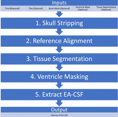
This research proposes a novel framework to measure brain EA-CSF volume. Auto EACSF is a user-friendly GUI based software tool for automatic computation of EA-CSF from T1w. The tool’s pipeline contains five steps: skull stripping, reference alignment, tissue
segmentation, ventricle masking, and extracting EA-CSF. Auto EACSF uses CMake to manage the build process, QT to run the GUI, and Python and XML scripts to produce the final output. The source code is available at https://github.com/hay318/auto_EACSF.
2. Methods
Auto EACSF runs five fundamental processes (Figure 3) to extract EA-CSF. Each process runs an open source software to achieve its purpose. Figure 3 shows that Auto EACSF requires a user to provide a T1-weighted image (T1w), then self-generates the necessary files from the given T1w. However, a user also has an option to provide a T2-weighted image (T2w), brain mask, tissue segmentation, and ventricle mask. Each procedure of five processes is
Figure 3. Pipeline Overview
2.1 Skull Stripping
Skull Stripping removes the skull and only extracts the whole brain including the cerebrospinal fluid. This process separates the brain tissues from non-brain image parts such as bone, skin, fat, etc. Skull stripping is an essential step of Auto EACSF. This process directly affects the accuracy of brain segmentation from non-brain tissue and measurement of brain fluid. Auto EACSF first performs rigid alignment and skull stripping on subject MRI scans. After performing a skull stripping process, Auto EACSF generates a new output file of skull stripped T1w, which will be used in following steps.
Output
Volume of EA-CSF
5. Extract EA-CSF
4. Ventricle Masking
3. Tissue Segmentation
2. Reference Alignment
1. Skull Stripping
Inputs
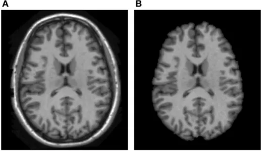
Figure 4. ABC based brain skull-stripping result. (A) the brain tissue in original MRI scan, (B) the skull-stripped brain; Jiahui Wang; March, 2014
This process generates the skull stripped image by using the Atlas Based Classification (ABC). (A) of Figure 4 represents the T1w that user will provide, and (B) of Figure 4 is the skull-stripped brain that will be achieved by running this process.
2.2 Reference Alignment
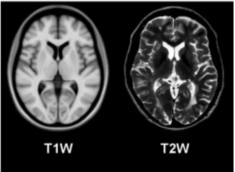
Figure 5. T1w vs. T2w; Nicolas Guizard; May, 2015
This process is achieved by utilizing the BRAINSfit Module within 3D Slicer. The BRAINSfit performs 3D rigid image registration of brain image and creates brain atlas, which is necessary for the next step, Tissue Segmentation.
2.3 Tissue Segmentation
Tissue segmentation identifies and labels the brain tissue sections. It identifies white matter, gray matter, and cerebrospinal fluid. From this process, we can figure out the total volume of cerebrospinal fluid, including both intra-axial and extra-axial cerebrospinal fluids.
2.4 Ventricle Masking
Keeping in mind that Auto EACSF only extracts the volume of EA-CSF fluid, we need to subtract the volume of the intra-axial cerebrospinal from the total volume of cerebrospinal fluid. Ventricle masking finds ventricles and removes intra-axial cerebrospinal fluid. This process computes a deformation field to the T1w vs. atlas, and it applies the ANTS warp to the ventricle mask and identifies the ventricles with a unique color.
2.5 Extract Extra-Axial Cerebrospinal Fluid
The extract extra-axial cerebrospinal fluid process calculates the exact volume of EA-CSF. After performing four procedures, now we know the precise location of EA-CSF fluid. This step uses the ImageMath Module and ImageStat Module within ANTS to produce quantitative data.
3. GUI Implementation
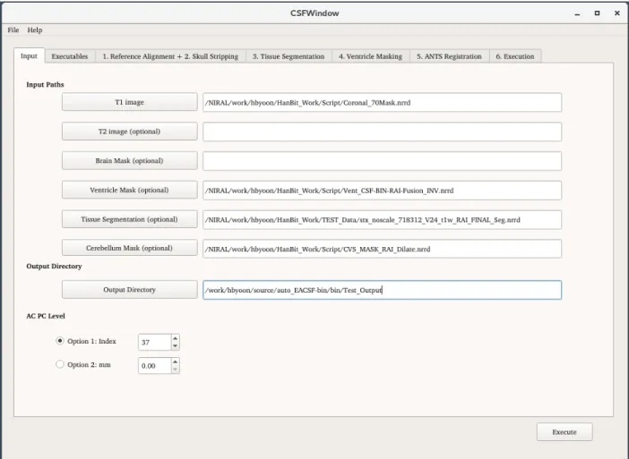
Figure 6. GUI of Auto EACSF
Figure 6 is the GUI of Auto EACSF; all the user interface files are all written in C++. Auto EACSF uses Qt and CMake to manage build process of the GUI. The tool also utilizes the Qt Resources System to have master scripts with replaceable keywords. When a user sets all input data and clicks the executing button, Qt writes customized Python and XML scripts with the input parameters provided by the user. Generated scripts will be used to run the pipeline. 4. Results
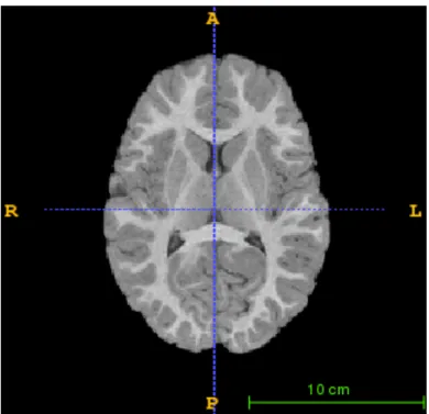
scans used to test the pipeline’s functionality. Figure 7 is the given T1w; Figure 8, Figure 9, and Figure 10 are generated by Auto EACSF.
Figure 8. Output after Skull Stripping and Reference Alignment
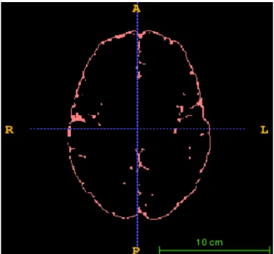
Figure 10. Preserved EA-CSF after the Ventricle Mask Removal
The tool removes all non-brain tissues from Figure 7, and it generates Figure 8 by performing skull stripping and reference alignment. Then, the tissue segmentation procedure identifies white matter, grey matter, and cerebrospinal fluid. Figure 9 represents the segmented brain tissues after the tissue segmentation. The tool performs the ventricle masking on Figure 9 and uses the
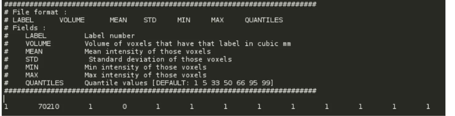
Figure 11. Calculated Volume of EA-CSF
Figure 11 is the text file generated by Auto EACSF. The last line in Figure 11 represents the calculated data after running all five procedures. The second value from the left represents the volume of EA-CSF; the tool disregards other values since it only measures one EA-CSF per execution. To compute this particular example, it took approximately 20 hours. In general, Auto EACSF takes 18 hours ~ 24 hours to execute the entire pipeline. Users can shorten the
processing time if they provide an already generated brain mask, tissue segmentation mask, and a ventricle mask.
4. Discussion and Conclusion
Auto EACSF presents a framework to build an interactive software tool with pipeline functionality. This tool automatically computes the volume of EA-CSF from the T1w; it
Acknowledgement
Works Cited
“Autism Spectrum Disorder (ASD).” Centers for Disease Control and Prevention, Centers for Disease Control and Prevention, 26 Feb. 2015, www.cdc.gov/ncbddd/autism/signs.html. “CDC Estimates 1 in 68 Children Has Been Identified with Autism Spectrum Disorder.” Centers
for Disease Control and Prevention, Centers for Disease Control and Prevention, 24 Mar.
2014, www.cdc.gov/media/releases/2014/p0327-autism-spectrum-disorder.html. Guizard, Nicolas, et al. “Rotation-Invariant Multi-Contrast Non-Local Means for MS Lesion
Segmentation.” NeuroImage: Clinical, vol. 8, no. May, The Authors, 2015, pp. 376–89, doi:10.1016/j.nicl.2015.05.001.
Rundo, Leonardo, et al. “Automated Prostate Gland Segmentation Based on an Unsupervised Fuzzy C-Means Clustering Technique Using Multispectral T1w and T2w MR Imaging.”
Information (Switzerland), vol. 8, no. 2, 2017, doi:10.3390/info8020049.
Shen, Mark D., et al. “Increased Extra-Axial Cerebrospinal Fluid in High-Risk Infants Who
Later Develop Autism.” Biological Psychiatry, vol. 82, no. 3, 2017, pp. 186–93,
doi:10.1016/j.biopsych.2017.02.1095.
Wang, Jiahui, et al. “Multi-Atlas Segmentation of Subcortical Brain Structures via the AutoSeg
Software Pipeline.” Frontiers in Neuroinformatics, vol. 8, no. March, 2014,