The Journal of Free Radicals and Antioxidants. Photon 142 (2015) 399-406
https://sites.google.com/site/photonfoundationorganization/home/the-journal-of-free-radicals-and-antioxidants Original Research Article. ISJN: 9102-2347: Impact Index: 5.13
The Journal of Free Radicals and Antioxidants
Ph ton
Co–Adminisration of Ascorbic acid and Alpha-Tocopherol Ameliorate
Oxidative Stress Parameters of Rams Transported by Road for Eight Hours
Adenkola Adeshina Yahaya*, Onyeberechi A.S.
Department of Physiology Pharmacology and Biochemistry, College of Veterinary Medicine, University of Agriculture, Makurdi, Nigeria
Adenkola Adeshina Yahaya and Onyeberechi A.S. receive Hans Adolf Krebs Research Award-2015 in Biochemistry
Article history: Received: 20 August, 2014 Accepted: 23 August, 2014 Available online: 17 March, 2015 Keywords:
Ascorbic acid, Alpha-Tocopherol, Oxidative Parameters, Rams, Road transportation
Corresponding Author: Yahaya A.A.*
DVM, M.Sc., Ph.D
Email: [email protected] Abstract
Road transportation exposes animals to unfamiliar environment with concurrent environmental stress, which elicit a stress response and cause increase in free radicals generation. This study was designed to investigate the effect of administration of combination of ascorbic acid + alpha- tocopherol and ascorbic acid alone on oxidative stress parameters of rams transported by road for eight hours. On the day of transportation, 7 rams (Group
1) were individually administered with ascorbic acid at the dose of 250 mg/kg dissolved in 10 ml of water and also alpha-tocopherol at the dose of 75 mg/kg, per os while another 7 rams (Group 2) was administered individually with ascorbic acid (250 mg/kg) only. The 3rd group (7 rams) was the control, and they were administered with only 10 ml of sterile water. Blood samples (5 ml) were taken in the morning a day before transportation, I h, 8 h journey time and 3 days post-transportation for full blood count and the harvested serum for analysis of oxidative parameters. In conclusion this study indicated that eight hours road transportation of ram is a source of stress and co-administration of ascorbic acid and alpha tocopherol ameliorate oxidative stress parameters due to synergistic effect.
Citation:
Yahaya A.A., Onyeberechi A.S, 2015. Co – Adminisration of Ascorbic acid and Alpha - Tocopherol Ameliorate Oxidative Stress Parameters of Rams Transported by Road for Eight Hours. The Journal of Free Radicals and Antioxidants. Photon 142, 399-406.
All Rights Reserved with Photon.
Photon Ignitor: ISJN91022347D743217032015 1. Introduction
Road transportation of animals disrupts normal pattern of feeding and drinking in animals and this is associated with exposure to novel environments, sometimes involving mixing with unfamiliar and closely confined animals, noise, vibration, and extremes of ambient temperature and humidity (Warris, 2004; Adenkola and Ayo, 2010) and these are likely to elicit a stress response which have been demonstrated to cause increase free radical generation (Nazifi et al., 2009) and erythrocyte haemolysis (Adenkola and Ayo, 2009) as well as decrease antioxidant absorption (Naziroglu et al., 2000). During stressful situation the tissue antioxidant vitamin decreases (Sahin et al., 2001), lipid peroxidation increases in the plasma and tissues leading to oxidative damage of erythrocyte cell membranes (Adenkola and Ayo, 2009).
1.1 Justification of Research
The free radical induce oxidative damage to macromolecules, cells, tissues (Altan et al., 2003) as well as haematological changes (Hulf et al., 2005) leading to increase morbidity and mortality, poor meat quality and decrease productivity (Franco – Jimemez and Beck, 2007) that consequently leads to substantial economic losses (St. Pierre et al., 2003). Thus the supplementation of the animal by exogenous antioxidants such as alpha tocopherol and ascorbic acid (Alvarado et al., 2006; Niki, 2010) improves the antioxidant status, and seems to be very helpful to fight free radicals, because exposure of animal to stress has been demonstrated to induce an increase in free radicals in the body (Sahin et al., 2001., Power and Jackson, 2008).
1.2 Objective of Research
Thus the aim of the present study was to investigate the effect of Co-administration of ascorbic acid and alpha tocopherol as well as singly administration of ascorbic acid on oxidative stress parameters of road transported rams for eight hours.
2. Materials and methods
2.1 Experimental Site
The study was conducted at Small Ruminant unit of the University of Agriculture Teaching and Research Farm Makurdi (070 41/ N, 080 37/ E) in the Southern Guinea Savannah Zone of Nigeria.
2.2 Experimental Design
On each experimental day, 7 rams (Group 1) were orally and individually administered with ascorbic acid (AA) (Juhel® Nigeria Ltd.) at the dose of 250 mg/kg (Chervyakov et al., 1977) dissolved in 10 ml of water and also alpha-tocopherol (α-T) (100 mg
DL-α-tocopherol) (Patterson Zoochonist Ltd.
Nigeria) at the dose of 75 mg/kg, while another 7 rams (Group 2) was administered orally and individually with AA (250 mg/kg) only. The 3rd group (7 rams) was the control, and they were administered orally and individually with only 10 ml of sterile water. The administrations were made immediately before loading the ram into the vehicle.
2.3 Vehicle Design, Loading, and the Journey
A standard Peugeot bus (J5), popularly used in the middle belt region of Nigeria for transportation of livestock was used to transport the rams. Other transportation procedures were carried out in accordance with the standard guidelines governing the welfare of livestock during road transportation (Warris, 1996). Food and water were withdrawn 12 hours before and throughout the journey period, which lasted 8 hours to and fro University of Agriculture, covering a total distance of 300 km. The speed of the vehicle was at a range of 40 - 50 km/h. The journey duration included stop-overs, to measure RT, very briefly (about 20 minutes) and for police-checking. After completing the journey, the rams were unloaded at the spot where they were originally loaded. The animals were given feed and water as they had been prior to the journey. Blood samples were taken early in the morning a day
before transportation, immediately after
transportation on arrival and 3 days post-transportation. Five millimeters of blood was taken aseptically from the jugular vein of each animal using 18 gauge x 11/2 inch sterile needle mounted on 5 ml syringe. Two millimeters of blood for determination of haematological parameters was immediately poured inside a sample bottle, containing an anticoagulant, disodium salt of ethylene diaminetetra-acetic acid (EDTA) at the
rate of 2 mg/ml of blood (Oyewale et al., 1992), while the remaining blood was immediately centrifuged 1,500 x g for 15 minutes and the resultant serum harvested and stored in the refrigerator.
2.4 Evaluation of Serum Malonaldehyde Concentration
Serum malonaldehyde (MDA) concentration as a marker of lipid peroxidation was determined by the double-heating method of Draper and Hadley (1990) as modified by Altuntas et al. (2002).
2.5 Determination of Cortisol Concentration
The stored serum was used to serum was then stored in the refrigerator until assayed for cortisol
using Elisa linked immunosorbent assay
(Diagnostic Automation, Inc.) method based on the principle of Cortisol (antigen) in the sample competing with horseradish peroxidase-Cortisol (enzyme-labelled antigen) for binding onto the limited number of anti-Cortisol (antibody) sites on
the microplates (solid phase). Cortisol
concentration in the sample is calculated based on a series by a set of standard. The colour intensity is inversely proportional to the cortisol concentration in the sample.
2.6 Determination of Haematological
Parameters
Packed cell volume (PCV) was determined using microhaematocrit method; total erythrocyte count (TRBC) and total leucocyte count was determined
(TWBC) using haemocytometeric, while
haemoglobin (Hb) concentration was determined using the cyanomethaemoglobin method (Schalm et al., 1975).
3. Results
3.1 Variation in Malondialdehyde
Concentration before, during and 3 Days after the Journey
The value obtained for serum MDA before transportation was not significantly different (P > 0.05) in all treatments before the journey. However 1 hour into the journey the value increased tremendously especially the control group without any antioxidants with a value of 2.13 ± 0.51 ng/ml in the first hour of the journey and 2.43 ± 0.22 ng/ml in the 8th hour which was significantly (P < 0.05) higher than the values obtained in treatment 1 and 2. The values rose slightly in all groups 3-days post-transportation above pre-transportation value which was not significantly (P > 0.05) different.
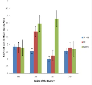
3.2 Fluctuation in Cortisol Concentration before, during and 3 Days after the Journey
The obtained value in group 1 was 1.85 ± 1.28 ng/ml which was not significantly (P > 0.05)
different from the recorded value of 1.88 ± 0.74 and 1.78 ± 0.36 recorded in groups 2 and 3 respectively. One hour into the journey the value rose significantly (P <0.05) in the control group to a value of 3.45 ± 1.40 ng/ml and 3.8 ± 0.74 ng/ml in the 8th hour which was significantly (P < 0.05) higher than the obtained values in groups 1 and 2, by the 3rd day post-transportation, the value came down to 1.77 ± 0.9, 1.57 ± 0.6 and 1.7 ± 0.6 in groups 1, 2 and 3 respectively, these values was not significantly (P < 0.05) different.
3.3 Variation in Packed Cell Volume before, during and 3 Days after the Journey
A lower value 23.5 ± 1.19 % was obtained in group II which was non significantly different (P<0.05) from a value of 22.5 ± 1.75 % and 25.75 ± 1.06 % obtained in group I and III respectively. In the eight hour of journey, the PCV dropped to 19.67 ± 1.67 % in the control group which was significantly different (P<0.05) from the values recorded in group I and II. The value was not significantly (P< 0.05) different in the groups three days post transportation (Figure 3).
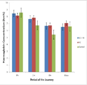
3.4 Variation in Haemoglobin Concentration before, during and 3 Days after the Journey
A non significant value (P<0.05) was obtained in all the groups before transportation. However the value dropped to 6.56 ± 1.38 gm % from the initial 8.59 ± 1.67 gm % in the eight hour of the journey in group III which was significantly (P<.05) lower than the recorded values in group I and II. The recorded values three days post transportation was not significantly different (Figure 5).
3.5 Variation in Total Erythrocyte Count Concentration before, during and 3 Days after the Journey
The Total Erythrocyte Count was not significantly different (P<0.05) in all the groups pre transportation. However a lowest value of 6.71 ± 1.39 x 10⁶/µl was obtained immediately after transportation of the journey in the control group. The recorded values 3 days post transportation was not significant (P<0.05) different (Figure 5).
3.6 Variation in Total Leucocyte Count Concentration before, during and 3 Days after the Journey
The value of Leucocyte increased in the entire group 1 hour post-transportation, but the highest significant (P<0.05) value of 10.55 ± 0.85 x 10³/µl was obtained in the control group. The recorded values 3 days post-transportation was not significantly (P<0.005) different (Figure 6).
3.7 Fluctuation in Catalase Enzyme
Concentration before, during and 3 Days after the Journey
The obtained value of catalase enzyme in control group was 59.25 ± 7.4 u/ml was not significantly different (P<0.05) from the recorded values in the experimental group pre transportation, however the value rose to 62.75 ± 4.86 u/ml in the first hour of the journey untill a value of 66.33 ± 3.51 u/ml in the eight hour of the journey which was statistically higher (P<0.05) than the value of 50.33 ± 5.04 u/ml recorded in group1. Three days post transportation the value of 62.33 ± 3.32 u/ml obtained in group I was not significantly different (P<0.05) from the values obtained in group I and II (Figure 7).
3.8 Fluctuation in Superoxide dismutase Enzyme Concentration before, during and 3 Days after the Journey
The values of SOD recorded pre transportation which is 2.33 ± 0.4 u/ml, 2.4 ± 0.62 u/ml and 2.35 ± 0.21 in group I, II and III respectively was not significantly (P<0.05) different. However a lower value of 2.1 ± 0.72 u/ml was recorded in group I which was significantly lower than a highest value of 2.63 ± 0.45 u/ml obtained in group III which is the control group. A non significantly value (P<0.05) was obtained in all the groups three days post transportation (Figure 8).
4. Discussion
MDA a biomarker for lipid peroxidation is the oxidative deterioration of polyunsaturated lipids which leads to the production of a degraded product called malondialdehyde (Belge et al., 2003). The increase in MDA concentration seen in this study during transportation especially in group III (control) which was not administered any antioxidant could be due to higher level of glucocorticoids and adrenaline- induces pathways of aerobic energy production associated with stress. This process was modulated possibly by the
antioxidant vitamins (AA + α-T) which have been
demonstrated to prevent or reduce considerably the ROS- induced damage to the body cells (Pregel et al., 2005) because this ROS have been shown to induce lipid lipoperoxidation of cytomembrane resulting in cell damage and destruction (Altan et al., 2003; Solichova et al., 2003). Also reduction of MDA in group 1 and II may be due to the enhancement of the fatty acid transport by antioxidants (AA + α-T) into mitochondria for
energy production, thereby lowering the
availability of lipid for peroxidation. This result of this present study agrees with that of Tokarzewski et al. (2004) who demonstrated a high level of MDA concentration in transported birds. Thus serum malondialdehyde concentration may be used
as biomarker of oxidative stress in rams subjected to road transportation stress.
The obtained result for cortisol concentration was considerably high in group III (control) animals. Cortisol is a glucocorticoids produced by the adrenal cortex as a result of the activation of the hypothalamic- pituitary adrenal axis (HPA) during a stressful situation. The result of this finding agrees with the work of Odore et al. (2011) who reported an increase in cortisol level of goat transported for a short- term. However, the obtained results in group 1 and 11 were lower due to the effect of administered antioxidants. These vitamins antioxidants possibly inhibit the release of cortisol, especially combined effect (AA + α-T) was more effective. This finding agrees with the earlier work of Karanth et al. (2000) and Powers and Jackson (2008). Cortisol release in this study was probably during the alarm stage which is characterized by mobilization of all defense mechanism in the body to combat adverse effect of stress as documented by Seyle (1977) which in this case is in the 1st hour of the journey.
The increase in leucocyte counts 1 h into the journey especially in group III (control) may be as a result of the presumed sudden release of glucocorticoids during handling and loading and possibly due to novel environment and the fear of the journey which is exaggerated in the control animals could be responsible for the release of leucocytes from the bone marrow (Kannan et al., 2002; Stanger et al., 2005; Adenkola et al., 2009). Group 111 (control) recorded the highest level of cortisol concentration in this study, which is in agreement with the finding of Khasmi et al. (2013) who recorded a higher cortisol level in blood samples collected after transportation of Moroccan dromedary camel than in samples collected before the journey. The results obtained in group 1 and II during the journey were lower than the control animal, this could possibly be due to the inhibitory role of the administered antioxidants especially AA which is known to be a chain- breaking antioxidant and has been reported to be involved in the prevention and restriction of free radical chain formation and propagation, consequently protecting blood cells from oxidative damage (Surai, 2002; Alok et al., 2003; Power and Jackson, 2008). Similarly, AA and α-T are shown to inhibit the release of corticosteroid, a hormone known to destroy immune cells (Liakakos et al., 1975; Balz, 2003).
Changes in blood picture of animal subjected to transportation stress have been established by many researchers (Adenkola et al., 2011; Saeb et al., 2010). Increase in PCV, erythrocyte count and haemoglobin concentration 1 h into the journey
especially the group III could be attributed to the effect of excitement as a result of increase in circulatory catecholamines and possibly
Figure 1: Variation in Malondialdehyde Concentration before, during and 3 Days after the Journey
Figure 2: Fluctuation in Cortisol Concentration before, during and 3 Days after the Journey
Figure 3: Variation in Packed Cell Volume before, during and 3 Days after the Journey
Figure 4: Variation in Haemoglobin Concentration before, during and 3 Days after the Journey
Figure 5: Variation in Total Erythrocyte Count Concentration before, during and 3 Days after the Journey
Figure 6: Variation in Total Leucocyte Count Concentration before, during and 3 Days after the Journey
Figure 7: Fluctuation in Catalase Enzyme Concentration before, during and 3 Days after the Journey
Figure 8: Fluctuation in Catalase Enzyme Concentration before, during and 3 Days after the Journey
haemoconcentration due to dehydration, this leads to splenic contraction and release of erythrocyte into the circulation (Fazio and Ferlazzo, 2003). Antioxidant mechanisms that protect metabolically active cells against free radical damage include enzymatic systems such as superoxide dismutase and catalase (Gurgoze et al., 2005). Under normal physiological condition, there is a balance between free radicals and antioxidant defence systems for
various reasons, lipid, protein, and DNA
components of cell may undergo oxidative damage
(Halliwell and Chirico, 1999). Superoxide
dismutase enzyme provided transformation of superoxide radical to hydrogen peroxide and molecular oxygen and thus reduces cellular
superoxide content exhibiting antioxidant
properties (Onmaz et al., 2011). Catalase is considered as an antioxidant enzyme found in nearly all the cells that are exposed to oxygen, where it catalyses decomposition of hydrogen
peroxide to water and oxygen. Hydrogen peroxide is a harmful by product of many normal metabolic processes. To prevent damage, it must be quickly converted into other, less dangerous substances (Maan and Kataria, 2012). Higher values of superoxide dismutase and calase activity seen in this study especially control group suggested the ability of the animals to provide defense against free radicals damage and biomarkers of lipid peroxidation and are considered the best indicators of oxidative stress (Georgieva, 2005) as measured by MDA concentration which was highest in the control group. These are enzymes of antioxidant defense system that eliminates and controls the toxic oxygen species (Maan and Kataria, 2012). However in the supplemented group with antioxidant the activity of catalse and superoxide dismutase was however reduced because the free radicals could have possibly been scavenged by the exogeneous antioxidant administered. Higher serum activities of the enzymes in stressed animals indicate the higher rate of hydrogen peroxide formation (Kataria et al., 2010a), therefore it is considered as one of the markers to assess oxidative stress in animals (Kataria et al., 2010b; Kataria et al., 2012).
Conclusion
In conclusion this study indicated that eight hours road transportation of ram is a source of stress and co-administration of ascorbic acid and alpha tocopherol ameliorate oxidative stress parameters of road transported rams for eight hours possibly due to synergistic effect.
Research highlights
The research demonstrated clearly that animals transported for eight hours are stressful due to production of enormous free radicals.
Limitations
The driver of the vehicle want to exceed the recommended constant speed of 45 km/hr, also some analysis was not done immediately as a result the sample has to be stored in the refrigerated condition until the analysis is done.
Recommendations
Transported animals should be administered with antioxidants prior to transportation by road in order to reduce the risk of adverse effects of transportation stress on their health. However co – administration of (AA + α-T) is better than singly administration of AA, possibly due to synergistic effect.
References
Adenkola A.Y., Ayo J.O., 2009. Effect of eight hours road transportation stress on erythrocyte osmotic fragility of pigs administered ascorbic acid during the harmattan season in Zaria Nigeria. Journal of Cell and Animal Biology, 3(1), 004 - 008.
Adenkola A.Y., Ayo J.O., Sackey A.K.B., Adelaiye A.B., 2009. Haematological changes in pigs administered with ascorbic acid and transported by road for four hours during the harmattan season. Journal of Cell and Animal Biology, 3 (2), 021- 028.
Adenkola A.Y., Ayo J.O., 2010. Physiological and behavioural responses of livestock to road transportation stress: A Review. African Journal of Biotechnology, 9(31), 4845 – 4856.
Adenkola A.Y., Ayo A.Y., Sackey A.K.B., Adelaiye A.B., 2011. Eight hours road transportation and ascorbic acid administration effects on haematological parameters of pigs during the harmattan season. Agriculture and Biology Journal of North America 2(8), 1143 – 1150. Alok K.B., Amrittal M., Dipanjan C., Sajal C., 2003. “Oxidants, antioxidants and physical exercise,” Molecular and Cellular Biochemistry, 253, 307 – 312. Altan O., Pabuccuoglu A., Konyaliogu S., Bayracktar H., 2003. Effect of heat stress on oxidative stress, lipid peroxidation and some stress parameters in broilers. British Poultry Science, 44, 545 - 550.
Altuntas I., Delibas N., Sutcu R., 2002. The effects of organophosphate insecticide methidathion on lipid peroxidation and anti-oxidation enzymes in rat erythrocyte: Role of vitamins E and C. Human and Experimental Toxicology. 21, 681 – 685.
Alvarado C., Alvares P., Puerto M., Gausseres N., Jimenez L., De la Funenete M., 2006. Diesary supplementation with autioxid outs meroves functions and deceases oxidative stress of leucocytes from prematurely aging mice. Nutrition, 22, 767 – 777. Balz C., 2003. Vitamin-C intake. Nutritional Disease, 14, 1 - 8. Belge F., Cinar A., Selcuk M., 2003. Effects of stress produced by adrenocorticotropin on lipid peroxidation and some antioxidants in vitamin C treated and nontreated chickens. South African Journal of Animal Science, 33, 201-205.
Chervyakov D.K., Yevdokimov P.D., Vishker A.S., 1977. Drugs in Veterinary Medicine. Kolos Publishing House, Moscow, 496pp (in Russian). Draper H., Hadley M., 1990. Malondialdehyde determination as index of lipid peroxidation, Methods in Enzymology, 186, 421 - 431.
Fazio E., Ferlazzo A., 2003. Evaluation of stress during transport. Veterinary Research Communication, 27 (suppl 1), 519 - 524.
Franco-Jimenez D.J., Beck M.M., 2007. Physiological changes to transient exposure to heat stress observed in laying hens. Poultry Science, 86, 538 - 544.
Georgieva N.V., 2005. Oxidative stress as a factor of disrupted ecological oxidative balance in biological systems – a review. Bulgarian Journal of Veterinary Medicine, 8, 1-11.
Gurgoze S.Y., Cetin H., Cen O., Yilmaz S., Atli M.O., 2005. Chnges in malondialdehyde concentrations and glutathione peroxidase activity in purebred Arabian mares with endometritis. Veterinary Journal, 170, 135 – 137.
Halliwell B., Chirico S., 1999. Lipid peroxidation. Its mechanism measurement and significance. American Journal of Clinical Nutrition, 57, 715 – 725.
Hulf G.R., Huff W.E., Balog J.M., Rath N.C., Anthony N.B., Nestor K.E., 2005. Stress response differences and disease susceptibility refected by heterophil to lymphocyte ratio in Turkeys selected for increased body weight. Poultry Science, 84, 709 -717.
Kannan G., Terril T.H., Kouokou B., Gelaye S., Amoah E.A., 2002. Simulated preslaughter holding and isolation effects on stress responses and live mass shrinkage in meat goats. Journal of Animal Science, 80, 1771 - 1780. Karanth S., Yu, W.H., Walczewska A., Mastronardi C., McCann S.M., 2000. Ascorbic acid acts as an inhibitory transmitter in the hypothalamus to inhibit stimulated luteinizing hormone-releasing hormone release by scavenging nitric oxide. Proceedings of the National Academy of Science of the United States of America, 97, 1891 - 1896.
Kataria N., Kataria A. K., Pandey N., Gupta P., 2010a Serum biomarkers of physiological defense against reactive oxygen species during environmental stress in Indian dromedaries. Human and Veterinary Medicine Bioflux, 2, 55 - 60.
Kataria N., Kataria A.K., Maan R., Gahlot, A.K., 2010b. Evaluation of oxidative stress in brucella infected cows. Journal of Stress Physiology and Biochemistry, 6, 19 – 25.
Kataria N., Kataria A. K., Joshi A., Pandey N., Khan S., 2012 Serum antioxidant status to assess oxidative stress in brucella infected buffaloes. Journal of Stress Physiology and Biochemistry, 8, 5 - 9.
Khasmi M.E.H., Chakir Y., Riad F., Safwate A., Tahir E.L.H., Farh M., EL Abbadi N., Abouhafs R., Faye B., 2013. Effect of transportation stress during the hot-dry season on some haematological and physiological parameters in Moroccan dromedary camels (Camelus dromedaries). Journal of Life Sciences, 7(1), 13 – 25. Powers S.K., Jackson M.J., 2008. Exercise- induced oxidative stress: Cellular mechanisms and impact on muscle force production. Physiology Review, 88, 1243 - 1276.
Liakakos D., Doulas N.L., Ikkos D., Anoussakis C., Viachos P., Jouramani G., 1975.“Inhibitory effect of ascobic acid on cortisol secretion following adrenal stimulation in children,” Clinica Chimica Acta, 65, 251 – 255.
Maan R., Kataria N., 2012. Evaluation of oxidative stress during adverse environmental conditions in Marwari sheep from arid tracts in India. Animal Biology and Animal Husbandry. International Journal of the BIOFLUX Society, 4(2), 38 – 42.
Nazifi S., Saeb M., Baghshani H., Saeb S., 2009 ‘Influence of road transportation during hot-summer condition on oxidative status biomarkers in Iranian dromedarian camels (Camelus dromedarius)’, African Journal of Biochemistry Research, 3(7), 282–287. Naziroglu M., Sahin K., Simsek H., Aydilek N., Ertas O.N., 2000. The effect of food withdrawals and darkening on lipid peroxidation of laying hens in high ambient temperatures, Deut Sch. Tierarzit. Wischr. 107: 199 - 202.
Niki E., 2010. Antioxidants in relation to lipid peroxidation. Chemistry of Physiology of Lipids, 44, 227 - 253.
Odore R., Badino P., Re G., Barbero R., Cuniberti B., D’Angelo A., Girardi C., Fraccaro E., Tarantola M., 2011. Effects of housing and short-term transportation on hormone and lymphocyte receptor concentrations in beef cattle. Research in veterinary Science, 90(2), 341-345. Onmaz A.C., Van Den Hoven R., Gunes V., Cinar M., Kucuk O.C., 2011. Oxidative stress in horses after a 12- hours transport period. Revue Medicine Veterinary, 162(4): 213 – 217.
Oyewale J.O., 1992. Changes in osmotic resistance of erythrocytes of cattle, pigs rats, and rabbits during variations in temperature and pH. Journal of Veterinary Medicine, A39, 98 - 104.
Pregel P., Bollo E., Cannzo F.T., Biolatti B., Contato E., Biolatti P.G., 2005. Antioxidants capacity as a reliable marker of stress in daily calves transported by road. Veterinary Records, 150, 53 – 54.
Saeb M., Baghshani H., Naziti S., Saeb S., 2010. Physiological response of dromedary camels to road transportation in relation to circulating levels of cortisol, thyroid hormones and some serum biochemical parameters. Tropical Animal Health and Production, 42, 55 - 63.
Sahin K., Sahin N., Onderci M., Yaraliogu S., Kucuk O., 2001. Protective role of supplemental vitamin E on lipid peropxidation. Vitamin E, A, and some mineral concentration of broilers reared under heat stress. Veterinary Medicine, (Czech) 46, 140 - 144.
Schalm O.W., Jain N.C., Caroll E.J., 1975. Textbook of veterinary Haematology, 2nd Edition, Published by Lea and Febiger, Philadelphia, pp. 129 - 250.
Selye H., 1977. The concept of stress as we understand it in 1976. In: Advances in Hormones and Mechanisms of their Action. (Naukova Dumka, Kiev, Pp. 27 – 51 (in Russian).
Solichova D., Korecka L., Svobodova I., Musli F., Blaha V., Zdansky P., Zadak Z., 2003. Development and
validation of HPLC method for the determination of α -tocopherol in human erythrocyte for clinical applications. Annual Biannual Chemistry, 376, 444 – 447.
Surai P.F., 2002. “Selenium in poultry nutrition 1. Antioxidant properties, deficiency and toxicity,” World’s Poultry Science Journal, 58, (3): 333–347.
Stanger K.J., Ketheesan N., Parker A.J., Coleman C.J., Lazzaroni S.M., Fitzpatrick L.A., 2005. “The effect of transportation on the immune status of Bos indicus steers,” Journal of Animal Science, vol. 83(11) 2632 – 2636.
St-Pierre N.R., Cobanov B., Schnitkey G., 2003. Economic losses from heat stress by US livestock industries. Journal of Dairy Science, 86, 52-77.
Tokarzewski S., Wernicki A., Kankofer M., Urban-Chimelli R., 2004. Lipid peroxidation as the additional indicator of transport stress in broilers. Poland Journal of Veterinary Science, 7(2), 109 – 112.
Warris P.D., 1996. The welfare of animals during transport. The Veterinary Annuals, 36, 73 – 85.
Warris P.D., 2004. The transport of animals: A long way to go. Veterinary Journal, 168, 213-214.