40
HISTO-ANATOMICAL STUDY OF THE ANNUAL OFFSPRING AND
LEAF’S LIMB OF VITEX NEGUNDO L. (VERBENACEAE) SPECIES
BEJENARU L. E.1, BEJENARU CORNELIA2, MOGOŞANU G. D.1, OANCEA CARMEN NICOLETA2 1 Department of Pharmacognosy & Phytotherapy, Faculty of Pharmacy, University of Medicine and
Pharmacy of Craiova, 2 Petru Rareş Street, 200349 Craiova.
2
Department of Vegetal & Animal Biology, Faculty of Pharmacy, University of Medicine and Pharmacy of Craiova, 2 Petru Rareş Street, 200349 Craiova, e-mail: [email protected].
Keywords: Vitex negundo L., annual offspring, leaf’s limb, histo-anatomy, study
ABSTRACT
This paper presents the histo-anatomical study of the annual offspring and leaf’s limb of Vitex negundo species, as an essential stage for the pharmacognostic expertise advancement. The pharmacological importance of the medicinal products (roots, leaves, seeds) obtained from V. negundo was also emphasized.
INTRODUCTION
Vitex negundo L., Chinese chaste tree, five-leaved chaste tree, Verbenaceae family,
is a shrub or small tree, 2–8 m tall, native to tropical Africa (Kenya, Tanzania, Madagascar) and Asia (India, Indonesia, Vietnam, Malaysia, Philippines, China, Japan), widely naturalized and cultivated as an ornamental plant (Vitex, 2013).
For therapeutic purposes, roots, leaves and seeds are used because of their content of active principles such as iridoid glycosides, e.g. negundoside and derivatives (Tasduq et al., 2008; Sharma et al., 2009; Huang et al., 2013), essential oil (Khokra et al., 2008; Nagarsekar et al., 2010), flavonoids (Sathiamoorthy et al., 2007), sesquiterpenoids and diterpenoids (Zheng et al., 2010; Zheng et al., 2012), lignans (Xin et al., 2013), fatty acids (Kannathasan et al., 2008), ecdysteroids, sterols, catechic tannin, simple carbohydrates, proteins, enzymes, organic acids, vitamins, mineral salts (Vishwanathan & Basavaraju, 2010).
The medicinal products harvested from V. negundo have some important properties: hepatoprotective (Tasduq et al., 2008; Kadir et al., 2013), antibacterial (Khokra et al., 2008; Nagarsekar et al., 2010; Kamruzzaman et al., 2013), antifungal (Sathiamoorthy et al., 2007), anti-inflammatory (Zheng et al., 2010; Chattopadhyay et al., 2012), apoptosis-inductive and antitumoral (Xin et al., 2013), larvicidal (Kannathasan et al., 2008), vermicide (Sahare & Singh, 2013), antiandrogenic (Das et al., 2004), anticonvulsant (Tandon & Gupta, 2005), antitussive (Haq et al., 2012), anti-hyperglycemic (Sundaram et al., 2013), antioxidant and anti-lipid peroxidation (Nagarsekar et al., 2011), prevention of selenite-induced oxidative stress and cataractogenesis (Rooban et al., 2012), inhibition of melanogenesis (Huang et al., 2012). Extractive preparations obtained from the flowers of some Vitex sp. are used for their contraceptive effect in Southeast Asia, India and Africa (Das et al., 2004).
Even considering its medicinal importance, in the specialty papers there are no data regarding V. negundo histo-anatomy. The aim of our paper was the histo-anatomical study of the annual offspring and leaf’s limb of the above-mentioned species, as an important step for the pharmacognostic expertise.
MATERIAL AND METHODS
The vegetal material was collected from V. negundo plants in blossom, in May 2013, from “Alexandru Buia” Botanical Garden, University of Craiova, Dolj County, Romania.
41
microscope at diverse objectives (×4, ×10, ×40) and then photographed on a Soligor SR 300 system adapted to the microscope.
Description of microscopic cross-sections was accomplished according to some classical authors (Toma & Rugină, 1998).
Using ×40 objective (corresponding on 0.037 mm2
area), the analysis of stomatal index was made on a Nikon Eclipse 55i binocular photon microscope coupled with a Nikon DS–Fi1 high definition video camera.
Image acquisition and processing were performed by means of ImageProPlus ver. 6.0 software package.
For each bottom, middle and upper area of the examined leaf’s limb, the average of 10 samples were taken into consideration.
RESULTS AND DISCUSSIONS
Structure of annual offspring
The annual offspring exhibits circular shape and secondary structure determined by libero-ligneous and subero-phellodermic cambium.
In cross-section, from the outside to the inside, in the lower third of annual offspring, the following histological sequence was observed:
Peridermis is made of 4–5 layers of suber, a single layer of subero-phellodermic cambium and 4–5 layers of cellulosic phelloderm.
Into the primary bark, numerous periphloemic sclerenchyma caps are observed. The conducting tissues are predominant secondary being generated by the libero-ligneous cambium.
An external ring of secondary phloem tissue includes sieve tubes, annex cells and phloem parenchyma.
The libero-ligneous cambium has a circular-winding shape.
The secondary xylem tissue is well represented, consisting of secondary xylem vessels of different diameters spread into a mass of libriform tissue. Xylem vessels have curly and reticular thickenings highlighted in longitudinal radial sections. The secondary xylem tissue is organized in the form of annual rings.
Medullary rays are multicellular, uniseriate, cellulosic into the phloem tissue and lignified at the level of xylem tissue.
42
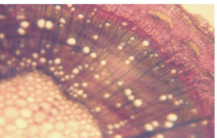
Figure 1. Cross-section through V. negundo annual offspring. Congo red–chrysoidine staining, ×100.
Overview.
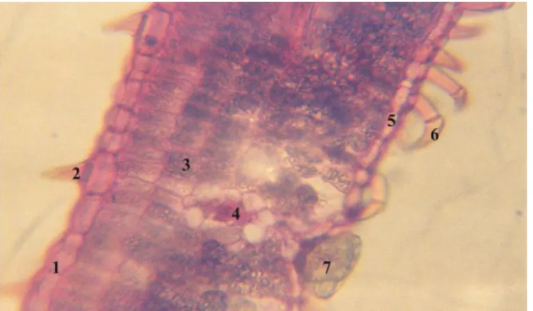
Figure 2. Cross-section through V. negundo annual offspring. Congo red–chrysoidine staining, ×100. 1 –
Epidermis, 2 – Periphloemic sclerenchyma cap, 3 – Phloem tissue, 4 – Libero-ligneous cambium, 5 – Xylem tissue, 6 – Medullary ray.
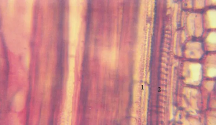
Figure 3. Cross-section through V. negundo annual offspring. Congo red–chrysoidine staining, ×400. 1 –
43
Figure 5. Longitudinal radial section through V. negundo annual offspring. Congo red–chrysoidine staining,
×400. 1 – Reticular xylem vessel, 2 – Curly xylem vessel.
Structure of leaf’s limb
In cross-section, from the outside to the inside, the leaf’s limb shows the following tissue sequence:
The upper epidermis is made of a single layer of flattened big cells, with thickened external and internal tangential walls and thin radial walls. Tangential external walls are covered by cutine. Unicellular sharp tector hairs are found from point to point. Stomata are missing at this level.
The homogenous mesophyll is made of three layers of palisade tissue with big prosenchymatous cells delineating small intercellular spaces and of 3–4 layers of smaller size prosenchymatous cells delineating much larger aeriferous spaces, which are also placed in palisade.
Numerous libero-ligneous conducting fascicles surrounded by an assimilatory sheath are found into the mesophyll. The plant is of C4 type. Some larger fascicles are attached to the epidermises through supporting tissue.
The lower epidermis is made of a single layer of small cells, closely linked together, with thin radial walls and thickened internal and external tangential walls. At this level, many flexed bicellular tector hairs and tetracellular glandular hairs are found. Into the lower epidermis, anisocytic stomata and cupuliform glandular hairs, in front of the mesophyll’ conducting fascicles, are highlighted.
The leaf’s limb has equifacial hypostomatic structure.
In cross-section, the median rib has circular shape. At this level, the epidermis is made of small heterodiametric cells with thickened external and internal tangential walls and thin radial walls. Here and there are found flexed bicellular tector hairs to the abaxial (dorsal) side, unicellular tector hairs to the adaxial (ventral) side, tetracellular glandular hairs and stomata.
Below the epidermis are found 2–3 layers of angular collenchyma and then the fundamental leaf’s parenchyma.
At the level of the median rib, one big centrally placed libero-ligneous conducting fascicle is found (Figures 6–8).
Our analyses show that the stomatal index of V. negundo leaf (15.9–16.4) is different
compared to V. agnus-castus (26.8–27.3, personal unpublished results). This fact is very
44
Figure 6. Cross-section through V. negundo leaf’s limb. Congo red–chrysoidine staining, ×400. 1 – Upper
epidermis, 2 – Unicellular tector hair, 3 – Palisade parenchyma, 4 – Libero-ligneous conducting fascicle, 5 – Lower epidermis, 6 – Flexed
bicellular tector hair, 7 – Cupuliform glandular hair.
Figure 7. Cross-section through V. negundo leaf’s median rib. Congo red–chrysoidine staining, ×400. 1 –
Upper epidermis, 2 – Tector hair, 3 – Tetracellular glandular hair, 4 – Angular collenchyma.
Figure 8. Cross-section through V. negundo leaf’s median rib. Congo red–chrysoidine staining, ×400. 1 – Fundamental leaf’s parenchyma, 2 – Xylem tissue.
45
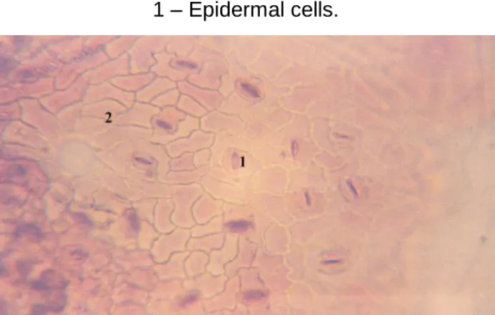
Figure 10. V. negundo lower epidermis. Congo red–chrysoidine staining, ×400. 1 – Anisocytic stomata, 2 – Epidermal cells.
CONCLUSIONS
The histo-anatomical study of the annual offspring and leaf’s limb of Vitex negundo
was achieved. The annual offspring exhibits circular shape and secondary structure. Many periphloemic sclerenchyma caps are observed into the primary bark. Produced by the libero-ligneous cambium, the conducting tissues are predominant secondary. The leaf’s limb has equifacial hypostomatic structure with unicellular/flexed bicellular tector hairs, cupuliform or tetracellular glandular hairs and anisocytic stomata.
REFERENCES
Andrei M., Paraschivoiu Roxana Maria, 2003, Microtehnică botanică, Edit.
Niculescu, Bucureşti, 222 pag.
Chattopadhyay P., Hazarika S., Dhiman S., Upadhyay A., Pandey A., Karmakar S. and Singh L., 2012, Vitex negundo inhibits cyclooxygenase-2 inflammatory cytokine-mediated inflammation on carrageenan-induced rat hind paw edema. Pharmacognosy Res. 4(3): 134–137.
Das S., Parveen S., Kundra C. P. and Pereira B. M., 2004, Reproduction in male rats is vulnerable to treatment with the flavonoid-rich seed extracts of Vitex negundo. Phytother. Res. 18(1): 8–13.
Haq R. U., Shah A. U., Khan A. U., Ullah Z., Khan H. U., Khan R. A. and Malik A., 2012, Antitussive and toxicological evaluation of Vitex negundo. Nat. Prod. Res. 26(5): 484–
488.
Huang H. C., Chang T. Y., Chang L. Z., Wang H. F., Yih K. H., Hsieh W. Y. and Chang T. M., 2012, Inhibition of melanogenesis versus antioxidant properties of essential oil extracted from leaves of Vitex negundo Linn. and chemical composition analysis by GC-MS. Molecules. 17(4): 3902–3916.
Huang J., Wang G. C., Wang C. H., Huang X. J. and Ye W. C., 2013, Two new glycosides from Vitex negundo. Nat. Prod. Res. Jan 29 (published online).
Kadir F. A., Kassim N. M., Abdulla M. A. and Yehye W. A., 2013, Hepatoprotective role of ethanolic extract of Vitex negundo in thioacetamide-induced liver fibrosis in male rats. Evid. Based Complement. Alternat. Med. 2013: 739850.
Kamruzzaman M., Bari S. M. and Faruque S. M., 2013, In vitro and in vivo bactericidal activity of Vitex negundo leaf extract against diverse multidrug resistant enteric bacterial pathogens. Asian Pac. J. Trop. Med. 6(5): 352–359.
Kannathasan K., Senthilkumar A., Venkatesalu V. and Chandrasekaran M., 2008, Larvicidal activity of fatty acid methyl esters of Vitex species against Culex quinquefasciatus. Parasitol. Res. 103(4): 999–1001.
46
Sci. 70(4): 522–526.
Nagarsekar K. S., Nagarsenker M. S. and Kulkarni S. R., 2010, Evaluation of composition and antimicrobial activity of supercritical fluid extract of leaves of Vitex
negundo. Indian J. Pharm. Sci. 72(5): 641–643.
Nagarsekar K. S., Nagarsenker M. S. and Kulkarni S. R., 2011, Antioxidant and antilipid peroxidation potential of supercritical fluid extract and ethanol extract of leaves of Vitex
negundo Linn. Indian J. Pharm. Sci. 73(4): 422–429.
Rooban B. N., Sasikala V., Gayathri Devi V., Sahasranamam V. and Abraham A., 2012, Prevention of selenite induced oxidative stress and cataractogenesis by luteolin isolated from Vitex negundo. Chem. Biol. Interact. 196(1–2): 30–38.
Sahare K. N. and Singh V., 2013, Antifilarial activity of ethyl acetate extract of Vitex
negundo leaves in vitro. Asian Pac. J. Trop. Med. 6(9): 689–692.
Sathiamoorthy B., Gupta P., Kumar M., Chaturvedi A. K., Shukla P. K. and Maurya R., 2007, New antifungal flavonoid glycoside from Vitex negundo. Bioorg. Med. Chem. Lett. 17(1): 239–242.
Sharma R. L., Prabhakar A., Dhar K. L. and Sachar A., 2009, A new iridoid glycoside from Vitex negundo Linn. (Verbenaceae). Nat. Prod. Res. 23(13): 1201–1209.
Sundaram R., Shanthi P. and Sachdanandam P., 2013, Effect of iridoid glucoside on plasma lipid profile, tissue fatty acid changes, inflammatory cytokines, and GLUT4 expression in skeletal muscle of streptozotocin-induced diabetic rats. Mol. Cell Biochem. 380(1–2): 43–55.
Tandon V. R. and Gupta R. K., 2005, An experimental evaluation of anticonvulsant activity of Vitex negundo. Indian J. Physiol. Pharmacol. 49(2): 199–205.
Tasduq S. A., Kaiser P. J., Gupta B. D., Gupta V. K. and Johri R. K., 2008, Negundoside, an iridoid glycoside from leaves of Vitex negundo, protects human liver cells against calcium-mediated toxicity induced by carbon tetrachloride. World J. Gastroenterol. 14(23): 3693–3709.
Toma C., Rugină Rodica, 1998, Anatomia plantelor medicinale. Atlas. Edit. Academiei Române, Bucureşti, 320 pag.
Vishwanathan A. S. and Basavaraju R., 2010, A review on Vitex negundo L. – a medicinally important plant. eJ. Biol. Sci. 3(1): 30–42.
Xin H., Kong Y., Wang Y., Zhou Y., Zhu Y., Li D. and Tan W., 2013, Lignans extracted from Vitex negundo possess cytotoxic activity by G2/M phase cell cycle arrest and apoptosis induction. Phytomedicine. 20(7): 640–647.
Zheng C. J., Huang B. K., Wang Y., Ye Q., Han T., Zhang Q. Y., Zhang H. and Qin L. P., 2010, Anti-inflammatory diterpenes from the seeds of Vitex negundo. Bioorg. Med. Chem. 18(1): 175–181.