© 2020 by the Serbian Biological Society How to cite this article: Mihailović M, Uskoković A, Arambašić Jovanović J, 233 Grdović N, Dinić S, Poznanović G, Franić A, Đorđević M, VidakovićM. Treatment of
streptozotocin-induced diabetic rats with Castanea sativa and Lactarius deterrimus extracts decreases liver damage by initiating activation of the Akt prosurvival kinase. Arch Biol Sci. 2020;72(2):233-42.
Treatment of streptozotocin-induced diabetic rats with
Castanea sativa
and
Lactarius
deterrimus
extracts decreases liver damage by initiating activation of the Akt prosurvival
kinase
Mirjana Mihailović1, Aleksandra Uskoković1, Jelena Arambašić Jovanović1, Nevena Grdović1, Svetlana Dinić1, Goran Poznanović1, Aida Franić2, Marija Đorđević1 and Melita Vidaković1,*
1Department of Molecular Biology, Institute for Biological Research “Siniša Stanković”, National Institute of the Republic of
Serbia, University of Belgrade, Bulevar despota Stefana 142, 11060 Belgrade, Serbia
2Clinical Hospital Center Rijeka, Department of Transfusion Medicine, Tome Stizica 3, 51000 Rijeka, Croatia
*Corresponding author: [email protected]
Received: April 10, 2020; Revised: April 18, 2020; Accepted: April 20, 2020; Published online: April 22, 2020
Abstract: Diabetes is the most important non-infectious disease affecting 5% of the general population. Different plant and mushroom extracts with hypoglycemic and antioxidant properties have been used traditionally as antidiabetic herbal medicines. The aim of this study was to study the in vivo effect of extracts obtained from the edible mushroom, Lactarius deterrimus (Ld), and chestnut, Castanea sativa (Cs), on the alleviation of liver damage in streptozotocin (STZ)-induced diabetic rats. The extracts were applied, either alone or in combination, for four weeks, starting from the last day of STZ administration. Diabetic rats treated with the extracts exhibited reduced hyperglycemia and lower hepatic oxidative stress. Extract treatment decreased the level of O-linkage of N-acetylglucosamine modified superoxide dismutase, catalase and NF-κB. Masson trichrome staining showed a decrease in collagen fiber deposition in the liver. Immunoblot analysis revealed the activation of the prosurvival Akt kinase after extract application. The obtained results revealed that the hyperglycemia-reducing and antioxidant effects of the Ld and Cs extracts suppressed cytotoxic signaling pathways, attenuating the nega-tive effects of diabetes on the liver. The examined extracts are beneficial in the prevention of liver damage and could be considered for prediabetes and diabetes management after a definitive phytochemical description of extract constituents and subsequent evaluation in preclinical and clinical studies.
Keywords: diabetes; Castanea sativa;Lactarius deterrimus; antioxidants; hepatoprotection
Abbreviations and acronyms: advanced glycation end product (AGE); Castanea sativa (Cs); catalase (CAT); CCAAT/ enhancer-binding protein β (C/EBPβ); diabetic animal group (DM); glutathione (GSH); glutathionylated proteins (GSSP); glycated Hb (GlyHb); hemoglobin (Hb); hexosamine biosynthetic pathway (HBP); intraperitoneal (i.p.); Lactarius deterrimus
INTRODUCTION
There is extensive experimental evidence that per-sistent hyperglycemia promotes the production of reactive oxidative species (ROS) that participate in the pathogenesis of diabetes [1]. The imbalance between ROS production and removal due to a surge in reactive chemical species and impaired antioxidant defenses leads to increased cell death in different organs [2]. Diabetes-induced oxidative stress in the liver plays an important role in the genesis of chronic liver disease in diabetes [1].
Insulin therapy is the only treatment for patients suffering from diabetes type 1 (T1D). There are six types of insulin available that are characterized by different acting times ranging from short-acting (such as regular insulin) to longer-acting (such as insulin glargine) insulin types [3]. Unfortunately, aside from its therapeutic value, insulin treatment can also have negative side effects. In addition, improper adminis-tration of insulin can result in transient and serious hypo- and hyperglycemia and wide glycemic excur-sions [3]. Reports have shown that natural antioxi-dants from higher plants and fungi can attenuate the complications of diabetes in experimental models and patients [4]. Natural plant extracts, either in the form of raw extracts or their chemical constituents, have been shown to be very efficient in the prevention of different pathologies linked to oxidative stress [5]. The flowers, fruits, leaves and skins of the sweet chestnut tree from the Fagaceae family, which grows in the Mediterranean region of Europe, have been widely used in folk medicine for treating various respiratory diseases [6,7]. Recent investigations have shown that the extract of the chestnut Castanea sativa possesses antiviral effects and the ability to lower oxidative stress and DNA damage [8,9]. Recent findings suggest that edible mushrooms are efficient antioxidants, possess-ing a radical scavengpossess-ing activity whose antioxidant properties are effective in diabetes management [10]. Prevention of hepatic necrosis and the hepatoprotective effects related to increased activity of the antioxidant defense system by mushroom preparations have been described in animal models [11]. Lactarius deterrimus, an edible mushroom from the family Russulaceae, mainly grows in northern, northeastern and central Europe. Lactarius species represent a prospective source of natural immunostimulatory constituents
with antitumor, antiviral and antimicrobial potential and activities [12,13].
We described the antioxidant properties of chest-nut C. sativa (Cs) and mushroom L. deterrimus (Ld) extracts. Our findings revealed the in vitro effects of the extract on the prevention of lipid peroxidation, and on improved cell viability due to increased DNA protection from oxidative damage in STZ-induced rat pancreatic β-cell death [8]. We also showed that Cs and Ld extracts have the capacity to attenuate the complications observed in the pancreas, liver and kidney in STZ-induced diabetic rats [14,15]. Considering the positive properties of Ld and Cs extracts, we assumed that the administration of these extracts to diabetic rats would reduce hepatic oxidative stress and subsequent DNA damage that underlies liver tissue dysfunction in diabetes. In the present study we examined the beneficial
in vivo effect of the application of Ld and Cs extracts on antioxidant protection and molecular mechanism of hepatoprotection in STZ-induced diabetic rats.
MATERIALS AND METHODS
Extraction procedures and characterization of the extracts
Spiny burrs of the sweet chestnut (C. sativa Mill.) (Cs) were collected from the middle of September to the end of October in the Bihać region, western Bosnia and Herzegovina. The mushroom L. deterrimus (Ld) was col-lected in the summer in Istria, Croatia. Cs and Ld extract preparation, their total phenolic and flavonoid contents and qualitative and quantitative analyses were performed using the HPLC/DAD method as described [9,14,15].
Animals
Experimental protocol
Diabetes was induced by intraperitoneal injection (i.p.) of 40 mg/kg STZ in 0.1 M sodium citrate buffer, pH 4.5 (MP Biomedicals, Solon, OH, USA), to male albino Wistar rats for five consecutive days. Blood glucose was measured 24 h after the last STZ injection (Accu-Chech Active, Roche Diagnostics Scientific Office, Cairo, Egypt) and rats were considered to have diabetes when the fasting blood glucose level exceeded 20 mmol/L. Rats were randomly divided into five groups as follows: (i) NDM – the non-diabetic group (n=7) which received a citrate buffer equivalent to the STZ injection for 5 consecutive days; (ii) DM – diabetic group (n=8) that was injected STZ for 5 consecutive days; (iii) DM+Cs – diabetic group (n=8) treated with the Cs extract daily for four weeks (60 mg/kg, i.p.), initiated on the last day of STZ administration; (iv) DM+Ld – diabetic group (n=8) treated with Ld daily for four weeks (60 mg/kg, i.p.), started on the last day of STZ administration; (v) DM+Cs/Ld – diabetic group (n=8) treated with the combination of Cs (60 mg/kg) and Ld (60 mg/kg) extracts daily for 4 weeks, started on the last day of STZ administration. Four weeks after diabetes induc-tion the rats fasted overnight and blood serum was collected. Fasting blood glucose was measured using a commercial kit (Gluco-quant Glucose/HK, Boehringer Mannheim, Germany). The animals were killed under light anesthesia (ketamine 80 mg/kg). For determi-nation of glutathionylated proteins (GSSP), protein sulfhydryl groups (SH), antioxidant enzyme activities (MnSOD, CuZnSOD and CAT), immunoblot analysis and purification of O-GlcNAc-modified proteins, a 10% homogenate of fresh liver tissue was prepared in sucrose buffer (0.25 M sucrose, 1 mM EDTA, 0.05 M Tris-HCl, pH 7.4), sonicated and centrifuged at 100,000 xg for 90 min. For GSH and GSSG determination, a 20% homogenate of fresh liver tissue was prepared in phosphate buffer (100 mM NaH2PO4, 1 mM EDTA, pH 7.5). Aliquots of the supernatant were stored at -80°C.
Determination of diabetes indicators in the serum
The serum was obtained after blood clotting and cen-trifugation at 2,000 xg for 10 min. Hemoglobin (Hb) was determined according to Drabkin and Austin [16]. Glycated Hb (GlyHb) was determined by the colorimetric assay according to Parker et al. [17].
Determination of glutathionylated proteins in liver
Acid-precipitated proteins in liver homogenates were thoroughly washed with the precipitating solution and the pellets were resuspended and brought to alkaline pH (pH 7.5-8) for 5-30 min. The reaction was stopped by the addition of trichloroacetic acid to a final 5% concentration. The amount of released GSH was determined enzymatically in the supernatants after centrifugation [18].
Determination of protein sulfhydryl groups in liver
Protein sulfhydryl groups (SH) were determined by Ellman’s method [19]. The amount of SH was calculated using the formula:
mol SH/g protein=[(A sample/14150 × dilution factor]/g proteins.
Determination of SOD and CAT activities in liver
Total SOD (tot SOD) activity was measured in liver homogenates by the epinephrine method) [20] and expressed as U/g of wet mass. CAT activity was deter-mined according to [21] by the rate of H2O2 decomposi-tion and expressed as μmol H2O2/min/g of wet mass.
Quantitative Real-time PCR (qRT-PCR)
CuZnSOD gene, respectively; 5΄-GCG AAT GGA GAG GCA GTG TAC-3΄, and 5΄-GAG TGA CGT TGT CTT CAT TAG CAC TG-3΄ forward and reverse primers for the rat CAT gene, respectively. To perform the real-time PCR reaction, 10 μL Maxima SYBR Green/ROX qPCR Master Mix (2x) were mixed with 400 ng of the cDNA template. The program of qRT-PCR comprised an initial step at 50°C for 2 min, followed by an initial denaturation step at 95°C for 10 min, and a subsequent two-step PCR program at 95°C for 15 s, and 58°C for 60 s for 40 cycles. PCR reactions were carried out in triplicate. The results are presented as 2-dCt, where dCt was the difference between Ct values of specific genes and endogenous control (β-actin).
Detection of O-GlcNAc glycosylated proteins in the liver
One hundred μg of liver homogenates in a total volume of 100 μL lysis buffer were pre-cleared with 50 μL of 50% (v/v) of non-conjugated agarose beads for elimi-nation of nonspecific interactions of proteins with the agarose beads [22]. The supernatant was transferred to a new reaction tube and incubated with 50 μL of 50% (v/v) wheat germ agglutinin (WGA)-conjugated agarose beads (Sigma-Aldrich, St. Louis, MO, USA) overnight. The WGA-conjugated agarose beads were collected by centrifugation and washed three times. Equal aliquots of eluted proteins of all four experimental groups were separated by 12% SDS-PAGE. The protein gels were subjected to silver staining or analyzed by immunoblot analysis.
SDS-PAGE and immunoblot analysis
Twenty μg of liver homogenates or eluted O-GlcNAc-containing glycoproteins were separated by electro-phoresis. Immunoblot analysis was performed using polyclonal antibodies to rat MnSOD, p65-NFκB, Akt, phosphorylated Akt, STAT3 and C/EBPβ (Santa Cruz Biotechnology, Santa Cruz, CA, USA) and CAT (Abcam, Cambridge, CB4 0FL, UK). Anti-β actin (Abcam, Cambridge, UK) and anti-lamin B (Santa Cruz Biotechnology, Santa Cruz, CA, USA) antibodies were used as loading controls. Immunoreactive bands were identified by an enhanced chemiluminescence (ECL) detection system (Santa Cruz Biotechnology, Santa Cruz, CA, USA) according to the manufacturer’s
instructions. The blots were normalized to the cor-responding loading controls (actin for the cytosol fraction and lamin B for the nuclear fraction) and quantified by TotalLab [Phoretix] electrophoresis software [ver. 1.10].
Masson trichrome staining
The livers from all experimental groups were removed, fixed in 10% buffered formalin and sectioned at 5 μm thickness. Sections of livers were stained with Masson trichrome stain for collagen fiber detection [23]. Tis-sue sections were observed under a light microscope (Leica DMLB; objective magnification was 40x).
Statistical analysis
Data were expressed as the means±SEM (standard er-ror of mean) for 7-8 rats in each group. For intergroup comparison between two means, one-way analysis of variance (ANOVA) was used; in significant cases, Duncan’s multiple range test was applied. The differ-ence was considered statistically significant at P<0.05.
RESULTS
Changes in body weight and biochemical parameters in STZ-induced diabetic rats treated with C. sativa and L. deterrimus extracts
The changes in body weight from initial values and four weeks after treatments with Cs and Ld extracts are presented in Table 1. In the control group, a 19.5±0.2% increase in body weight was observed, which was in contrast to the diabetic group that displayed a 6.9±0.2% decrease in body weight (P<0.05). The decreases in body weights were lower in rats that were
adminis-Table 1. Net body weights of the experimental groups at the beginning and at the end of the experiment.
Experimental groups
Body weight (g) Beginning of the
experiment experimentEnd of the
NDM 230±9.2 274.6±9.8
DM 236.7±9.4 223.3±8.7
DM+Cs 223.3±8.9 213.3±8.5
DM+Ld 240±7.2 230±7
tered the extracts, with rats treated with Ld, Cs and Cs/Ld exhibiting decreases of 4.4±0.1%, 4.1±0.1% and 3.2±0.1%, respectively.
The blood glucose concentration was significantly increased (P<0.05) in the diabetic group (30.1±1.12 mmol/l) (Supplementary Table S1). Treatment of dia-betic animals with the Cs extract significantly reduced (P<0.05) the glucose concentration to 6.2±0.25 mmol/L, whereas treatment with the Ld extract reduced the glucose concentration to 22.7±0.94 mmol/L. Treating diabetic rats with the Cs/Ld mix also significantly re-duced (P<0.05) blood glucose concentration (19.6±0.91 mmol/L). The concentration of GlyHb was 1.8-fold higher under diabetic conditions (Supplementary Table S1). Treating diabetic rats with the Cs extract reduced GlyHb to the control level. Treating diabetic rats with the Ld extract and with the Cs/Ld mix significantly reduced (P<0.05) the concentration of GlyHb to levels that were increased 1.4- and 1.3-fold, respectively, as compared to the control value (Supplementary Table S1). The serum levels of the markers of liver dysfunc-tion, AST and ALT, increased 1.4- and 1.9-fold in the diabetic group, respectively, as compared to the intact control group (Supplementary Table S1). Treatment with the Cs extract slightly decreased the level of AST, while Ld and the combination of extracts further lowered the level of AST (P<0.05). Surprisingly, ALT remained unaffected by the Ld treatment; while Cs extract administration reduced ALT to the control level, and the Cs/Ld combination of extracts lowered the level of ALT to 1.5-fold above the control value (P<0.05) (Supplementary Table S1).
Parameters of oxidative stress in the livers of
Castanea sativa- and Lactarius deterrimus-treated
diabetic rats
As can be seen in Fig. 1A, the level of GSSPs was 1.6-fold higher in the livers of diabetic rats as compared to the control (P<0.05). Treatment with the Cs extract completely restored the GSSPs, while the administration of Ld and the Cs/Ld mix lowered GSSPs 1.4- and 1.2-fold, respectively; which was above the intact control level. As can be seen in Fig. 1B, the SH content was significantly lower (P<0.05) in the livers of diabetic rats. Treatments with all three extracts restored SH, with the best result observed with the Ld extract.
Antioxidative protection in the liver of Castanea
sativa- and Lactarius deterrimus-treated diabetic
rats
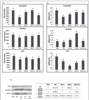
The results of examination of liver CuZnSOD, MnSOD and CAT enzyme activities are presented in Fig. 2A. The STZ treatment provoked a decline (P<0.05) of all examined antioxidant enzymes whereas treatment with Cs, Ld and the Cs/Ld mix recovered the activities of CuZnSOD, MnSOD and CAT (Fig. 2A). The effects of the treatments on antioxidant enzyme transcrip-tion levels in the liver are presented in Fig. 2B. In the diabetic state when compared to the control, the levels of expression of genes encoding for hepatic CuZnSOD, MnSOD and CAT decreased (P<0.05) 1.9-, 1.3- and 2.4-fold, respectively. The transcriptional activities of CuZnSOD and MnSOD increased the most after ad-ministration of the Ld extract, whereas adad-ministration of the Cs/Ld mix caused CAT transcription to increase more than other treatments (Fig. 2B).
One of the most frequent types of intracellular enzymatic glycosylation reactions is through the
O-Fig. 1. The effect of C. sativa and L. deterrimus extracts and their combination on the content of the level of glutathionylated proteins (GSSPs) (A) and the content of free sulfhydryl groups (SH) (B) in the liver of diabetic rats four weeks after administration of the last dose of STZ. NDM – control rats; DM – diabetic rats; DM+Cs
– C. sativa extract treated diabetic rats; DM+Ld – L. deterrimus
linkage of N-acetylglucosamine (O-GlcNAc), which impairs the activities of antioxidant enzymes. Immu-noblot analysis with anti-CAT revealed the presence of O-GlcNAc-modified CAT in the eluted fractions of diabetic rats after the Ld treatment (Fig. 2C). Treat-ments with Cs and the Cs/Ld mix decreased the level of glycosylated CAT (Fig. 2C). O-GlcNAc-modified MnSOD was observed in the liver of diabetic rats. O-GlcNAc-modified MnSOD was lower after treatment with the Cs/Ld mix (1.4-fold lower compared to the diabetic group), whereas O-GlcNAc-modified MnSOD was completely absent after Cs or Ld administration
(Fig. 2C). The redox-sensitive nuclear transcription factor NF-κB was increased in the livers of diabetic rats and modified by O-GlcNAcylation (which was 5.2-fold higher than in the control) (Fig. 2C). In rats treated with either the Cs or Ld extracts, the fraction of NF-κB containing O-GlcNAc was decreased 1.7-fold, remaining 3.4-fold higher than in the control. The lowest level of O-GlcNAc addition to NF-κB was observed after administration of the Cs/ Ld mix (Fig. 2C).
Akt, STAT3 and C/EBPβ protein distribution and expression in the liver of Castanea sativa- and Lactarius
deterrimus-treated diabetic rats
Immunoblot analysis (Fig. 3) revealed the presence of phosphoinositide-3-kinase (PI-3K)/protein kinase B (Akt), an important mediator of cellular prosurvival signals linked to proliferation and differentiation, and its active, phosphorylated form (pAkt). The ratio of pAkt to Akt decreased in the nuclear fraction of diabetic rats (P<0.05). The administration of Cs, Ld and the Cs/ Ld mix increased the presence of pAkt in the nuclear fraction. The increased level of STAT3, associated with signal trans-duction in hepatic injury, was observed in the cytosol and its translocation to the nucleus under diabetic conditions (Fig. 3) (P<0.05) were decreased after administration of Cs, Ld and the Cs/Ld mix (Fig. 3) (P<0.05). STAT3 protein has an important function in interleukin 6 (IL-6)-mediated transcription of the C/EBP-β gene, which is expressed during liver differentiation and which regulates the acute phase response in liver cells. As can be seen in Fig. 3, the active 35 kD isoform of transcription factor C/EBP-β was increased in the liver of diabetic rats (P<0.05). Administration of Cs, Ld and the Cs/Ld mix of extracts reduced the level of C/EBP-β (Fig. 3) (P<0.05).
Masson trichrome staining of the livers of non-diabetic and non-diabetic rats treated with Castanea
sativa and Lactarius deterrimus extracts
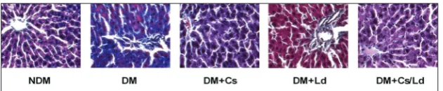
Masson trichrome staining of livers for collagen fiber deposition is presented in Fig. 4B. In the livers of con-trol rats, minimal collagen deposition was observed around the central vein (Fig. 4, NDM). Hepatocytes formed long strips of tissue radiating from the periph-ery towards the central vein, with no inflammatory infiltration and fatty degeneration. In the livers of untreated diabetic controls, lobular architecture was disordered and a prominent increase in collagen fiber deposition was detected around the central vein and between hepatocytes (Fig. 4, DM). Collagen fibers were decreased around the central vein and in the
perisinusoidal region in the livers of Cs-, Ld- and Cs/Ld-treated diabetic rats. The hepatocytes remained po-lygonal and comparable to normal liver hepatocytes (Fig. 4, DM+Cs, DM+Ld and DM+Cs/Ld).
DISCUSSION
The present study revealed that treatments with Cs and Ld extracts, either individually or in combi-nation (Cs/Ld), reduced the con-centrations of blood glucose and glycated hemoglobin, O-GlcNAc-modified antioxidant enzymes and NF-κB, and decreased proinflamma-tory signaling through restoration of the nuclear/cytosol redistribution of STAT3 and C/EBPβ, activated pro-survival Akt kinase and decreased collagen fiber deposition in the liver.
Diabetes-related liver disorders include inflammation, fibrosis, cir-rhosis, hepatocellular carcinoma and hepatitis [24]. The data pre-sented herein describe a decrease in AST and ALT after administration of Cs and the Cs/Ld combination of extracts. A decrease in collagen fiber deposition that was observed in the liver in Cs-, Ld- and Cs/Ld-treated diabetic rats indicated that the fibrotic process, defined as excessive deposition of collagens, was reduced. Enhancement of biochemical markers as well as the decrease of the fibrotic process after extract treatment pointed to improved protection of liver cells/tissue to diabetes-induced damage.
Hyperglycemia induces the overproduction of ROS that leads to oxidative damage of biomolecules such as DNA, proteins and lipids. The decrease in free SH and increased formation of GSSPs in the livers of diabetic rats revealed increased oxidative stress, which the antioxidant capacity of Cs and Ld was ca-pable of significantly improving (i.e. the recovery of these parameters). Oxidative stress in diabetes lowers
Fig. 3. The effect of C. sativa and L. deterrimus and their combination on the distribution between cytosol and nuclear fraction of Akt, pAkt, STAT3 and C/EBPβ in the liver of diabetic rats four weeks after administration of the last dose of STZ. Immunoblot analysis was performed on the cytosol (c) and the nuclear protein fraction (n). NDM – control rats; DM – diabetic rats; DM+Cs – C. sativa extract treated diabetic rats; DM+Ld –L.
deterrimus extract treated diabetic rats; DM+Cs/Ld – diabetic rats treated with the
com-bination of Cs and Ld. The blots were normalized to the corresponding loading controls (actin for cytosol fraction and lamin B for nuclear fraction) and quantified by TotalLab [Phoretix] electrophoresis software [ver. 1.10]. A representative blot of three independent experiments is shown. The values are presented as the means±SEM; values not sharing a common superscript letter differ significantly at P<0.05.
Fig. 4. The effects of individual application of C. sativa and L. deterrimus extracts and their combination on collagen fiber deposition by Masson trichrome staining in the liver of diabetic rats at four weeks after administration of the last dose of STZ. NDM – control rats; DM – diabetic rats; DM+Cs – C. sativa extract treated diabetic rats; DM+Ld – L.
deterrimus extract treated diabetic rats; DM+Cs/Ld – diabetic rats treated with the
the antioxidant status and antioxidant defenses. The present study revealed that the activity of antioxidant enzymes, SOD and CAT, was decreased in diabetic animals, whereas treatment with Cs and Ld extracts recovered the activities of the antioxidant enzymes. The lower level of antioxidant enzyme activities in diabetic rats was accompanied by decreased expression of the corresponding genes. Treatments with extracts were followed by an increase in transcriptional activities of the Mn- and CuZnSOD genes, particularly after treatment with the Ld extract. The transcriptional activity of CAT was increased only in diabetic rats and after treatment with Cs/Ld; the profile of CAT transcriptional activity was not accompanied by a proportional increase in CAT enzyme activity. It has been reported that oxidative stress inactivates enzymes through protein glycation [6]. In diabetes, increased O-GlcNAcylation is directly linked to hyperglycemia-induced glucose toxicity and diabetic complications, through the enzymatic addi-tion of the N-acetyl glucosamine (GlcNAc) moiety to the hydroxyl groups of serine and/or threonine in nuclear and cytoplasmic proteins [24]. O-GlcNAc is recognized as a stress-responsive modification which affects the stability, differential targeting and activity of proteins [26]. We observed that the decrease in antioxidant enzyme activities (MnSOD and CAT) in diabetic rats was accompanied by their increased O-GlcNAc modification. In addition, the increase in O-GlcNAc modification was also observed for redox-sensitive nuclear transcription factor (NF-κB).
Aside from the improvement of the antioxidant response in the livers of animals treated with Cs, Ld or Cs/Ld, the activation of the prosurvival Akt pathway is a very important aspect of the observed repair of liver damage in diabetic rats. Akt is a crucial regula-tor of cell proliferation and differentiation. Dietary polyphenols attenuate hyperglycemia, dyslipidemia and insulin resistance, alleviate oxidative stress and affect important cell signaling pathways involving NF-κB, activator protein-1 DNA binding, extracellular signal-regulated protein kinase (ERK), Akt, mitogen-activated protein kinases and nuclear factor erythroid 2 related factor 2 (Nrf2) [27]. The upregulation of this pathway in the liver of extract-treated diabetic rats was observed as phosphorylation of Akt. In addition to the slight disturbance in the nuclear/cytosol distribution of pAkt, the livers of diabetic rats displayed an altered nuclear/cytosol distribution of STAT3 and C/EBPβ
which is characteristic of the proinflammatory state. The interplay of cytokine signaling pathways, which include tumor necrosis factor alpha (TNFα)/NF-κB, IL-6/STAT3, AP-1 and mitogen-activated protein kinase (MAPK)/extracellular signal-regulated protein kinase (ERK), as well as the prosurvival/antiapoptotic phosphatidylinositol 3-kinase (PI3K)/Akt pathway, assume important roles in cell/tissue regeneration [28]. In the abovementioned interplay, IL-6, which regulates STAT3 transcription by tethering STAT3 to liver-enriched activator protein (LAP)/C/EBPβ [29], assumes an important role. In addition, the constitutive activation of Akt and STAT3 are essential in cell survival [30]. Inflammatory signals contribute to the regulation of the acute phase response in the liver through the activities of transcription factors of the C/EBP family. Members of this family have also been implicated in liver regeneration where they regulate the process of hepatocyte proliferation and differentiation [29].
The anti-hyperglycemic potential of Cs described herein could be attributed to its high phenolic and fla-vonoid contents. Total phenols, polyphenols and many bioactive metabolites contained in mushroom extracts have been used in anti-hypoglycemic applications [10]. The important biological activities of mushrooms could also be attributed to cell-wall components such as β-glucans and polysaccharides, which possess im-portant immunomodulatory activities and the ability to improve the systemic redox disturbance in diabetes through antioxidant and anti-inflammatory activities [10]. Synergistic effects of combinations of extracts in vivo have been observed to provide better protection than a single extract or compound [31]. However, the results in this study showed that Cs and Ld provided better results in some parameters that the Cs/Ld mix, however, the combination of Cs and Ld afforded the best effect in the prevention of body weight decrease, reduced GSH content, liver DNA damage and col-lagen fiber deposition. Although the combination of extract provided an additive beneficial effect, the lack of the synergistic outcome that was observed for some parameters in this study, could be the result of the reactivity of compounds present in the mixtures and their structural modifications that resulted in altered antioxidant activities [32-34].
examined extracts in the prevention of liver damage, revealing their considerable potential in prediabetes and diabetes management. However, the ultimate benefit of these extracts in a clinical context can only be properly evaluated in humans, in preclinical and clinical studies. Furthermore, the complex nature of natural plant extracts and variations in their constituents in different species, localities and preparations, render it essential that a consistent description of the product that can be further used in clinical trials is obtained.
Funding: this work was supported by the Ministry of Education, Science and Technological Development of the Republic of Serbia, Contract No. 451-03-68/2020-14/200007.
Author contributions: MM and MV designed the study. MM, AU, JAJ, NG, SD and MĐ performed the experiments, analyzed and interpreted the results. AF provided the extracts. MM wrote the manuscript. MV and GP critically reviewed the manuscript. Conflict of interest disclosure: The authors declare no conflict of interest.
REFERENCES
1. Lucchesi AN, Tavares de Freitas N, Cassettari L, Marques SFG, Spadella CT. Diabetes mellitus triggers oxidative stress in the liver of alloxan-treated rats: a mechanism for diabetic chronic liver disease. Acta Cir Bras. 2013;28:502-8.
2. Oter S, Jin S, Cucullo L, Dorman HJD. Oxidants and antioxi-dants: friends or foes? Oxid Antioxid Med Sci. 2012;1:1-4. 3. Trief PM, Cibula D, Rodriguez E, Akel B, Weinstock RS.
Incorrect Insulin Administration: A Problem That Warrants Attention. Clin Diabetes. 2016;34(1):25-33.
4. Mukherjee PK, Maiti K, Mukherjee K, Houghton PJ. Leads from Indian medicinal plants with hypoglycemic potentials. J Ethopharm. 2006;106:1-28.
5. Saeed N, Khan RM, Shabbir M. Antioxidant activity, total phenolic and total flavonoid contents of whole plant extracts Torilis leptophylla L. BMC Complement Altern Med. 2012;12:221.
6. Sanz M, Cadahìa E, Esteruelas E, Muñoz AM, de Simón F, Hernández T, Estrella I. Phenolic compounds in chestnut (Castanea sativa Mill.) heartwood. Effect of toasting at coo-perage. J Agric Food Chem. 2010;58:9631-40.
7. Mujić A, Grdović N, Mujić I, Mihailović M, Živković J, Poznanović G, Vidaković M. Antioxidative effects of phe-nolic extracts from chestnut leaves, catkins and spiny burs in streptozotocin-treated rat pancreatic β-cells. Food Chem. 2011;125:841-9.
8. Lupini C, Cecchinato M, Scagliarini A, Graziani R, Catelli E. In vitro antiviral activity of chestnut and quebracho woods extracts against avian reovirus and metapneumovirus. Res Vet Sci. 2009;87:482-7.
9. Grdović N, Dinić S, Arambašić J, Mihailović M, Uskoković A, Marković J, Poznanović G, Vidović S, Zeković Z, Mujić A, Mujić I, Vidaković M. Protective effect of Lactarius deter-rimus and Castanea sativa extract mix(MIX Ld/Cs) on STZ-induced oxidative stress and pancreatic beta-cell death. Brit J Nutrit. 2012;108:1163-76.
10. Lo HC, Wasser SP. Medicinal mushrooms for glycemic con-trol in diabetes mellitus: history, current status, future per-spectives, and unsolved problems. Int J Med Mushrooms. 2011;13:401-426.
11. Jayakumar T, Ramesh E, Geraldine P. Antioxidant activity of the oyster mushroom, Pleurotus ostreatus, on CCL4-induced liver injury in rats. Food Chem Toxicol. 2006;44:1989-96. 12. Hou Y, Ding X, Hou H, Song B, Wang T, Wang F, Zhong J.
Immunostimulant activity of a novel polysaccharide isolated from Lactarius deliciosus (l. ex fr.) gray. Ind J Pharmacol. 2013;75:393-9.
13. Athanasakis G, Aligianni N, Gonou-Zagou Z, Skaltsounis AL., Fokialakis N. Antioxidant properties of the wild edible mushroom Lactarius salmonicolor. J Med Food. 2013;16:760-4.
14. Mihailović M, Arambašić Јovanović J, Uskoković A, Grdović N, DinićS, VidovićS, PoznanovićG, MujićI, Vidaković M. Protective Effects of the Mushroom Lactarius deterrimus Extract on Systemic Oxidative Stress and Pancreatic Islets in Streptozotocin-Induced Diabetic Rats. J Diabet Res. 2015;2015:576726.
15. Arambašić Jovanović J, Mihailović M, Uskoković A, Grdović N, Dinić S, Poznanović G, Mujić I, Vidaković M. Evaluation of the Antioxidant and Antiglycation Effects of Lactarius deterrimus and Castanea sativa Extracts on Hepatorenal Injury in Streptozotocin-Induced Diabetic Rats. Front Phar-macol. 2017;8:793.
16. Drabkin D, Austin H. Spectrophotometric studies prepara-tions from washed blood cells. J Biolog Chem 1935;112:51-5. 17. Parker KM, England JD, Da Costa J, Hess EL, Goldstein DE. Improved colorimetric assay for glycosylated haemoglobin. Clin Chem. 1981;27:669-72.
18. Sedlak J, Lindsay RH. Estimation of total, protein bound and non-protein bound sulfhydryl groups in tissue with Elman’s reagent. Anal Biochem. 1968;25:192-205.
19. Ellman CL. Tissue sulfhydryl groups. Arch Biochem Biophys. 1959;82:70-7.
20. Misra HP, Fridovich I. The role of superoxide anion in the autooxidation of epinephrine and simple assay for superoxide dismutase. J Biolog Chem. 1972;247:3170-5.
21. Beutler E. Catalase. In: Beutler E, editor. Red cell metabolism. A manual of biochemical methods. New York: Grune and Stratton Inc.; 1982. p. 105-16.
22. Gandy JC, Rountree AE, Bijur GN. Akt1 is dynamically modi-fied with O-GlcNAc following treatments with PUGNAc and insulin-like growth factor-1. FEBS Lett. 2006;580:3051-8. 23. Drury RA, Wallington EA, Cancerson R. Carlton’s
Histo-pathological Techniques. 4 th ed. Oxford, London, New York: Oxford University press; 1976.
24. Ma J, Hart GW. Protein O-GlcNAcylation in diabetes and diabetic complications. Expert Rev Proteom. 2013;10:365-80. 25. Dinić S, Arambašić J, Mihailović M, Uskoković A, Grdović
N, Marković J, Karadžić B, Poznanović G, Vidaković M. Decreased O-GlcNAcylation of the key proteins in kinase and redox signaling pathways is a novel mechanism of the beneficial effect of alpha-lipoic acid in diabetic liver. Brit J Nutrit. 2013;110:401-12.
26. Zhang B, Kang M, Xie Q, Xu B, Sun C, Chen K, Wu Y. Antho-cyanins from Chinese bayberry extract protect β cells from oxidative stress-mediated injury via HO-1 upregulation. J Agric Food Chem. 2011;59:537-45.
27. Jackson LN, Larson SD, Silva SR, Rychahou PG, Chen LA, Qiu S, Rajaraman S, Evers BM. PI3K/Akt activation is critical for early hepatic regeneration after partial hepatectomy. Am J Physiol. 2008;294:1401-10.
28. Niehof M, Streetz K, Rakemann T, Bischoff SC, Manns MP, Horni F, Trautwein C. Interleukin-6-induced tethering of STAT3 to the LAP/C/EBPb promoter suggests a new mecha-nism of transcriptional regulation by STAT3. J Biolog Chem. 2011;276:9016-27.
29. Choudhari SR, Khan MA, Harris G, Picker D, Jacob GS, Block T, Shailubha K.. Deactivation of Akt and STAT3 signaling promotes apoptosis, inhibits proliferation, and enhances the
sensitivity of hepatocellular carcinoma cells to an anticancer agent Atiprimod. Mol Cancer Ther. 2007;6:112-21.
30. Patra K, Jana S, Mandal DP, Bhattacharjee S. Evaluation of the antioxidant activity of extracts and active principles of commonly consumed Indian spices. J. Environ. Pathol Toxicol Oncol. 2016;35:299-315.
31. Pinelo M, Manzocco L, Nunez MJ, Nicoli MC. Interaction among phenols in food fortification: negative synergism on antioxidant capacity. J Agric Food Chem. 2004;54:1177-80. 32. Vieira V, Marques A, Barros L, Barreira JCM, Ferreira I.
Insights in the antioxidant synergistic effects of combined edible mushrooms: phenolic and polysaccharidic extracts of Boletus edulis and Marasmius oreades. J Food Nutr Res. 2012;51:109-16.
33. Nedamani ER, Mahoonak AS, Ghorbani M, Kashanine-jad M. Antioxidant properties of individual vs. combined extracts of rosemary leaves and oak fruit. J Agric Sci Technol. 2014;16:1575-86.
Supplementary Material