Research Article
a
October
2018
Computer Science and Software Engineering
ISSN: 2277-128X (Volume-8, Issue-10)
Analysis of Breast Cancer Cell Based on Hyperchromatic
Crowded Group Using Multiple Techniques of Data Mining
Chayanika Sarmah
M.Tech, Department of Information Technology, Gauhati University, Guwahati, Assam, India
Abstract: In this paper, an effort is made to analyze MGG stain images of breast cell in FNAC which will help in early detection of malignant breast cancer cell. There are many morphological features based on which MGG stain test smear images can be categorized into normal and abnormal classes. Some of them are area, perimeter and presence of hyperchromatic crowded (HCG) group etc. So, in this approach we analyse the breast cell based on a presence in the malignant cell using multiple techniques of data mining (Images are examined). The proposed approach is implemented in WEKA, a java based data mining tool
Keywords: breast cancer, HCG, MGG, FNAC.
I. INTRODUCTION
Cancer is a term used for diseases in which abnormal cells divide without control and are able to invade other tissues. Cancer cells can spread to other parts of the body through the blood and lymph systems [1].Cancer is not just one disease but many diseases. There are more than 100 different types of cancer. Most cancers are named for the organ or type of cell in which they start - for example, cancer that begins in the colon is called colon cancer; cancer that begins in melano cytes of the skin is called melanoma.Cancer types can be grouped into broader categories. The main categories of cancer include:
Carcinoma - cancer that begins in the skin or in tissues that line or cover internal organs. There are a number of subtypes of carcinoma, including adeno carcinoma, basal cell carcinoma, squamous cell carcinoma, and transitional cell carcinoma.
Sarcoma - cancer that begins in bone, cartilage, fat, muscle, blood vessels, or other connective or supportive tissue.
Leukemia - cancer that starts in blood-forming tissue such as the bone marrow and causes large numbers of abnormal blood cells to be produced and enter the blood.
Lymphoma and myeloma - cancers that begin in the cells of the immune system.
Central nervous system cancers - cancers that begin in the tissues of the brain and spinal cord.
Origins of Cancer
All cancers begin in cells, the body's basic unit of life. To understand cancer, it's helpful to know what happens when normal cells become cancer cells.
The body is made up of many types of cells. These cells grow and divide in a controlled way to produce more cells as they are needed to keep the body healthy. When cells become old or damaged, they die and are replaced with new cells. However, sometimes this orderly process goes wrong. The genetic material (DNA) of a cell can become damaged or changed, producing mutations that affect normal cell growth and division. When this happens, cells do not die when they should and new cells form when the body does not need them. The extra cells may form a mass of tissue called a tumor. One the methods to identify breast cancer which is used more than the others, is Mammography. But it is frequently seen that different interpretation of radiologists about images is obtained from this way. Another method is Fine needle aspiration cytology [2] (FNAC) and its accuracy is 90%. Therefore, it is better to discover another accurate method.
ISSN(E): 2277-128X, ISSN(P): 2277-6451, pp. 7-20 In our approach we try to classify the breast cells into normal and abnormal classes based on analysis of HCG features.
II. OVERVIEW OF BREAST CANCER
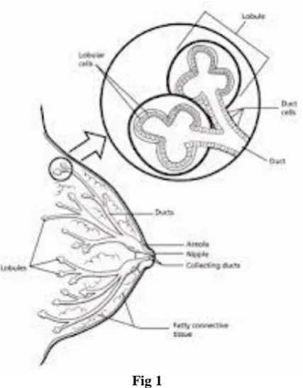
Definition of breast cancer: Cancer that forms in tissues of the breast. The most common type of breast cancer [4] is ductal carcinoma, which begins in the lining of the milk ducts (thin tubes that carry milk from the lobules of the breast to the nipple). Another type of breast cancer is lobular carcinoma, which begins in the lobules (milk glands) of the breast. Invasive breast cancer is breast cancer that has spread from where it began in the breast ducts or lobules to surrounding normal tissue. Breast cancer occurs in both men and women, although male breast cancer is rare.
Estimated new cases and deaths from breast cancer in the United States in 2014: New cases: 232,670 (female); 2,360 (male)
Deaths: 40,000 (female); 430 (male)
Fig 1
The first symptom of breast cancer that can be noticed is a lump that feels different from the rest of breast tissue [5]. 80% of breast cancer cases are discovered when a woman feels a lump. Earliest breast cancers can detected by a mammogram. Lumps found in the lymph located in armpitscan also indicate breast cancer [6].
III. PROPOSED METHOD
The input of the image is taken in .jpg format. The images Sample collected from Dr B. Barooh Cancer Institute, Guwahati –
Normal sample = 14 Abnormal sample = 34
Above MGG stain samples are categorized into two main classes Normal and Abnormal.
We analyzed 200 MGG stain samples and tried to draw a conclusion on how a threshold can be generated which will help in distinguishing the Normal Classes and Abnormal Classes. This identified threshold can be fitted in an automatic system to identify the percentage abnormality of a MGG stain test sample. But the quality of the Images matters in this approach. MGG stain images collected from liquid based technology is better for our approach.
3.1 Proposed method for Hyperchromasia identification
For detecting the hyper chromatic Crowded Groups following measures are taken into consideration. 1. Select the image for which HCG to be identified.
2. Crop the particular region of interest (ROI), i.e. the portion which is dark and crowded. 3. Find the number of cells inside the ROI.
ISSN(E): 2277-128X, ISSN(P): 2277-6451, pp. 7-20 Presence of Hyperchromasia leads to abnormality in breast cells.
Fig 2 Above figure shows the presence of Hyperchromatic Crowded Group (HCG).
IV. EXPERIMENTAL RESULTS
The breast cancer database comes from the Dr. B.Barooah Cancer Institute and contains real observations of 200 pathological instances. The conditional attributes describe information gained from the digitalized images of the breast mass. Datasets reveals that there are 4 classes of attribute of breast cancer data to prove the Hyperchromatic crowded group. Breast cancer datasets in arff (attribute relation file format).
No of instances: 200
No of benign cases: 63
No of malignant cases: 137
No of attribute: 4 @relation breast_cancer_2013 @attribute no_of_cell numeric @attribute image_no numeric @attribute HCG {Present,Absent} @attribute class {Benign,Malignant}
ISSN(E): 2277-128X, ISSN(P): 2277-6451, pp. 7-20
4.1 Classification - 10 fold cross validation on breast-cancer dataset
First we use the data mining tools WEKA to do the training data prediction. In here, we will use 10 fold cross validation on training data to calculate the machine learning rules their performance. The results are as follows:
4.1.1 Results for: Naive Bayes
=== Run information ===
Scheme:weka.classifiers.bayes.NaiveBayes Relation: breast_cancer_2013
Instances: 200 Attributes: 4
no_of_cell image_no HCG class
Test mode: 10-fold cross-validation
=== Classifier model (full training set) ===
Naive Bayes Classifier
Class
Attribute Benign Malignant (0.32) (0.68)
=================================== no_of_cell
mean 7.0769 34.5255 std. dev. 5.3113 17.7487 weight sum 63 137 precision 2.2632 2.2632
image_no
Mean 34.9711 75.0426 std. dev. 23.185 42.4206 Weight sum 63 137 Precision 1.0139 1.0139
HCG
Present 4.0 138.0 Absent 61.0 1.0 [Total] 65.0 139.0
Time taken to build model: 0.02 seconds
=== Stratified cross-validation === === Summary ===
Correctly Classified Instances 197 98.5 % Incorrectly Classified Instances 3 1.5 % Kappa statistic 0.9648
ISSN(E): 2277-128X, ISSN(P): 2277-6451, pp. 7-20 === Detailed Accuracy By Class ===
TP Rate FP Rate Precision Recall F-Measure ROC Area Class
0.952 0 1 0.952 0.976 0.99 Benign
1 0.048 0.979 1 0.989 0.99 Malignant Weighted Avg. 0.985 0.033 0.985 0.985 0.985 0.99
=== Confusion Matrix ===
a b <-- classified as 60 3 | a = Benign 0 137 | b = Malignant
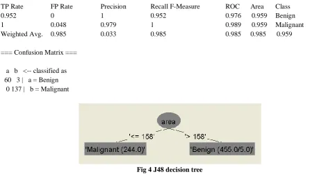
4.1.2 Results for: J48 decision tree (implementation of C4.5)
=== Run information ===
Scheme: weka.classifiers.trees.J48 -C 0.25 -M 2 Relation: breast_cancer_2013
Instances: 200 Attributes: 4
no_of_cell image_no HCG class
Test mode:10-fold cross-validation
=== Classifier model (full training set) ===
J48 pruned tree ---
HCG = Present: Malignant (140.0/3.0) HCG = Absent: Benign (60.0)
Number of Leaves: 2
Size of the tree: 3
Time taken to build model: 0.06 seconds
=== Stratified cross-validation === === Summary ===
Correctly Classified Instances 197 98.5 % Incorrectly Classified Instances 3 1.5 % Kappa statistic 0.9648
ISSN(E): 2277-128X, ISSN(P): 2277-6451, pp. 7-20 === Detailed Accuracy By Class ===
TP Rate FP Rate Precision Recall F-Measure ROC Area Class
0.952 0 1 0.952 0.976 0.959 Benign
1 0.048 0.979 1 0.989 0.959 Malignant
Weighted Avg. 0.985 0.033 0.985 0.985 0.985 0.959
=== Confusion Matrix ===
a b <-- classified as 60 3 | a = Benign 0 137 | b = Malignant
Fig 4 J48 decision tree
4.1.3Results for: JRip (implementation of the RIPPER rule learner)
=== Run information ===
Scheme:weka.classifiers.rules.JRip -F 3 -N 2.0 -O 2 -S 1 Relation: breast_cancer_2013
Instances: 200 Attributes: 4
no_of_cell image_no HCG Class
Test mode: 10-fold cross-validation
=== Classifier model (full training set) ===
JRIP rules: ===========
(no_of_cell <= 14) => Class=Benign (60.0/0.0) => Class=Malignant (140.0/3.0)
Number of Rules: 2
Time taken to build model: 0.02 seconds
=== Stratified cross-validation === === Summary ===
Correctly Classified Instances 197 98.5 % Incorrectly Classified Instances 3 1.5 % Kappa statistic 0.9648
ISSN(E): 2277-128X, ISSN(P): 2277-6451, pp. 7-20 Relative absolute error 6.8175 %
Root relative squared error 26.2275 % Total Number of Instances 200
=== Detailed Accuracy By Class ===
TP Rate FP Rate Precision Recall F-Measure ROC Area Class
0.952 0 1 0.952 0.976 0.959 Benign
1 0.048 0.979 1 0.989 0.959 Malignant
Weighted Avg. 0.985 0.033 0.985 0.985 0.985 0.959
=== Confusion Matrix ===
a b <-- classified as 60 3 | a = Benign 0 137 | b = Malignant
4.2 Classification – Compare with test and training data set
Above machine learning tools will use in this section to diagnose cancer dataset. To construct a dataset with 200 instances. The first 121 instances in the dataset are chosen as the training data, and the remaining 79 as the test data.
4.2.1 Results for training data: Naive Bayes
=== Run information ===
Scheme:weka.classifiers.bayes.NaiveBayes Relation: breast_cancer_2013 Instances: 121
Attributes: 4
no_of_cell image_no HCG class
Test mode:evaluate on training data
=== Classifier model (full training set) ===
Naive Bayes Classifier
Class
Attribute Benign Malignant (0.28) (0.72)
=================================== no_of_cell
mean 7.9211 34.5457 std. dev. 5.5232 18.3201 weight sum 34 87 precision 2.2632 2.2632
image_no
mean 22.2328 48.0225 std. dev. 20.7536 26.8276 weight sum 34 87 precision 1.0215 1.0215
ISSN(E): 2277-128X, ISSN(P): 2277-6451, pp. 7-20 Present 3.0 88.0
Absent 33.0 1.0 [total] 36.0 89.0
Time taken to build model: 0 seconds
=== Evaluation on training set === === Summary ===
Correctly Classified Instances 119 98.3471 % Incorrectly Classified Instances 2 1.6529 % Kappa statistic 0.9583
Mean absolute error 0.0254 Root mean squared error 0.1267 Relative absolute error 6.2507 % Root relative squared error 28.1847 % Total Number of Instances 121
=== Detailed Accuracy By Class ===
TP Rate FP Rate Precision Recall F-Measure ROC Area Class
0.941 0 1 0.941 0.97 0.991 Benign 1 0.059 0.978 1 0.989 0.991 Malignant Weighted Avg. 0.983 0.042 0.984 0.983 0.983 0.991
=== Confusion Matrix ===
a b <-- classified as 32 2 | a = Benign 0 87 | b = Malignant
4.2.2 Results for test data: Naive Bayes
=== Run information ===
Scheme:weka.classifiers.bayes.NaiveBayes Relation: breast_cancer_2013 Instances: 79
Attributes: 4
no_of_cell image_no HCG class
Test mode:user supplied test set: size unknown (reading incrementally)
=== Classifier model (full training set) ===
Naive Bayes Classifier
Class
Attribute Benign Malignant (0.37) (0.63)
=================================== no_of_cell
ISSN(E): 2277-128X, ISSN(P): 2277-6451, pp. 7-20 weight sum 29 50
precision 2.6875 2.6875
image_no
mean 50.2069 121.9782 std. dev. 15.9091 14.8706 weight sum 29 50 precision 1.4545 1.4545
HCG
Present 2.0 51.0 Absent 29.0 1.0 [total] 31.0 52.0
Time taken to build model: 0 seconds
=== Evaluation on test set === === Summary ===
Correctly Classified Instances 78 98.7342 % Incorrectly Classified Instances 1 1.2658 % Kappa statistic 0.9726
Mean absolute error 0.0127 Root mean squared error 0.1125 Relative absolute error 2.7207 % Root relative squared error 23.341 % Total Number of Instances 79
=== Detailed Accuracy By Class ===
TP Rate FP Rate Precision Recall F-Measure ROC Area Class
0.966 0 1 0.966 0.982 0.994 Benign
1 0.034 0.98 1 0.99 0.994 Malignant Weighted Avg. 0.987 0.022 0.988 0.987 0.987 0.994
=== Confusion Matrix ===
a b <-- classified as 28 1 | a = Benign 0 50 | b = Malignant
4.2.3 Results for training data: J48 decision tree (implementation of C4.5)
=== Run information ===
Scheme:weka.classifiers.trees.J48 -C 0.25 -M 2 Relation: breast_cancer_2013
Instances: 121 Attributes: 4
no_of_cell image_no HCG class
ISSN(E): 2277-128X, ISSN(P): 2277-6451, pp. 7-20 === Classifier model (full training set) ===
J48 pruned tree ---
HCG = Present: Malignant (89.0/2.0) HCG = Absent: Benign (32.0)
Number of Leaves : 2
Size of the tree : 3
Time taken to build model: 0.03 seconds
=== Stratified cross-validation === === Summary ===
Correctly Classified Instances 119 98.3471 % Incorrectly Classified Instances 2 1.6529 % Kappa statistic 0.9583
Mean absolute error 0.0325 Root mean squared error 0.1284 Relative absolute error 8.0055 % Root relative squared error 28.5509 % Total Number of Instances 121
=== Detailed Accuracy By Class ===
TP Rate FP Rate Precision Recall F-Measure ROC Area Class
0.941 0 1 0.941 0.97 0.947 Benign
1 0.059 0.978 1 0.989 0.947 Malignant
Weighted Avg. 0.983 0.042 0.984 0.983 0.983 0.947
=== Confusion Matrix ===
a b <-- classified as 32 2 | a = Benign 0 87 | b = Malignant
4.2.4 Results for test data: J48 decision tree (implementation of C4.5)
=== Run information ===
Scheme:weka.classifiers.bayes.NaiveBayes Relation: breast_cancer_2013
Instances: 79 Attributes: 4 no_of_cell image_no HCG class
Test mode:user supplied test set: size unknown (reading incrementally)
=== Classifier model (full training set) ===
ISSN(E): 2277-128X, ISSN(P): 2277-6451, pp. 7-20 Class
Attribute Benign Malignant (0.37) (0.63)
=================================== no_of_cell
mean 5.931 34.2925 std. dev. 4.9595 16.5106 weight sum 29 50 precision 2.6875 2.6875
image_no
mean 50.2069 121.9782 std. dev. 15.9091 14.8706 weight sum 29 50 precision 1.4545 1.4545
HCG
Present 2.0 51.0 Absent 29.0 1.0 [total] 31.0 52.0
Time taken to build model: 0 seconds
=== Evaluation on test set === === Summary ===
Correctly Classified Instances 78 98.7342 % Incorrectly Classified Instances 1 1.2658 % Kappa statistic 0.9726
Mean absolute error 0.0127 Root mean squared error 0.1125 Relative absolute error 2.7207 % Root relative squared error 23.341 % Total Number of Instances 79
=== Detailed Accuracy By Class ===
TP Rate FP Rate Precision Recall F-Measure ROC Area Class
0.966 0 1 0.966 0.982 0.994 Benign
1 0.034 0.98 1 0.99 0.994 Malignant Weighted Avg. 0.987 0.022 0.988 0.987 0.987 0.994
=== Confusion Matrix ===
a b <-- classified as 28 1 | a = Benign 0 50 | b = Malignant
4.2.5. Results for training data: JRip (implementation of the RIPPER rule learner)
=== Run information ===
Scheme:weka.classifiers.rules.JRip -F 3 -N 2.0 -O 2 -S 1 Relation: breast_cancer_2013
ISSN(E): 2277-128X, ISSN(P): 2277-6451, pp. 7-20 no_of_cell
image_no HCG class
Test mode:10-fold cross-validation
=== Classifier model (full training set) ===
JRIP rules: ===========
(no_of_cell <= 14) => class=Benign (32.0/0.0) => class=Malignant (89.0/2.0)
Number of Rules : 2
Time taken to build model: 0.03 seconds
=== Stratified cross-validation === === Summary ===
Correctly Classified Instances 117 96.6942 % Incorrectly Classified Instances 4 3.3058 % Kappa statistic 0.9182
Mean absolute error 0.0487 Root mean squared error 0.1741 Relative absolute error 11.9902 % Root relative squared error 38.7096 % Total Number of Instances 121
=== Detailed Accuracy By Class ===
TP Rate FP Rate Precision Recall F-Measure ROC Area Class
0.941 0.023 0.941 0.941 0.941 0.946 Benign
0.977 0.059 0.977 0.977 0.977 0.946 Malignant
Weighted Avg. 0.967 0.049 0.967 0.967 0.967 0.946
=== Confusion Matrix ===
a b <-- classified as 32 2 | a = Benign 2 85 | b = Malignant
4.2.6 Results for test data: JRip (implementation of the RIPPER rule learner)
=== Run information ===
Scheme:weka.classifiers.rules.JRip -F 3 -N 2.0 -O 2 -S 1 Relation: breast_cancer_2013
Instances: 79 Attributes: 4
no_of_cell image_no HCG class
ISSN(E): 2277-128X, ISSN(P): 2277-6451, pp. 7-20 === Classifier model (full training set) ===
JRIP rules: ===========
(no_of_cell <= 14) => class=Benign (28.0/0.0) => class=Malignant (51.0/1.0)
Number of Rules : 2
Time taken to build model: 0.05 seconds
=== Evaluation on test set === === Summary ===
Correctly Classified Instances 78 98.7342 % Incorrectly Classified Instances 1 1.2658 % Kappa statistic 0.9726
Mean absolute error 0.0248 Root mean squared error 0.1114 Relative absolute error 5.3314 % Root relative squared error 23.111 % Total Number of Instances 79
=== Detailed Accuracy By Class ===
TP Rate FP Rate Precision Recall F-Measure ROC Area Class
0.966 0 1 0.966 0.982 0.983 Benign
1 0.034 0.98 1 0.99 0.983 Malignant Weighted Avg. 0.987 0.022 0.988 0.987 0.987 0.983
=== Confusion Matrix ===
a b <-- classified as 28 1 | a = Benign 0 50 | b = Malignant
4.2.7 Summary
This section presents summary tables for scheme accuracy and running times.
Table 4.2.7.1: Accuracy and running time summary table for 10 fold cross validation
Model Running time 10 fold cross val.
Naive Bayes .02 seconds 98.5%
J48 decision tree (C4.5) .06 seconds 98.5%
JRip (RIPPER rule learner) .02 seconds 98.5%
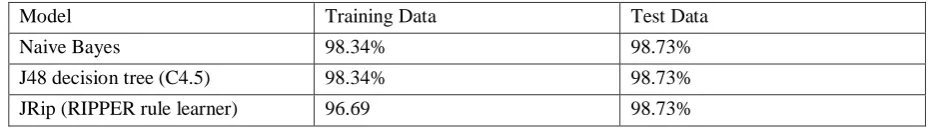
Table 4.2.7.2: Accuracy for training and test data between different models
Model Training Data Test Data
Naive Bayes 98.34% 98.73%
J48 decision tree (C4.5) 98.34% 98.73%
ISSN(E): 2277-128X, ISSN(P): 2277-6451, pp. 7-20
V. CONCLUSION
Medical images have various limitations such as low quality, presence of noise and human error in interpretation .Digital image processing can help the pathologists to a great extent. So this type of automatic detection of breast cancer can help in early detection and diagnosis which can save patients.
REFERENCES
[1] http://www.cancer.gov/cancertopics/cancerlibrary
[2] Alkhair Abd Almahmoud Idris, Muhammed Sidahmed Hussain, ―Comparison of the efficacy of three stains used for the detection of cytological changes in Sudanese females with breast lumps‖Sudanese journal of public health. 2009,vol.4, No.2.
[3] www.polysciences.com
[4] American Cancer Society, Nov 2013
[5] Sariego J (2010). "Breast cancer in the young patient". The American surgeon 76 (12): 1397–1401.
[6] Florescu A, Amir E, Bouganim N, Clemons M (2011). "Immune therapy for breast cancer in 2010—hype or hope?". Current Oncology18 (1): e9–e18. .
[7] N. Lassouaoui, L. Hamami, and N. Nouali, 2007,Morphological Description of Cervical Cell Images for the Pathological Recognition, World Academy of Science, Engineering and Technology,pg 49-51