109
© 2018 by the Serbian Biological Society How to cite this article: Zou S, Liu Y, Min G, Liang Y. Trs20, Trs23, Trs31 and Bet5 participate in autophagy through GTPase Ypt1 in Saccharomyces cerevisiae. Arch Biol Sci. 2018;70(1):109-118.
Trs20, Trs23, Trs31 and Bet5 participate in autophagy through GTPase Ypt1 in
Saccharomyces cerevisiae
Shenshen Zou*, Yan Liu, Gaoyi Min and Yongheng Liang
College of Life Sciences, Key Laboratory of Agricultural Environmental Microbiology of Ministry of Agriculture, Nanjing Agricultural University, Nanjing 210095, China
*Corresponding author: [email protected]
Received: April 8, 2017; Revised: June 20, 2017; Accepted: July 29, 2017; Published online: August 28, 2017
Abstract: TRAPP (transport protein particle) is a large, highly conserved, multi-subunit complex. Four types of TRAPP
complexes (I, II, III and most recently IV) have been identified in Saccharomyces cerevisiae. Studies on the roles of TRAPP
II, TRAPP III and TRAPP IV specific subunits (Trs130, Trs85 and Trs33) have demonstrated that TRAPP II, TRAPP III and TRAPP IV activate the small GTPases that regulate autophagy. Up to now, the roles of the common TRAPP subunits have been well studied in vesicle transport. However, the roles of the common TRAPP subunits and their relationship to Ypt/Rab GTPases in autophagy are not clear. In this paper, we examined Trs20, Trs23, Trs31, and Bet5 (the common TRAPP subunits), which are required for starvation-induced autophagy and the cytoplasm-to-vacuole targeting (Cvt) pathway. During autophagy, GFP-Atg8 accumulates as single or multiple dots and is not recruited into the pre-autophagosomal
structures (PAS) in trs20ts, trs23ts, trs31ts and bet5ts mutant cells. Furthermore, these dots are linked to the endoplasmic
reticulum in mutant cells. Additionally, overexpression of Ypt1, but not Ypt31, suppresses the autophagy defect in trs20ts,
trs23ts, trs31ts and bet5ts mutant cells. Based on these results, we concluded that Trs20, Trs23, Trs31, and Bet5 are required for autophagy, and that these common TRAPP subunits regulate autophagy partially through GTPase Ypt1, but not Ypt31.
Key words: autophagy; TRAPP; common TRAPP subunits; Ypt1; Ypt31
Abbreviations: Atg − autophagy-related gene; Cvt − cytoplasm-to-vacuole targeting; ER − endoplasmic reticulum; GEF − guanine nucleotide exchange factor; GFP − green fluorescent protein; PAS − phagophore assembly site/pre-autophagosomal structure; RFP, red fluorescent protein; TRAPP − transport protein particle; Ypt − yeast protein transport
INTRODUCTION
Vesicle transport among different organelles in yeast is regulated by molecular switches known as Ypt/Rab GTPases, which are activated by guanine-nucleotide exchange factors (GEFs) [1,2]. The TRAPP (transport protein particle) complex is a large, highly conserved, multi-subunit complex [3]. Four forms of TRAPP complexes have been identified in yeast with GEF activities toward GTPases Ypt1 and Ypt31/32 [4-12]. Five common subunits (Bet3, Bet5, Trs20, Trs23 and Trs31) form the TRAPP I complex, which regulates endoplasmic reticulum (ER)-to-Golgi trafficking by activating Ypt1 [13,14]. TRAPP II, which contains components of TRAPP I plus five specific subunits (Trs120, Trs130, Trs65, Trs33 and Tca17), functions in trans-Golgi trafficking [10,15-17]. However, TRAPP
II has also been shown to play important roles in au-tophagy by activating Ypt31/32 [18]. TRAPP I and a specific subunit, Trs85, form the TRAPP III com-plex, is involved in autophagy and vesicle traffick-ing by activattraffick-ing Ypt1 [6,7]. Accordtraffick-ing to a recent study, TRAPP I and a specific subunit, Trs33, form the TRAPP IV complex, which is also required for autophagy by activating Ypt1 [12].
of autophagy. Vacuolar proteases, the precursor forms of Ape1 (hydrolase aminopeptidase I; prApe1) are synthesized in the cytoplasm and transported to the vacuole for further processing through a selective au-tophagy pathway named the cytoplasm-to-vacuole targeting (Cvt) pathway, which is a model for selec-tive autophagy [20]. In contrast, during nonselecselec-tive autophagy, portions of the cytoplasm, such as proteins and organelles, are recycled when the cell is under stress [22,23]. Autophagy usually refers to nonselec-tive autophagy.
Findings from studies of specific subunits of Trs130, Trs85 and Trs33 show that TRAPP II, TRAPP III and TRAPP IV are required for autophagy [7,12,18]. However, the role of the common TRAPP subunits in autophagy is unclear. The common subu-nits, Bet3 and Trs20, were recently shown to maintain the stability of the TRAPP III complex and are in-volved in autophagy [24,25]. In addition, according to the results from a previous study, Bet3 affects both the TRAPP III complex and the function of the TRAPP I complex during autophagy through the GTPase Ypt1 [26]. However, researchers have not clearly identified the roles of the other common TRAPP subunits (Bet5, Trs20, Trs23 and Trs31) in autophagy or whether Ypt1 or Ypt31 suppress the autophagy defects in strains with mutations in these subunits.
As will be shown in this paper, Bet5, Trs20, Trs23 and Trs31, the common subunits of the TRAPP com-plex, are required for nonselective and selective au-tophagy. The trs20ts, trs23ts, trs31ts and bet5ts mu-tant cells grow at the permissive temperature of 26°C but do not grow at 37°C. However, a defect in the autophagy phenotype is observed in these strains at the permissive temperature of 26°C. Furthermore, the Atg8 protein, a protein required to form the autophagosome, is not transported to the pre-au-tophagosomal structures (PAS) in the trs20ts, trs23ts,
trs31ts and bet5ts mutant cells. Finally, Ypt1, but not Ypt31, suppresses the autophagy defect in trs20ts,
trs23ts, trs31ts and bet5ts mutant cells under starva-tion condistarva-tions. Therefore, in this work, the common subunits of the TRAPP complex, Bet5, Trs20, Trs23 and Trs31, partially regulate autophagy through the GTPase Ypt1.
MATERIALS AND METHODS
Strains, plasmids and reagents
The yeast strains used in this study are listed in Table S1. The common TRAPP subunit mutants used here included trs20ts, trs23ts, trs31ts and bet5ts [27]. The desired genes were amplified from the background of the common TRAPP subunit mutants using polymer-ase chain reaction (PCR) and transferred to the strain SEY6210 for this study. For gene deletion, the entire gene coding region was replaced with a drug-resist-ance cassette using PCR amplification. Plasmids ex-pressing GFP-ATG8 (URA3), RFP-APE1 (LEU2) and
ATG9-3XGFP (URA3) under the control of the endog-enous promoter were described in a previous study [18]. For the genetic interaction experiments, pRS425 plasmids (2 μg, LEU2) expressing YPT1 and YPT31
were used [18]. The open reading frames (ORFs) of
TRS20, TRS23, TRS31 and BET5, along with 1000 bp of the 5’-promoter regions were cloned into the pRS425 vector.
The antibodies used in this study are as follows: mouse anti-GFP (Santa Cruz), rabbit anti-Ape1 (a gift from Y. Ohsumi), rabbit anti-pGK1 (Acris, Herford, Germany), and rabbit anti-glucose-6-phosphate de-hydrogenase (G6PDH, Sigma-Aldrich, MO, United States). A horseradish peroxidase-conjugated goat-anti-rabbit IgG antibody and an ECL kit were obtained from Millipore Corporation (MA, United States).
All chemical reagents were purchased from AM-RESCO (OH, United States), unless indicated other-wise. SynaptoRed, also known as FM4-64, was pur-chased from Molecular Probes (OR, United States). Geneticin was obtained from Gibco Laboratories (NY, United States). Restriction enzymes and buffers were from Takara Biotechnology (Dalian, China).
Yeast culture conditions and the induction of autophagy
For live-cell fluorescence microscopy, yeast cul-tures were grown at 26°C in YPD or SD media to log phase. If the cells were subjected to nitrogen starva-tion, they were washed and transferred to SD-N for 2 h at 26°C. If necessary, FM4-64 was added to the liquid medium to a concentration of 1.6 μM to stain the vacuoles during the last hour of incubation. Slides were visualized using a Nikon Eclipse Ti inverted re-search microscope (Tokyo, Japan). The results were quantified using Photoshop CS.
For the genetic interaction analysis, cells were grown in SD media overnight at 26°C, normalized to the same density and spotted onto agar plates in 10-fold serial dilutions. Plates were incubated at different temperatures for the genetic interaction assays.
GFP-Atg8 and Ape1 processing assays
The GFP-Atg8 and Ape1 processing assays were used to detect the level of autophagy in different strains. The concrete steps were as follows: strains were grown at 26°C to an early log phase in YPD media. Autophagy was then induced by SD media and cells were kept at 26°C. Culture aliquots were collected at intervals of 2 h and GFP-Atg8 cleavage or pr-Ape1 matura-tion were determined by Western blot analysis of the cell extracts. Western blot analysis was performed as previously described [17] and repeated at least twice.
RESULTS
Common TRAPP subunits are required for both the Cvt pathway and starvation-induced autophagy
In yeast cells, four types of TRAPP complexes have been identified [7,12,14,28], and by studying the spe-cific TRAPP subunits (Trs130 and Trs85), TRAPP II, TRAPP III and TRAPP IV complexes were shown to regulate autophagy by activating Ypt31/32 and Ypt1, respectively [7,12,18]. According to the results of our recent study, Bet3, a common TRAPP subunit, regu-lates autophagy by activating Ypt1 [26]. However, as it is not clear whether the other common subunits of TRAPP (Trs20, Trs23, Trs31 and Bet5) are involved in autophagy, we examined their involvement in this process.
We visualized the localization of Atg8 in wild-type (WT), trs20ts, trs23ts, trs31ts and bet5ts strains under non-starvation and starvation conditions to directly monitor the autophagy phenotype. The Cvt pathway, a model of selective autophagy, was analyzed using a nutrient enriched medium [20]. Thus, we treated all strains with YPD liquid medium to detect the Cvt pathway. About 22% of WT cells displayed a single GFP-Atg8 dot that colocalized with the PAS marker RFP-Ape1 (Fig. 1A and B), indicating a normal Cvt
Fig. 1. The Cvt pathway and starvation-induced autophagy are impaired in trs20ts, trs23ts, trs31ts and bet5ts mutant cells. A. Atg8 and Ape1 were mislocalized in common TRAPP subunit mutant cells, trs20ts, trs23ts, trs31ts and bet5ts. WT and mutant cells were transformed with GFP-Atg8 and RFP-Ape1 integration plasmids. The cells were grown in YPD medium to mid-log phase at 26°C; if the cells were subjected to nitrogen starvation, they were washed and transferred to SD-N at 26°C for 2 h. The experiments were repeated twice and representative results are presented. Bar – 5 μm. B. Percentage of cells that had at least one GFP-Atg8 dot that was quantified. At least 300 cells from each strain were counted in at least three fields. Error bars represent the standard deviation.
C. Ape1 maturation was blocked and GFP-Atg8 degradation was
pathway. GFP-Atg8 was dispersed in the cytoplasm in four common TRAPP subunit mutant cells (Fig. 1A and B), suggesting a defective Cvt pathway. We also used the Ape1 processing assay to detect mature Ape1 in YPD medium. As expected, WT cells con-tained the mature form of Ape1 (mApe1) and the precursor form of Ape1 (prApe1) (Fig. 1C), suggest-ing a normal Cvt pathway. In contrast, trs20ts, trs23ts,
trs31ts and bet5ts mutant cells accumulated prApe1, but mApe1 was not detected, suggesting that the Cvt pathway was blocked (Fig. 1C), which was consistent with the findings in Fig. 1A. Autophagy was induced when cells were grown under starvation conditions (in SD-N medium), and GFP-Atg8 and RFP-Ape1 were normally delivered to the vacuole in WT cells (Fig. 1A). In contrast, GFP-Atg8 and RFP-Ape1 both accumulated as dots in the cytoplasm and were not delivered to the vacuole in four common TRAPP subunit mutant cells (Fig 1A and B), suggesting an autophagy defect. Meanwhile, the GFP-Atg8 dots were not colocalized with RFP-Ape1 (Fig. 1A). We used the GFP-Atg8 processing assay and Ape1 processing assay to monitor autophagy and extend our analysis. These results were consistent with the fluorescence pheno-type, showing that most of the Atg8 was not degraded and most of the Ape1 had not matured in the mutant cells (Fig. 1C). Thus, Trs20, Trs23, Trs31 and Bet5 are required for the Cvt pathway and autophagy.
Trs20, Trs23, Trs31 and Bet5 are common subu-nits of the TRAPP complex, and their loss of func-tion will affect the funcfunc-tions of the TRAPP I, II, III and IV complexes. Thus, we wanted to determine the functions of these common subunits of the TRAPP complex in autophagy. We compared the GFP-Atg8 phenotype in trs85Δ (TRAPP III specific subunit) mu-tant, trs130ts (TRAPP II specific subunit) mutant and
trs130ts trs85Δ double-mutant cells (Fig. S1A). Be-cause trs130ts is a temperature-sensitive mutant, nor-mal autophagy is observed in this mutant at permis-sive temperatures (26°C) (Fig. S1A) [18]. When cul-tured at a high temperature (37°C), GFP-Atg8 formed multiple dots in more than 82% of trs130ts mutant cells through both the Cvt pathway and autophagy (Fig. S1A). Therefore, we focused on the different phe-notypes observed in different TRAPP-specific subu-nit mutants under high-temperature conditions. In
trs85Δ mutant cells, GFP-Atg8 was dispersed in the cytoplasm through the Cvt pathway (~97% of cells)
and was present in vacuoles and the cytoplasm as a result of autophagy (~92% of cells) (Fig. S1A). In con-trast, GFP-Atg8 distribution in trs130ts trs85Δ double-mutant cells was similar to trs20ts, trs23ts, trs31ts and
bet5ts mutant cells through the Cvt pathway (~97% of cells); upon the induction of autophagy, GFP-Atg8 dots were formed in the cells, and one of the multiple GFP-Atg8 dots was colocalized with the PAS marker RFP-Ape1 in trs130ts trs85Δ cells (~72% of cells) (Fig. S1B). Because GFP-Atg8 is not transported to the PAS in trs20ts, trs23ts, trs31ts and bet5ts mutant cells, the GFP-Atg8 phenotype in mutants of specific subunits of the TRAPP complex is different fromthat in trs20ts,
trs23ts, trs31ts and bet5ts mutant cells (Figs. 1A, S1A and B). In addition, Trs33 (TRAPP IV-specific subu-nit) is also involved in autophagy, and the GFP-Atg8 phenotype in the trs33Δ mutant is similar to trs130ts, as GFP-Atg8 formed multiple dots [18]. Thus, Trs20, Trs23, Bet5 and Trs31 are involved in autophagy, but these subunits may participate in autophagy but not through the TRAPP II, III and IV complexes.
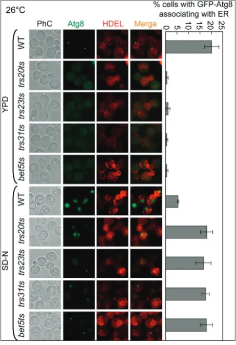
GFP-Atg8 accumulates in a region in contact with the ER in trs20ts, trs23ts, trs31ts and bet5ts
mutant cells under starvation-induced autophagy
The endoplasmic reticulum (ER) has been shown to play an essential role in the formation of autophago-somes [29-31]. The autophagosomal structures formed during the processes used to form autophagosomes, PAS, phagophores, and autophagosomes are linked to the ER, which provides a double-lipid membrane for autophagosomes [29]. Atg8, a protein related to autophagy, is involved in the elongation and establish-ment of the autophagosome [32,33]. Since we showed that in trs20ts, trs23ts, trs31ts and bet5ts mutants Atg8 did not colocalize with the PAS marker RFP-Ape1 (Fig. 1A), we wanted to test whether Atg8 is located in a re-gion in contact with the ER in these common TRAPP subunit mutants. We created strains expressing GFP-Atg8 together with the ER marker DsRed-HDEL. As shown in Fig. 2, a GFP-Atg8 dot was linked to the ER in WT cells in YPD liquid culture medium, but no GFP-Atg8 dots were observed in the trs20ts, trs23ts,
to the ER. In contrast, in all mutant cells, the transport of GFP-Atg8 to the vacuole was blocked, and green dots linked to the ER accumulated. Thus, based on these data, GFP-Atg8 remained connected to the ER and was not transported to the vacuole in the trs20ts,
trs23ts, trs31ts and bet5ts mutant cells.
GFP-Atg8 dot formation depends on Atg1 in
trs20ts, trs23ts, trs31ts and bet5ts mutant cells
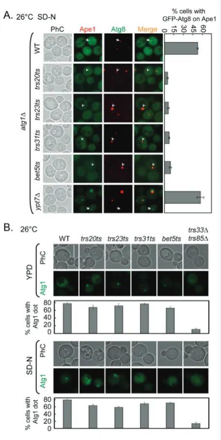
Atg1 is required for autophagy. The PAS is formed in cells lacking the ATG1gene, but the
autophago-somal structures stop at this step; phagophores and autophagosomes are not formed in atg1Δ mutant cells [34,35]. In atg1Δ mutant cells, GFP-Atg8 dots accu-mulate and are believed to be the PAS [18,35]. Accord-ing to the result shown in Fig. 1A, GFP-Atg8 was not delivered to the PAS. Thus, we wanted to determine whether the functions of Trs20, Trs23, Trs31 and Bet5 are upstream of Atg1. We deleted the ATG1 gene in WT, trs20ts, trs23ts, trs31ts, bet5ts and ypt7Δ strains to examine the localization of Atg8. If the common TRAPP subunits are upstream of Atg1, the deletion of ATG1 would not change the GFP-Atg8 phenotype in trs20ts, trs23ts, trs31ts and bet5ts mutant cells. As shown in Fig. 3A, GFP-Atg8 dots accumulated and colocalized with RFP-Ape1, the PAS marker, in atg1Δ cells. The Rab GTPase Ypt7 functions downstream of Atg1 and Ypt7 participates in the fusion of the autophagosomes with the vacuole [36,37]. Multiple GFP-Atg8 dots accumulate in cells lacking YPT7. In control ypt7Δatg1Δ mutant cells, GFP-Atg8 accumu-lated at a single dot that colocalized with RFP-Ape1. However, in trs20ts, trs23ts, trs31ts and bet5ts mutant cells in which ATG1 had been deleted, GFP-Atg8 was dispersed in the cytoplasm and the dot did not form. This phenotype is not similar to atg1Δ mutant cells or common TRAPP subunit mutant cells. To extend the analysis of the relationship between common TRAPP subunits and Atg1, we examined the location of Atg1 in trs20ts, trs23ts, trs31ts and bet5ts mutant cells un-der both non-starvation and starvation conditions. Specifically, in >78% of WT cells Atg1 localized to a single dot per cell and TRAPP III and IV regulated the localization of Atg1, which is diffuse in trs33Δtrs85Δ
mutant cells (Fig. 3B) [12]. When compared to WT cells, Atg1 also localized to a single dot per cell in
trs20ts, trs23ts, trs31ts and bet5ts mutant cells (Fig. 3B). Based on this result, the relationship between the four common TRAPP subunits and Atg1 is not a strict upstream or downstream relationship, but is most likely parallel, regulating GFP-Atg8 transport.
Trs20, Trs23, Trs31 and Bet5 are required for the localization of Atg9 to the PAS
Atg9 is a transmembrane protein in yeast that is in-volved in autophagosome formation [38]. Atg9 is located in several intracellular organelles, including the mitochondria, and Golgi apparatus, and it
Fig. 4. Atg9 anterograde movement is less efficient in the trs20ts, trs23ts, trs31ts and bet5ts mutant cells. A. WT and trs20ts, trs23ts, trs31ts and bet5ts mutant cells were transformed with Atg9-3XGFP integration plasmids. The cells were grown in YPD medium to mid-log phase at 26°C; if the cells were subjected to nitrogen star-vation, they were washed and transferred to SD-N medium for about 2 h to induce starvation. The experiments were repeated twice and representative results are presented. Bar – 5 μm. B.
WT and trs20ts, trs23ts, trs31ts and bet5ts mutant cells tagged with Atg9-3XGFP and RFP-Ape1 in the atg1Δ background. The cells were grown and treated as described in A. The results shown in this figure are representative of at least two independent ex-periments. Bar – 5 μm. Quantitative analysis of the brightness of the Atg9-3XGFP dot in each of the cells in (B) using ZEN 2012 software (Zeiss); the brightness of Atg9 in the WT is taken as 1. At least 300 cells from each strain were counted in at least three fields. Error bars represent the standard deviation.
mally cycles between the PAS and these organelles [39,40]. This process also requires several other Atg proteins; for example, the recycling of Atg9 from the PAS depends on the Atg1-Atg13 complex [34]. Thus, if cells lack ATG1, the recycling of Atg9 from the PAS is blocked and Atg9 remains trapped in the PAS [34]. Because Atg8 transport to the PAS depends on Atg9 and Atg8 is mislocalized in allmutant cells, we screened for the recycling of Atg9 in WT cells and the four common TRAPP subunits in mutant cells. Multiple Atg9-3XGFP dots were formed in all strains under non-starvation and starvation conditions (Fig. 4A), indicating that normal retrograde trafficking of Atg9 from the PAS occurred. When the ATG1 dele-tion was present in background strains, under non-starvation and non-starvation conditions Atg9-3XGFP was concentrated as a single dot that colocalized with RFP-Ape1, the PAS marker, in WT cells. In addition, the localization of Atg9-3XGFP in all mutant cells was similar to WT cells. Atg9-3XGFP accumulated at a single dot and colocalized with RFP-Ape1 (Fig. 4B). However, the brightness of the Atg9-3XGFP dot in WT cells was greater than in the mutant cells under starvation conditions. The quantitative analysis of the results revealed that the brightness of Atg9-3XGFP in the mutant was only 50~60% of that in the WT (Fig. 4B), suggesting that anterograde trafficking of Atg9 to the PAS is not completely blocked.
Ypt1 suppresses the autophagy defect in common subunits of TRAPP mutants
Four TRAPP complexes in yeast have been reported to exert GEF activity toward the Rab GTPases Ypt1 and Ypt31/32 [4-12]. In addition, the overexpression of YPT1,but not YPT31, suppressed the temperature sensitivity of trs20ts, trs23ts, trs31ts and bet5ts mutant cells (Fig. S2), indicating that all plasmids used in this study were functional. Thus, we tested whether Ypt1 and Ypt31 rescued the autophagy defects observed in all mutant cells. First, we visualized the transport of GFP-Atg8 in all mutant cells overexpressing Ypt1 or Ypt31 under starvation conditions. When WT cells were transformed with the vector, GFP-Atg8 was transported to the vacuole normally (Fig. 5A and B). Additionally, overexpression of the gene missing in the mutant reversed the defect in GFP-Atg8 transport, whereas the overexpression ofYpt1, but not Ypt31,
specifically suppressed the intracellular accumulation of GFP-Atg8 in all mutant cells. Second, we performed the GFP-Atg8 processing assay and the results were
Fig. 5. Ypt1, but not Ypt31, suppresses the GFP-Atg8 transport defect in the trs20ts, trs23ts, trs31ts and bet5ts mutant cells. Empty plasmids (Ø) and plasmids expressing the gene lost in the mutant, Ypt1 and Ypt31 (all genes in pRS425 vectors) were transformed into WT and trs20ts, trs23ts, trs31ts and bet5ts mutant cells. A.
Ypt1, not Ypt31, suppressed the autophagy defect in trs20ts, trs23ts, trs31ts and bet5ts mutant cells. The cells were grown as described in Fig. 4A and stained with FM4-64 during the last hour to label the vacuoles. The cells were washed twice and observed under a fluorescence microscope. Bar – 5 μm. B. Quantitation of the percentage of cells in which GFP-Atg8 was not observed in vacuoles. At least 300 cells from each strain were counted in at least three fields. Error bars represent the standard deviation.
consistent with the fluorescence phenotype, showing that Ypt1, but not Ypt31, can suppress the autophagy defect in all mutant cells (Fig. 5C). Thus, Trs20, Trs23, Trs31 and Bet5 regulate autophagy through Ypt1.
DISCUSSION
Trs20, Trs23, Trs31 and Bet5 are the common subu-nits of the TRAPP complex [13,14]. In this study, we show that these four subunits were also involved in autophagy. We took advantage of the transport of GFP-Atg8 in trs20ts, trs23ts, trs31ts and bet5ts mutant cells to directly monitor the autophagy phenotype. GFP-Atg8 was not transported to the vacuole and ac-cumulated into one or more dots in the cytoplasm in all mutant cells; these Atg8 dots were not located in the PAS. However, the common subunits belong to four TRAPP complexes, I, II, III and IV, and we compared the defects in autophagy among strains expressing different TRAPP complexes to further define the functions of the common subunits. When the strain lacked the TRAPP III complex (trs85Δ), TRAPP II complex (trs130ts) or both the TRAPP III and TRAPP II complex (trs85Δtrs130ts), GFP-Atg8 was located in the PAS. Thus, Trs20, Trs23, Trs31, and Bet5 do not affect the function of the TRAPP II or TRAPP III complex in autophagy. In addition, we observed a slight impairment in the anterograde trafficking of Atg9 in these mutant cells, but the Atg9-3XGFP phenotype differed among these common subunit mutants and the specific subunit mutants. In
trs85Δ and trs130ts mutant cells in the atg1Δ back-ground, Atg9-3XGFP accumulated in multiple dots [18,41], and the phenotypes observed in trs85Δ and
trs130ts mutants were different from those in trs20ts,
trs23ts, trs31ts and bet5ts mutant cells, which is con-sistent with the GFP-Atg8 phenotype. The TRAPP IV complex was recently shown to be required for autophagy, but multiple GFP-Atg8 dots accumulated in trs33Δ (specific subunit of TRAPP IV) mutant cells in our previous study [18]. This GFP-Atg8 phenotype in trs33Δ mutant cells differs from the phenotypes observed in trs20ts, trs23ts, trs31ts and bet5ts mutant cells, suggesting that the defect in autophagy was not mediated by the TRAPP IV complex in these common subunit mutants. These phenotypic differences be-tween common subunit and specific subunit mutants suggested that autophagy may be regulated by the
TRAPP I complex, but not the TRAPP II, TRAPP III and TRAPP IV complexes, in trs20ts, trs23ts, trs31ts
and bet5ts mutant cells.
In yeast, autophagosome biogenesis depends on Atg8 located in the PAS [32]. In this study, Atg8 was not located in the PAS in trs20ts, trs23ts, trs31ts or
bet5ts mutant cells, suggesting that the functions of Trs20, Trs23, Trs31 and Bet5 are to regulate the forma-tion of autophagosomes. This result is consistent with the findings obtained with the bet3ts mutant (a com-mon subunit of TRAPP complex mutant) [26]. The transport of Atg8 to the PAS depends on Atg9. When we examined the localization of Atg9, we found that Atg9 was located in the PAS, as the fluorescence signal was detected in the PAS in trs20ts, trs23ts, trs31ts and
bet5ts mutants, although the intensity was obviously decreased. This decrease in the localization of Atg9 to the PAS may partially block the transport of Atg8 to the PAS. Previous studies have shown that COPII vesicles are not only involved in vesicle transport from the ER to the Golgi but also play roles in the transport from the ER to the PAS during autophagy [5]. Thus, Trs20, Trs23, Trs31 and Bet5 function as coregula-tors of COPII vesicle and Atg9-mediated transport pathways and are required for the transport of Atg8 to the PAS.
Four TRAPP complexes function as GEFs to ac-tivate the GTPases Ypt1 and Ypt31, thus regulating vesicle trafficking and autophagy [7,10-12,14,18]. The functions of all TRAPP complexes require the common subunits, Trs20, Trs23, Trs31 and Bet5. Overexpression of Ypt1, but not Ypt31, suppressed the autophagy defects in trs23ts, trs20ts, trs31ts and
bet5ts mutant cells. Based on these results, we con-clude that Trs20, Trs23, Trs31 and Bet5 are required for Ypt1-mediated autophagy, but not Ypt31-mediated autophagy. However, Ypt1 is involved in multiple steps in autophagy, and deletion of the common subunits did not lead to the complete loss of Ypt1 function. Thus, in the future, we will focus on determining which step of the autophagy process is regulated by the common subunits through Ypt1.
genetic analysis suggested that autophagy defects in these common subunit mutants were partially caused by the reduced activity of Ypt1. These results contrib-ute towards a comprehensive understanding of the roles of TRAPP and their regulation mechanisms in autophagy.
Acknowledgments: We are grateful to Dr. P. Hieter (Max Planck Institute for Biophysical Chemistry) for providing the trs20ts, trs23ts, trs31ts and bet5ts mutant strains, Dr. N. Segev (University of Illinois) for yeast plasmids and Dr. Y. Ohsumi (Tokyo Institute of Technology) for providing the anti-Ape1 antibody. This work was supported by grants from the Natural Science Foundation of China (31301173 to SZ; 31671479 and 31271520 to YL), the Fun-damental Research Funds for Central Universities (KYZ201215 to YL; Y0201300236 to SZ), the Project sponsored by the Scientific Research Foundation for the Returned Overseas Chinese Scholars, State Education Ministry ([2011]508), and Nanjing Agricultural University (680-804094-521) to Y. Liang.
Author contributions: Shenshen Zou, Yan Liu and Gaoyi Min contributed equally to this work. ZSS performed the experiments, analyzed the data and wrote the manuscript, LY performed the experiments, MGY performed the experiments, LYH coordinated the research.
Conflict of interest disclosure: The authors declare that there is no conflict of interest.
REFERENCES
1. Segev N. Ypt and Rab GTPases: insight into functions through novel interactions. Curr Opin Cell Biol. 2001;13(4):500-11. 2. Stenmark H. Rab GTPases as coordinators of vesicle traffic.
Nat Rev Mol Cell Biol. 2009;10(8):513-25.
3. Cai H, Reinisch K, Ferro-Novick S. Coats, tethers, Rabs, and SNAREs work together to mediate the intracellular destina-tion of a transport vesicle. Dev Cell. 2007;12(5):671-82. 4. Pinar M, Arst HN, Jr., Pantazopoulou A, Tagua VG, de los
Rios V, Rodriguez-Salarichs J, Diaz JF, Penalva MA. TRAP-PII regulates exocytic Golgi exit by mediating nucleotide exchange on the Ypt31 ortholog RabERAB11. Proc Natl Acad Sci U S A. 2015;112(14):4346-51.
5. Tan D, Cai Y, Wang J, Zhang J, Menon S, Chou HT, Ferro-Novick S, Reinisch KM, Walz T. The EM structure of the TRAPPIII complex leads to the identification of a require-ment for COPII vesicles on the macroautophagy pathway. Proc Natl Acad Sci U S A. 2013;110(48):19432-7.
6. Zou S, Liu Y, Zhang XQ, Chen Y, Ye M, Zhu X, Yang S, Lipatova Z, Liang Y, Segev N. Modular TRAPP complexes regulate intracellular protein trafficking through multiple
Ypt/Rab GTPases in Saccharomyces cerevisiae. Genetics.
2012;191(2):451-60.
7. Lynch-Day MA, Bhandari D, Menon S, Huang J, Cai H, Bartholomew CR, Brumell JH, Ferro-Novick S, Klionsky DJ. Trs85 directs a Ypt1 GEF, TRAPPIII, to the
phago-phore to promote autophagy. Proc Natl Acad Sci U S A. 2010;107(17):7811-6.
8. Barrowman J, Bhandari D, Reinisch K, Ferro-Novick S. TRAPP complexes in membrane traffic: convergence through a common Rab. Nat Rev Mol Cell Biol. 2010;11(11):759-63. 9. Cai Y, Chin HF, Lazarova D, Menon S, Fu C, Cai H,
Scla-fani A, Rodgers DW, De La Cruz EM, Ferro-Novick S, Reinisch KM. The structural basis for activation of the Rab Ypt1p by the TRAPP membrane-tethering complexes. Cell. 2008;133(7):1202-13.
10. Morozova N, Liang Y, Tokarev AA, Chen SH, Cox R, Andrejic J, Lipatova Z, Sciorra VA, Emr SD, Segev N. TRAPPII sub-units are required for the specificity switch of a Ypt-Rab GEF. Nat Cell Biol. 2006;8(11):1263-9.
11. Jones S, Newman C, Liu F, Segev N. The TRAPP complex is a nucleotide exchanger for Ypt1 and Ypt31/32. Mol Biol Cell. 2000;11(12):4403-11.
12. Lipatova Z, Majumdar U, Segev N. Trs33-containing TRAPP IV: A Novel Autophagy-Specific Ypt1 GEF. Genetics. 2016;204(3):1117-28.
13. Sacher M, Jiang Y, Barrowman J, Scarpa A, Burston J, Zhang L, Schieltz D, Yates JR, 3rd, Abeliovich H, Ferro-Novick S. TRAPP, a highly conserved novel complex on the cis-Golgi that mediates vesicle docking and fusion. EMBO J. 1998;17(9):2494-503.
14. Sacher M, Barrowman J, Wang W, Horecka J, Zhang Y, Pypaert M, Ferro-Novick S. TRAPP I implicated in the specificity of tethering in ER-to-Golgi transport. Mol Cell. 2001;7(2):433-42.
15. Choi C, Davey M, Schluter C, Pandher P, Fang Y, Foster LJ, Conibear E. Organization and assembly of the TRAPPII com-plex. Traffic. 2011;12(6):715-25.
16. Tokarev AA, Taussig D, Sundaram G, Lipatova Z, Liang Y, Mulholland JW, Segev N. TRAPP II complex assembly requires Trs33 or Trs65. Traffic. 2009;10(12):1831-44. 17. Liang Y, Morozova N, Tokarev AA, Mulholland JW, Segev N.
The role of Trs65 in the Ypt/Rab guanine nucleotide exchange factor function of the TRAPP II complex. Mol Biol Cell. 2007;18(7):2533-41.
18. Zou S, Chen Y, Liu Y, Segev N, Yu S, Min G, Ye M, Zeng Y, Zhu X, Hong B, Bjorn LO, Liang Y, Li S, Xie Z. Trs130 participates in autophagy through GTPases Ypt31/32 in Sac-charomyces cerevisiae. Traffic. 2013;14(2):233-46.
19. Klionsky DJ, Baehrecke EH, Brumell JH, Chu CT, Codogno P, Cuervo AM, Debnath J, Deretic V, Elazar Z, Eskelinen EL, Finkbeiner S, Fueyo-Margareto J, Gewirtz D, Jaattela M, Kro-emer G, Levine B, Melia TJ, Mizushima N, Rubinsztein DC, Simonsen A, Thorburn A, Thumm M, Tooze SA. A com-prehensive glossary of autophagy-related molecules and pro-cesses (2nd edition). Autophagy. 2011;7(11):1273-94. 20. Lynch-Day MA, Klionsky DJ. The Cvt pathway as a model for
selective autophagy. FEBS Lett. 2010;584(7):1359-66. 21. Feng Y, He D, Yao Z, Klionsky DJ. The machinery of
macro-autophagy. Cell Res. 2014;24(1):24-41.
22. Klionsky DJ. The molecular machinery of autophagy: unan-swered questions. J Cell Sci. 2005;118(Pt1):7-18.
24. Brunet S, Shahrzad N, Saint-Dic D, Dutczak H, Sacher M. A trs20 mutation that mimics an SEDT-causing mutation blocks selective and non-selective autophagy: a model for TRAPP III organization. Traffic. 2013;14(10):1091-104.
25. Taussig D, Lipatova Z, Segev N. Trs20 is required for TRAPP III complex assembly at the PAS and its function in autoph-agy. Traffic. 2014;15(3):327-37.
26. Zou S, Liu Y, Zhang C, Yu S, Liang Y. Bet3 participates in autophagy through GTPase Ypt1 in Saccharomyces cerevisiae. Cell Biol Int. 2015;39(4):466-74.
27. Ben-Aroya S, Coombes C, Kwok T, O’Donnell KA, Boeke JD, Hieter P. Toward a comprehensive temperature-sensitive mutant repository of the essential genes of Saccharomyces cerevisiae. Mol Cell. 2008;30(2):248-58.
28. Sacher M, Barrowman J, Schieltz D, Yates JR, 3rd, Ferro-Novick S. Identification and characterization of five new subunits of TRAPP. Eur J Cell Biol. 2000;79(2):71-80. 29. Graef M, Friedman JR, Graham C, Babu M, Nunnari J. ER
exit sites are physical and functional core autophagosome biogenesis components. Mol Biol Cell. 2013;24(18):2918-31. 30. Ge L, Zhang M, Schekman R. Phosphatidylinositol 3-kinase
and COPII generate LC3 lipidation vesicles from the ER-Golgi intermediate compartment. Elife. 2014;3:e04135. 31. Hamasaki M, Furuta N, Matsuda A, Nezu A, Yamamoto A,
Fujita N, Oomori H, Noda T, Haraguchi T, Hiraoka Y, Amano A, Yoshimori T. Autophagosomes form at ER-mitochondria contact sites. Nature. 2013;495(7441):389-93.
32. Xie Z, Nair U, Klionsky DJ. Atg8 controls phagophore expansion during autophagosome formation. Mol Biol Cell. 2008;19(8):3290-8.
33. Nakatogawa H, Ichimura Y, Ohsumi Y. Atg8, a ubiquitin-like protein required for autophagosome formation, mediates membrane tethering and hemifusion. Cell. 2007;130(1):165-78.
34. Reggiori F, Tucker KA, Stromhaug PE, Klionsky DJ. The Atg1-Atg13 complex regulates Atg9 and Atg23 retrieval transport from the pre-autophagosomal structure. Dev Cell. 2004;6(1):79-90.
35. Suzuki K, Kubota Y, Sekito T, Ohsumi Y. Hierarchy of Atg proteins in pre-autophagosomal structure organization. Genes Cells. 2007;12(2):209-18.
36. Kirisako T, Baba M, Ishihara N, Miyazawa K, Ohsumi M, Yoshimori T, Noda T, Ohsumi Y. Formation process of autophagosome is traced with Apg8/Aut7p in yeast. J Cell Biol. 1999;147(2):435-46.
37. Chen Y, Zhou F, Zou S, Yu S, Li S, Li D, Song J, Li H, He Z, Hu B, Bjorn LO, Lipatova Z, Liang Y, Xie Z, Segev N. A Vps21 endocytic module regulates autophagy. Mol Biol Cell. 2014;25(20):3166-77.
38. Noda T, Kim J, Huang WP, Baba M, Tokunaga C, Ohsumi Y, Klionsky DJ. Apg9p/Cvt7p is an integral membrane pro-tein required for transport vesicle formation in the Cvt and autophagy pathways. J Cell Biol. 2000;148(3):465-80. 39. Reggiori F, Shintani T, Nair U, Klionsky DJ. Atg9 cycles
between mitochondria and the pre-autophagosomal structure in yeasts. Autophagy. 2005;1(2):101-9.
40. Mari M, Griffith J, Rieter E, Krishnappa L, Klionsky DJ, Reg-giori F. An Atg9-containing compartment that functions in the early steps of autophagosome biogenesis. J Cell Biol. 2010;190(6):1005-22.
41. Shirahama-Noda K, Kira S, Yoshimori T, Noda T. TRAPPIII is responsible for vesicular transport from early endosomes to Golgi, facilitating Atg9 cycling in autophagy. J Cell Sci. 2013;126(Pt21):4963-73.