DigitalCommons@UNMC
DigitalCommons@UNMC
Theses & Dissertations Graduate Studies
Spring 5-5-2018
The Effect of Dust Mite extract on Esophageal Tight Junctions in
The Effect of Dust Mite extract on Esophageal Tight Junctions in
Eosinophilic Esophagitis
Eosinophilic Esophagitis
Kyle HinzUniversity of Nebraska Medical Center
Follow this and additional works at: https://digitalcommons.unmc.edu/etd
Part of the Immune System Diseases Commons, and the Medical Anatomy Commons
Recommended Citation Recommended Citation
Hinz, Kyle, "The Effect of Dust Mite extract on Esophageal Tight Junctions in Eosinophilic Esophagitis" (2018). Theses & Dissertations. 274.
https://digitalcommons.unmc.edu/etd/274
This Thesis is brought to you for free and open access by the Graduate Studies at DigitalCommons@UNMC. It has been accepted for inclusion in Theses & Dissertations by an authorized administrator of DigitalCommons@UNMC. For more information, please contact digitalcommons@unmc.edu.
The Effect of Dust Mite extract on Esophageal Tight Junctions in
Eosinophilic Esophagitis
by
Kyle Kenyu Hinz
A THESIS
Presented to the Faculty of
the University of Nebraska Graduate College
in Partial Fulfillment of the Requirements
for the Degree of Master of Science
Genetics, Cell Biology, and Anatomy
Graduate Program
Under the Supervision of Dr. Samantha Simet
University of Nebraska Medical Center
Omaha, Nebraska
May, 2018
Advisory Committee:
Karen Gould, PhD.
Shantaram Joshi, PhD.
Travis McCumber, PhD.
Samantha Simet, PhD.
Acknowledgments
I would like to thank my advisor, Dr. Samantha Simet for her guidance and supervision throughout this project. I would also like to thank Dr. Karen Gould, Dr. Shantaram Joshi, Dr. Travis McCumber, for being members of my thesis committee. I also thank Jacqueline Pavlik for her help during my time in lab. Lastly, I would like to thank the Genetics, Cell Biology, and Anatomy department for use of their facilities for my project. Lastly, I am very grateful for everyone else who has helped me throughout my thesis and appreciate all the support they have given me.
The Effect of Dust Mite extract on Esophageal Tight Junctions in
Eosinophilic Esophagitis
Kyle Hinz, M.S. University of Nebraska, 2018 Advisor: Samantha Simet, PhD
Eosinophilic Esophagitis (EE) is a chronic inflammatory disease that effects individuals of all ages. EE is mediated by an allergen response, causing the release of thymic stromal lymphopoietin (TSLP), and subsequent inflammation and eosinophil migration and infiltration of the esophagus through the activation of the JAK/STAT pathway and CD4+ T-cells. EE has typically been
associated with food allergies, but studies have shown that aeroallergens can cause EE as well. Current treatments of EE have primarily focused on nonspecific methods, though anti-TSLP is beginning to be tested as a possible treatment for EE. The aim of this study was to see how house dust mites affected tight junction function in esophageal cells and determine if application of anti-TSLP will prevent dust mite’s effects. This was accomplished through a
transwell cell culture model. Dust mites along with experimental conditions Gö 6976, interferon-gamma, eosinophils, and TSLP synthetic peptide (anti-TSLP) was used to determine how these conditions affected esophageal cell tight junctions. Transepithelial electrical resistance (TEER) was recorded to measure epithelial integrity. Cells were analyzed through Prism and imaged by immunofluorescence staining. Results showed that dust mites caused a decrease in epithelial resistance through a 24-hour period. Interactions between eosinophils and dust mites caused further decreases in epithelium resistance. When esophageal cells are treated with anti-TSLP, the addition of dust mites did not cause a fall in electrical resistance; however, it did not stop eosinophils disruption of epithelial resistance. Immunofluorescence was used to show changes
in tight junction function for each condition. These findings indicate that dust mites do affect esophageal cell function and anti-TSLP negates the disruptive actions of dust mites. Further studies have to be conducted to understand aeroallergens role in EE and how treatments can be tailored for aeroallergens.
List of Figures
Fig. 1: 2
Histology of normal esophagus and EE. A. Normal histology of the esophagus. B. EE biopsy showing infiltration of eosinophils in the epithelium. C. Shows superficial layering of surface eosinophils through black arrow. D. An example of an eosinophilic microabscess (Liacouras et al., 2011)
Fig 2. 3
Age distribution of patients inflicted with EE. Majority of patients that are diagnosed with EE each year are in the age range of adults, primarily young to middle-aged adults. (Van Rhijin, 2012)
Fig. 3. 3
Prevalence trends across multiple cities around the world. Prevalence of EE has increased markedly since the 1990s, through the use of better diagnostic techniques and understanding of EE. (Dellon & Hirano, 2018)
Fig. 4: 4
Incidence rates of EE in multiple cities around the world. Just like prevalence rates, incidence rates have increased significantly since the 1990s and is expected to continue to increase. (Dellon & Hirano, 2018)
Fig 5. 7 Pathways activated by TSLP binding. TSLP activates many pathways when binding to its receptor. Activation of STAT, MAPK, NFKBIA, and others cause an inflammatory response in both airway diseases and EE. (Zhong et al., 2014)
Fig. 6: 8
Electron microscopy of junctional complexes. At the apical level, tight junctions can be seen. Underneath the tight junctions, adherens junctions and desmosomes can be seen as well. (Fawcett DW, The Cell: An Atlas of Fine Structure, WB Saunders, Philadelphia, 1966, p. 367.)
Fig. 7: 9
Barrier function proteins in adjacent cells. A. Shows junctional complexes and their interactions with the cytoskeleton/ basement membrane. B. Shows tight and adheren junctions mediate cell-cell contact and interact with actin cytoskeleton. (Loxham & Davies, 2017)
Fig 8: 12
Type 2 cell immune stimuli, such as dust mite protease activity, leads to an immune response. Activation of receptors on the epithelium leads to release of innate pro-Th2 cytokines. (Hammad & Lambrecht, 2015)
Fig 9. 19 Changes in epithelial resistance based on various concentrations of dust mites. Dust mites decreased epithelial resistance at each concentration level used. The combination of dust mites and eosinophils lead to even greater decreases in epithelial integrity
Fig 10. 22
Changes in epithelial resistance based on different experimental conditions applied to each well. Over the course of the 48 hours, decreased epithelial resistance was seen in wells containing EOS, TSLP EOS, and TSLP DM EOS
Fig. 11 25
Immunofluorescence imaging of ZO-1. A) Control shows a ZO-1 present between two different cells B) TSLP showed ZO-1 similar to the control, with defined ZO-1 between cells C) Eosinophil: Decreased number of ZO-1 is signified with decreased TEER readings D) Interferon-Gamma: Had limited effect on TEER readings and ZO-1 is still present between esophageal cells
Fig. 12 26
Immunofluorescence imaging of ZO-1. E) Gö 6976 show tight junctions separating two esophageal cells F) TSLP EOS show dispersed tight junction proteins, leading to decreased epithelial resistance G) TSLP DM shows retained epithelial resistance, however, imaging of TSLP
DM showed limited number of ZO-1 H) TSLP DM EOS: Very dispersed ZO-1 with increased staining by the nucleus of the cells, showing increased intracellular ZO-1
List of Abbreviations
EE Eosinophilic Esophagitis TSLP: Thymic stromal lymphopoietin GERD Gastroesophageal reflux disorder DM Dust mites
EOS Eosinophils IFN-Gamma Interferon-Gamma Anti-TSLP TSLP synthetic Peptide PPI Proton Pump Inhibitor
Table of Contents
Acknowledgments ... i
The Effect of Dust Mite extract on Esophageal Tight Junctions in Eosinophilic Esophagitis ... ii
List of Figures ... iv
List of Abbreviations ... viii
Chapter 1: Introduction ...1
What is Eosinophilic Esophagitis ... 1
Statistics on Eosinophilic Esophagitis ... 2
Gastroesophageal Reflux vs. Eosinophilic Esophagitis ... 4
Mechanism and signaling pathways involved in Eosinophilic Esophagitis ... 5
Tight Junctions ... 7
Treatments of Eosinophilic Esophagitis ... 9
Thymic stromal lymphopoietin ... 10
Asthma and TSLP connections ... 11
Dust mites and aeroallergens ... 12
Hypothesis: ... 13
Chapter 2: Materials and Methods ... 14
Cell Culture ... 14
Transwell Cell culture ... 15
Measuring Epithelial Integrity by Trans Epithelial Electrical Resistance (TEER) ... 15
Experimental Conditions ... 15
Immunofluorescence ... 16
Statistical Analysis ... 17
Chapter 3: Results ... 18
Determining Dust Mite Concentration ... 18
Effect of Dust Mites and Eosinophils on Epithelial Resistance ... 20
ZO-1 function impaired by eosinophils and dust mites ... 23
Chapter 4: Discussion ... 27
Chapter 1: Introduction
What is Eosinophilic EsophagitisEosinophilic Esophagitis (EE) is a chronic inflammatory disease of the esophagus effecting children and adults. EE is an allergy-mediated disease characterized by infiltration of eosinophils in the esophageal epithelium, which can lead to dysfunction of the esophagus (Fig 1) (Dellon, 2013). As a result of the increased infiltration of eosinophils into the esophagus, chronic inflammation occurs, which can lead to a variety of symptoms including dysphagia (food
impaction), development of esophageal rings, linear furrows, white plaques, exudates, high eosinophil number, and the presence of eosinophil degranulation (Dellon, 2009, Lucendo, 2017). Prominent symptoms of this disorder change based on the age of the patient. If these symptoms are left unchecked, they will lead to esophageal remodeling and fibrosis. To be able to diagnosis EE in a patient, an esophageal biopsy is taken after eight weeks on a proton pump inhibitor (PPI). This step is used to show that gastric acid is not the cause of the inflammation. Even though more current studies have recognized PPI-responsive esophageal eosinophilia (PPI-REE), PPIs are still required in order to formally diagnosis EE. Biopsies that shows greater than 15 eosinophils per high power field (eos/hpf) will signify EE in the patient (Hill, 2014). Currently, the number of people diagnosed with EE is approximately 1 in 1000 and is the number one diagnosis for patients that come in for complaints for food dysphagia. That number is currently expected to increase even with the increased knowledge of EE and better diagnosing techniques (Dellon, 2014).
Statistics on Eosinophilic Esophagitis
EE is capable of existing in all ages, with a majority of patients being middle aged adults, 30-50 (Fig 2), and men are being three times more likely to be diagnosed with EE compared to women. EE is a global disease, but it is far more prevalent in western countries, such as the United States and European countries due to differences in diet compared to other countries. (Hruz, 2014). Difficulty in diagnosing EE has made determining what the correlation between onset of symptoms and diagnosis of EE. According to a 2003 study conducted by Croese et al., patients would find strategies to alleviate EE symptoms, such as changing eating habits or avoiding certain foods that would aggravate EE symptoms. As a result, patients would be able to undergo long periods of time before diagnosis was determined, resulting in the mean time between patient reported onset of symptoms and diagnosis to be around 54 months (Croese, 2003). Other studies have shown a similar correlation between onset of symptoms and delayed diagnoses (Hruz, 2011). By modifying diet and developing strategies to limit food dysphagia and or EE symptoms, patients can prolong diagnosis of EE, creating a delay in identification of the disease. These ongoing issues signify the difficultly in diagnosing EE. (Hruz, 2011).
Fig. 1: Histology of normal esophagus and EE. A. Normal histology of the esophagus. B. EE biopsy showing infiltration of eosinophils in the epithelium. C. Shows superficial layering of surface eosinophils through black arrow. D. An example of an eosinophilic microabscess (Liacouras et al., 2011)
Fig. 3. Prevalence trends across multiple cities around the world. Prevalence of EE has increased markedly since the 1990s, through the use of better diagnostic techniques and understanding of EE. (Dellon & Hirano, 2018)
Overall, the prevalence of EE has been increasing since the 1990s, due in part of the increased research into EE and better diagnostic techniques to differentiate the disease from other related GI atopic diseases (Fig 3). In the 1980s and 90s, EE was not fully understood and as a result, was misdiagnosed, often confused with GERD, due to similar symptoms. Both adult and pediatric cases of EE have been increasing with current US adult prevalence rates of .05 to 1 patients per 1000 inhabitants in the United States, and pediatric prevalence rates of 50.5 patients per 10,000 inhabitants (Dellon, 2018). Which is statistically similar to pediatric inflammatory bowel disease (Cianferoni, 2015).
Fig 2. Age distribution of patients inflicted with EE. Majority of patients that are diagnosed with EE each year are in the age range of adults, primarily young to middle-aged adults. (Van Rhijin, 2012)
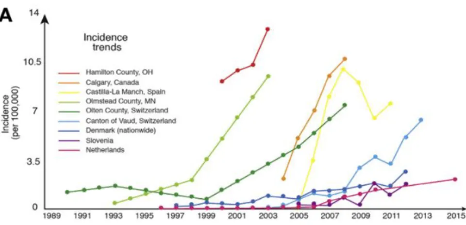
Fig. 4: Incidence rates of EE in multiple cities around the world. Just like prevalence rates, incidence rates have increased significantly since the 1990s and is expected to continue to increase. (Dellon & Hirano, 2018)
Incidence rates of EE have also increased (Fig 4). A longitudinal study revealed incidence rates were 6.37 cases reported per 100,000 inhabitants during the six-year period Disease incidence was also shown to be 19 times higher in males compared to females (Hruz, 2014). Gastroesophageal Reflux vs. Eosinophilic Esophagitis
Due to the similar symptoms between gastroesophageal reflux disease (GERD) and EE, it has been difficult to distinguish between the two diseases. GERD is a gastrointestinal disorder that can cause mucosal damage in the esophagus, oral cavity, and lungs. This is caused by abnormal reflux of gastric acid into the esophagus causing damage to the epithelial cells of the mucosa (Badillo, 2014). Multiple symptoms are similar between the two diseases. Heartburn, dysphagia (or food impaction), and chest pain are all commonly seen in patients with these conditions. Biopsies are often needed to determine the difference between the two (Molina-Infante, 2008). Inflammation of the two diseases is induced by different stimuli. Acid reflux into the lower esophagus is main cause for GERD while EE inflammation is induced by antigen-mediated inflammation that corresponds to eosinophil release of type-2 inflammatory
treated through the use of proton pump inhibitors (PPI) and EE being treated through topical corticosteroids and changes in diet. However, EE patients can also receive relief from symptoms through the use of PPIs, making the overlap between the diseases difficult to differentiate. EE and GERD are not mutually exclusive diseases.The relationship between the two diseases, along with the symptoms they both present, show a complex interaction and possible comorbidity between EE and GERD. However, researchers are still determining whether both diseases can coexist. Two studies have shown that about 50 percent of patients with EE had acid reflux. However, the number of patients that have both EE and GERD is still under debate, as there are inconsistent criteria in defining EE (Cheng, 2014).
New types of diagnostic techniques are always being employed to better differentiate the two diseases. One such method is measuring the difference in mucosal impedance (MI) by measuring the degree of spongiosis or epithelial integrity. By measuring MI along various parts of the esophagus, EE and GERD can be distinguished from each other with great accuracy and through minimally invasive methods (Choksi, 2017).
Mechanism and signaling pathways involved in Eosinophilic Esophagitis
EE is caused when antigens from allergens bind to receptors in the mucosa of the esophagus. When the antigen binds the esophageal mucosa, it activates a type 2 (Th2) helper T-cell immune response. Thymic Stromal Lymphopoietin (TSLP) is secreted from the epithelial T-cell as a result of antigen binding. TSLP is thought to be the main regulator and cause of the Th2 reaction in EE. TSLP promotes cytokine secretion and also induces maturation of dendritic cells, which leads to the infiltration of CD4+ T cells and their differentiation into Th2 cells, which mediate activation of cytokines against allergens. (Cianferoni, 2015). TSLP that is secreted from the esophageal epithelial cells will bind to TSLP receptor cells on dendritic cells, increasing the secretion of OX40 ligand. OX40 ligand induces CD4+ T-cells into secreting inflammatory
cytokines, such as interleukin (IL)-4, IL-5, and IL-13 (Shi, 2017). The binding of TSLP to TSLP receptors forms a complex that leads to activation of various signal transduction pathway. One pathway of note is the JAK/STAT pathway (Fig. 5) (Cheng, 2016). The JAK/STAT pathway is responsible for principal signaling for cytokines and growth factors. Activation of this pathway will lead to cell proliferation, differentiation, migration, and apoptosis. Inhibition of this signaling pathway will ultimately lead to decreases of these changes. Unwarranted activation of the pathway can ultimately lead to inflammation (Rawlings, 2004). TSLP will induce the
phosphorylation of janus kinases (JAK) and lead to the activation of the kinase. The activation of JAK will lead to the activation of signal transducers and activators of transcription (STAT) factors, as well as other proteins. JAK phosphorylates a tyrosine residue leading to dimerization of STAT, subsequent nuclear translocation, and binding to gene promoters for the activation or inhibition of various target genes (Zhong, 2014). In EE, JAK will specifically phosphorylate STAT 6, which increases expression of eotaxin and will lead to activation esophageal epithelial cells and fibroblasts to secrete Eoxtaxin-3. Eoxtaxin-3 is a powerful eosinophil chemoattractant responsible for the migration of the inflammatory cell to the esophagus (Cheng, 2016). The cytotoxins secreted from the eosinophils increase the permeability of esophagus, weakening the tight junctions and dilate intercellular spaces. Due to the inflammation caused by the cytokines, fibrotic changes occur, causing the symptoms seen within EE patients (Cheng, 2014).
Fig 5. Pathways activated by TSLP binding. TSLP activates many pathways when binding to its receptor.
Activation of STAT, MAPK, NFKBIA, and others cause an inflammatory response in both airway diseases and EE.
(Zhong et al., 2014)
Tight Junctions
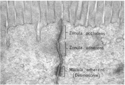
Tight junctions (Fig 6) are important to maintain barrier function between the internal and external environments. Tight junctions, along with adherens junctions, connect epithelial cells together, forming a physical barrier and sealing off of the paracellular space (Fig 7) (Abdulnour-Nakhoul, 2013). In epithelial cells, they connect adjacent cells to one another and regulate the passage of foreign molecules that come into contact with the cells. Tight are found at the most apical section between two adjacent cells. Tight junctions are made up of
transmembrane and intracellular proteins, which link up with the actin cytoskeleton. The function of tight junctions is to seal adjacent cells and regulate paracellular passages of molecules. Two major proteins that make up tight junctions are occludins and claudins.
Occludins are transmembrane proteins that regulate passage of macromolecules through the tight junction. Claudins, another transmembrane protein, are more specific and give tight junctions size and charge selective properties to regulate macromolecules that enter paracellularly (Loxham, 2017). Occludins and claudins are anchored intracellularly by cell junction complexes known as zonula occludens. Zonula occludens are scaffold proteins that connect junctional protein complexes to the cytoskeleton. (Steelant, 2016). Additional junctional protein complexes are present, such as adherens junctions and desmosomes are found along the basolateral cell surface. These junction complexes provide adhesion forces that help
maintain epithelial integrity (Loxham, 2017). In EE, studies have shown that these tight junctions and adherens junctions are affected; expression of E-cadherin and claudin-1 proteins decreased while occludins expression increased. Decreases in claudin-1 expression impairs tight junction barrier function. The significance of increased expression of occludins was not determined (Abdulnour-Nakhoul, 2013).
Fig. 6: Electron microscopy of junctional complexes. At the apical level, tight junctions can be seen. Underneath the tight junctions, adherens junctions and desmosomes can be seen as well. (Fawcett DW, The Cell: An Atlas of Fine Structure, WB Saunders, Philadelphia, 1966, p. 367.)
Treatments of Eosinophilic Esophagitis
Current treatment of EE typically involves dietary restrictions. The use of elimination diets has been used with some success. The treatment revolves around systematically removing certain foods known to cause an allergic response or inflammation. However, despite the success of these diets, they are difficult to maintain for long periods of time. Studies have found poor adherence to these diets, with under 50% of patients maintaining the lifestyle change (Kliewer, 2017). The difficulty in maintaining these diets is likely due to financial and logistical barriers. These ultimately limit the success of this treatment despite the increased resolution of symptoms.
Topical corticosteroids are also used to treat inflammatory symptoms of. Steroids decreased the amount of spongiosis and increased intracellular fluid by strengthening the tight Fig. 7: Barrier function proteins in adjacent cells. A. Shows junctional complexes and their
interactions with the cytoskeleton/ basement membrane. B. Shows tight and adheren junctions mediate cell-cell contact and interact with actin cytoskeleton. (Loxham & Davies, 2017)
junctions in the esophagus. Specifically, filaggrin levels are restored in treated EE patients, as untreated EE patients have a decrease in the protein. With the increase of filaggrin, spongiosis is decreased, preventing leakage between the tight junctions (Katzka, 2014). While both diet and corticosteroid treatments lead to an initial decrease in the symptoms of EE, combination treatment of both diet and steroids has been shown to significantly decrease relapse of EE (Reed, 2018).
Despite the success of these treatments, they have been limited in the success of treating EE completely. The main issue with these treatments are that they are nonspecific. Targeting specific immune pathways may provide treatment options to help resolve EE symptoms and treat the disease more effectively. TSLP is one such target. Initial studies conducted have shown that by neutralizing TSLP, symptoms of EE decreased as well as resolution of EE histologically (Noti, 2013).
Thymic stromal lymphopoietin
TSLP is an inflammatory interleukin (IL)-7-like cytokine that is secreted from multiple cell types, such as epithelial cells, epithelial keratinocytes, dendritic cells, and mast cells in response to antigen binding. TSLP has many connections to many types of allergic disorders, such asthma, atopic dermatitis, allergic rhinitis, and EE. Two isoforms, long and short, of TSLP have been described, each with different effects in the body. The two types of TSLP have protective and deleterious effects based on which one is secreted. Long form TSLP leads to deleterious effects via barrier dysfunction and inflammatory factors. The short form of TSLP leads to protective effects of barrier function and is the predominate form of TSLP in healthy individuals. In EE, long form TSLP is the primary form secreted due to the increase of inflammatory factors caused by the disorder (Dong, 2016). TSLP is produced by epithelial cells in the esophagus, promoting migration of immune cells to the area. TSLP binds to TSLP receptors on dendritic cells (DC),
producing a Th-2 favorable environment (Ito, 2012). TSLP will activate multiple signal
transduction pathways as a result of binding to the TSLP receptor in DCs, such as the JAK/STAT pathway. As a result of TSLP binding, DCs secrete more OX40 ligand, which regulates DC to T-cell interaction, and initiate the Th2 cellular response. OX40 ligand will prime CD4+ T cells to secrete inflammatory cytokines IL-4, 5, and 13, along with tumor necrosis factor alpha (Shi, 2017). Asthma and TSLP connections
Due to inflammation caused by Th2 cells, EE has pathologic connections with asthma and related disorders, such as allergic rhinitis and atopic dermatitis. In both diseases, Th-2 inflammation causes structural damage to each of the relevant pathways. In EE, esophageal epithelium is affected and allows migration of eosinophils to the esophagus. This has parallels to asthma, in which airway remodeling occurs due to the inflammation caused by a related Th2 reaction. Chronic inflammation in both cases causes structural changes in their associated organs (Nhu, 2017). Coexistence or comorbidity of EE and Asthma does exist as studies have found that 23-37.5 percent of patients with EE also have asthma as well (Durrani, 2018). EE patients are also more likely to have asthma than control patients (Gonzalez-Cervera, 2017). Treatment options for both of these diseases exist as well. Studies on the use of anti-TSLP has also been seen to increase resolution of symptoms of both diseases by limiting the amount of type 2 inflammation caused by the effects TSLP on inflammatory cells. By neutralizing TSLP, chronic inflammation is inhibited, preventing airway and esophageal remodeling in asthma and EE respectively (Chen, 2013).
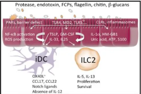
Fig 8: Type 2 cell immune stimuli, such as dust mite protease activity, leads to an immune response. Activation of receptors on the epithelium leads to release of innate pro-Th2 cytokines. (Hammad & Lambrecht, 2015)
Dust mites and aeroallergens
Dust mites and other aeroallergens have been found to promote tight junction barrier dysfunction (Fig 8). Dust mites are an environmental allergen that will produce a Th2 response, typically causing chronic inflammation. Inflammation caused by exposure to aeroallergens, leads to structural remodeling and alterations to the epithelial cells as well (Chen, 2013). A study looking at the effects of dust mites on patients with allergic rhinitis determined that tight junctions were affected, causing barrier dysfunction. The application of dust mites to the nasal epithelium resulted in tight junctions becoming more permeable, with increased exposure to dust mites. Protein and mRNA levels are markedly decreased compared to other tight junction proteins, specifically occludins and zonula occludens-1. Overall, dust mites have a significant impact on barrier function in airway pathway diseases (Steelant, 2016). Exposure of dust mites to the epithelial cells has also been shown to increase the amount of TSLP secreted due to the damage they cause to the epithelium. Like other types of respiratory diseases associated with
TSLP, dust mites will lead to overexpression of TSLP, and an increased cascade of inflammatory factors (Hu, 2017).
Research has recently investigated the role of aeroallergens and EE. A 2003 case study has shown that a patient with EE had symptoms become more severe during pollen season compared to winter seasons. The patient had no food allergies and no other significant medical issues other than asthma. Inflammation become significantly worse during the pollen seasons, with biopsies during this period showing increased inflammation response and symptoms. During the winter months, there was less inflammation caused by eosinophils. This case study hypothesized that aeroallergens have a role in EE. (Fogg, 2003) More recent studies have found similar results in pediatric patients, with increased esophageal eosinophilia during certain seasons depending on the season of the aeroallergen (Ram, 2015). A retrospective chart review of patients with EE also showed a greater number of patients with environmental allergies (58%) compared to food allergies (28%) (Patel, 2014). Other case studies have shown that aeroallergen sensitization might also increase with age, as older patients with EE are typically seen with only aeroallergens and no food allergies compared to pediatric patients (Sugnanam, 2007). These studies show that aeroallergens might play a greater role in EE than previously thought. Hypothesis:
Aeroallergens, such as dust mites, impact the development and severity of eosinophilic esophagitis through thymic stromal lymphopoietin (TSLP) signaling pathway.
Chapter 2: Materials and Methods
Cell CultureEPC2 cells, a gift from Hiroshi Nakagawa at the University of Pennsylvania, were harvested through a 55-year-old male who suffered from Barrett’s esophagus. The cells were
collected from a morphological normal site with testing concluding that they were normal and had no traces of cancer. Cells were cultured as monolayers in a Corning filtered cap chambered flask. Esophageal cells were grown in Keratinocyte-SFM medium supplemented with Bovine Pituitary Extract and human recombinant epidermal growth factor (Invitrogen). Cells were grown to 80% confluency, at which they were passed to another flask for continued growth or transferred to a Transwell epithelium plate for experimentation. Cells were passed by adding 5 mL of trypsin-EDTA to the flask to unadhered the cells from the flask surface. After incubations period of 5 minutes, soybean trypsin inhibitor (STI) (SigmaT9128) was added to deactivate to trypsin since EPC2 cells are sensitive to trypsin. The contents of the flask are centrifuged for 3 minutes at 1000 rpm at 40C. The cells are resuspended in specific media to needed cell concentration.
Eosinophils were cultured as well, cells were taken from the cell line AML
14.3D10/CCCKR3 Clone 16 (ATCC CRL-12079). These cells were acquired from the peripheral blood. Cells were stored in a nitrogen-gas freezer until they were thawed and used. Eosinophils were grown in RPMI 1640 Gibco (A10-491) media supplemented with .05 mM
2-mercaptoethanol, 2 mg/mL G418, 1 mM sodium pyruvate, 2 mM L-Glutamate, and 10% FBS. Flasks were incubated at 37oC to allow for growth.
Transwell Cell culture
For experimentation, esophageal cells were placed in Corning™ Transwell Multiple Well Plate with Permeable Polycarbonate Membrane Inserts. Inserts separate the well into two compartments to signify a basolateral and luminal compartment. Esophageal cells were placed inside the inserts (Luminal compartment) with media (200 uL) and 1 mL esophageal media was placed inside the bottom of each well (basolateral compartment). Plates were placed in a 37oC incubator to allow esophageal cells to grow to confluence and form monolayer. Cell media was changed every 2 days to allow for continued growth.
Measuring Epithelial Integrity by Trans Epithelial Electrical Resistance (TEER)
Integrity of the epithelial membrane electrical resistance was measured using the Millicell ERS-2 Voltohmmeter to measure transepithelial electrical resistance (TEER).
Measurements were acquired at 24 hours after addition of eosinophils and 1, 3, 6, 24, and 48-hour time periods. The probe was sanitized with 70 percent ethanol and calibrated using
esophageal media. The probe was then placed in the transwell, with one side of the probe in the luminal compartment and the other in the basal compartment. Readings were allowed to stabilize before being recorded. All readings were recorded in Ohms.
Experimental Conditions
Once esophageal cells were confluent on the epithelial plates, experimental conditions could be applied to each of the wells. The cells in each well were measured by TEER prior to addition of experimental conditions. Eosinophils (100 eos/ml/ per well) were then added to basal compartment of specific wells and were allowed 24 hours to equilibrate. Eosinophil concentration were determined by using the TC20 automatic cell counter (Biorad). Eosinophil concentration was kept low to limit their effect on the epithelium resistance. Cell TEER was measured again after the 24-hour period and other experimental conditions were then added to
each well. All conditions were applied to the basolateral compartments of each well. Conditions were added individually to each cell or in combination with another experimental condition. TSLP synthetic peptide (Invitrogen, PEP-049) (20 ng/mL), house dust mites (1 ug/mL), Interferon-Gamma (ThermoFisher Scientific) (100 ug/mL), and Gö 6976 (Cayman Chemical) (100 ug/mL), a PKC inhibitor, to strengthen tight junctions, were used in each experiment. Each condition was repeated at least twice with anti-TSLP, anti-TSLP and eosinophils (anti-TSLP EOS), TSLP and dust mites (TSLP DM), and TSLP, dust mites and eosinophils (TSLP DM EOS), run in triplet or more. TSLP synthetic peptide or anti-TSLP, interferon-gamma (IFN-G), and Gö 6976 (G0) was added after measurement of 24 hours after addition of eosinophils and allowed to incubate three hours. After the three hours, dust mites were then added to the appropriate wells. Once the conditions were applied, cells were stored in an incubator at 370C until measurements were conducted. Plates were allowed to equilibrate for 30 minutes before they were measured. TEER measurements were taken at 24 hours after added eosinophils, 1, 3, 6, 24, and 48 hours. Immunofluorescence
Changes in tight junction protein, ZO-1, were observed via immunofluorescence. Imaging of each experimental condition was taken from eight different plates. Once
experimentation was completed, each well was gently washed 3-5x with PBS. The cells were then placed in paraformaldehyde for 10 minutes. The wells were then rinsed again with 3-5x phosphate-buffered saline (PBS) and blocking solution was placed in each for 15 minutes. Blocking solution was made from 5% bovine serum albumin (Fisher Scientific), sodium azide (Fisher scientific), triton x-100 (Fisher Scientific), and 1x PBS. Cells were rinsed 3-5 times with PBS and then placed in the primary antibody Rabbit anti ZO-1 (Thermo Fisher Scientific, #617300). The cells and antibody were allowed to incubate for 1 hour. The primary antibody was then rinsed off lightly with PBS and the secondary antibody, Alexa 488 Goat anti-rabbit
(Thermo Fisher Scientific) was applied to the cells and allowed to incubate for 1 hour at room temperature. Once the secondary anti-body incubated, the cells were lightly washed with PBS and prepped for transfer to a microscope slide. The cells were cut away from the wells with a scalpel and placed onto the slide. Pro-Long Gold antifade with DAPI (Invitrogen) applied to the cells and a microscope slide cover sealed the cell sample. The slides were then stored in at 4oC until they could be analyzed through microscopy.
Microscopy
Esophageal cells were observed through the Zeiss Axio Observer Z1 Research
Microscope. Images were captured with the Axiocam 105 Digital Color camera via the ZEN 2.3 Pro imaging software. After images were captured, Adobe Photoshop was used for post processing to adjust contrast and brightness of the images to highlight ZO-1.
Statistical Analysis
Statistical analysis was completed using Graphpad Prism 7 software. Data was analyzed using a one-way ANOVA test to compare significant differences among each of the experimental conditions. T-tests were also used to compare conditions to the control. The data was
Chapter 3: Results
Determining Dust Mite ConcentrationDust mite concentration was based on initial experimental information prior to the project starting. Five different dust mite concentrations were used to determine the dosage of dust mites for the project’s experiments. The concentrations used in this experiment were 1 ul/mL, 10 ug/mL, 25 ug/mL, 50 ug/mL, and 100 ug/mL. Measurements were taken over a 24-hour period. The distribution obtained at each concentration of dust mites suggest that dust mites trigger a response in esophageal cells even at a low dosage. As seen in Fig. 9, over the course of 24-hour period, addition of dust mites leads to decreased epithelial resistance with exception at hour 0. Hour 0 increased in resistance due to the added esophageal media added when dust mites were added to the wells. All concentrations of dust mites show a decrease in epithelial resistance after 1 hour of application when compared to the control. After the 24 hours, dust mites extract caused a decrease of epithelial resistance by 61 ohms in 1ug/mL, 80 ohms in 10 ug/mL, 58 ohms in 25 ug/mL, 74.33 ohms in 50 ug/mL and 100.5 ohms in 100 ug/mL. The application of both eosinophils and dust mites lead to a further decrease in resistance (Fig 9 A-E). By comparing the mean differences between DM and DM EOS after 24 hours, DM EOS lead to a 2 times epithelial resistance decrease when compared to dust mites alone (Fig 9 A-E). This suggest that dust mites cause an additive effect when applied to eosinophils or they can cause damage to the epithelium directly, independent of the actions of eosinophils. Though only 50 ug/mL DM and 100 ug/mL DM EOS showed significance by ANOVA, decreases caused by DM and DM EOS is seen at all concentrations. Due to the response and trends observed from the data, the 1 ug/mL dust mite concentration was chosen for the experiment. This low dosage was selected as the minimal threshold response to limit the number of confounding variables.
Fig 9. Changes in epithelial resistance based on various concentrations of dust mites (A-D). Dust mites decreased epithelial resistance at each concentration level used. The combination of dust mites and eosinophils lead to even greater decreases in epithelial integrity.
[Grab your reader’s attention with
a great quote from the document or use this space to emphasize a key point. To place this text box anywhere on the page, just drag it.]
A
B
C
D
D
E
Eosinophil (EOS): 100 eos/mL/well **= ANOVA should significance for 50 ug DM and 100ug DM.
* = significant vs control p-value <.05 *
**
Effect of Dust Mites and Eosinophils on Epithelial Resistance
Based on the trends of decreased epithelial resistance seen in prior experiments by dust mites and eosinophils, we sought to see if application of anti-TSLP would counteract the effects of dust mites and eosinophils. Throughout the experiment, there were changes seen based on the conditions applied to the esophageal cells. During the first and third hours of the experiment on each plate, there were no changes seen in any of the experimental conditions compared to the control. Alterations in TEER begin to occur at six-hours. Compared to the control, there was a significant decrease in epithelial resistance in the anti-TSLP DM EOS treated cells (Fig. 10C, p<.05). The difference in mean between this condition and the control was approximately 15 ohms. At the 24-hour condition, anti-TSLP EOS along with anti-TSLP DM EOS were seen to significantly decrease epithelial resistance compared to the control (Figure 10D, p<.05). TSLP EOS mean decrease compared to the control was 44.3 ohms. Anti-TSLP DM EOS had a mean decrease of 55.72 ohms compared to the control. At the 48-hour measurement, three different condition saw significant changes compared to the control. Anti-TSLP EOS and anti-TSLP DM EOS saw significant decreases in epithelial resistance, while G0 saw a significant increase in
resistance, which was expected (Fig 10E, p<.05). anti-TSLP EOS decreased by a mean of 28.65 ohms and anti-TSLP DM EOS decreased by a mean of 48.16 ohms. Gö 6976, was the only
condition to increase epithelial resistance, increased by a mean of 38.5 compared to the control. Gö 6976 was also used as an additional control as well, by strengthening tight junction function. The Gö 6976 control was used to ensure esophageal cells were normal and were not mutated through multiple passages of cells during cell culture. Gö 6976 works by being a PKC inhibitor, preventing activation of the JAK STAT pathway. While not significant, the graphs show an additional decrease in resistance when added to the TSLP EOS condition compared to
anti-TSLP EOS alone. Application of dust mites also lead to a decrease in epithelial resistance as well as seen in Fig. 9.
Fig 10. Changes in epithelial resistance based on different experimental conditions applied to each well (A-E). Over the course of the 48 hours, decreased epithelial resistance was seen in wells containing EOS, anti-TSLP EOS, and anti-anti-TSLP DM EOS
Anti-TSLP: 20 ng/mL
Eosinophil (EOS): 100 eos/mL/well Dust mite (DM): 1ug/mL
Gö 6976 (G0): 100 ug/ mL
Interferon-Gamma (IFN-Gamma): 100 ug/mL * = Data is significant compared to the control
A
D
C
D
B
E
D
D
ZO-1 function impaired by eosinophils and dust mites
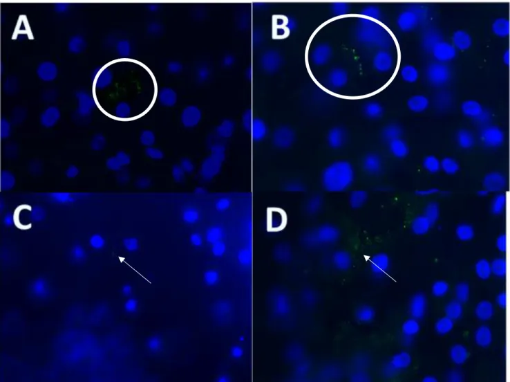
After seeing the decreased epithelium values for wells containing dust mites and eosinophils, tight junctions were imaged to determine dust mites and eosinophils effects on these proteins. Figure 11 shows differences between the control, Eosinophils, anti-TSLP, and interferon-gamma. The control shows intact tight junctions with ZO-1 forming a clear line between two adjacent cells. The nuclei of the cells are shown through the use of DAPI. TSLP shows a similar look to the control in Figure 11B. anti-TSLP slides had TSLP synthetic peptide added to the well. Tight junctions remained intact as well. Figure 11C shows wells applied with eosinophils. Tight junctions were decreased due to the cytotoxins released from eosinophils. As a result, immunofluorescence of ZO-1 was diminished compared to the control. Figure 12 shows the other four conditions used in the experiment. G0 was a control that would strengthen tight junctions. As the image shows, ZO-1 is very prominently displayed in the microscope slide with the proteins appearing between two esophageal cells. This was expected due to the increase in tight junction function that G0 adds. Imaging of TSLP DM shows multiple ZO-1 protein staining. Anti-TSLP limited the affect dust mites cause on tight junction proteins. This phenomenon was also seen in the epithelial resistance as well. In figure 12G, anti-TSLP EOS lead to a decrease in the amount of ZO-1 between esophageal cells. As seen in the image, very few ZO-1 proteins showed up and epithelial resistance confirms the decrease in tight junction proteins as seen in Fig. 10. Lastly, anti-TSLP DM EOS showed the most major changes in tight junction proteins. Large segments of the image show many ZO-1 proteins throughout the imaged area. Many of these proteins are close to the nuclei of esophageal cells as shown by DAPI. There is also no discernable pattern to ZO-1 proteins around the nuclei and not clear barrier is seen between esophageal cells. Each of these images indicate the changes to tight junction proteins under
different experimental conditions. Most of the changes seen are conditions that lead to a decrease in epithelial cell resistance, causing leaky tight junction barriers.
Fig. 11 Immunofluorescence imaging of ZO-1. A) Control shows a ZO-1 present between two different cells B) TSLP showed ZO-1 similar to the control, with defined ZO-1 between cells C) Eosinophil: Decreased number of ZO-1 is signified with decreased TEER readings D) Interferon-Gamma: Had limited effect on TEER readings and ZO-1 is still present between esophageal cells. Tight junction protein ZO-1 in green and esophageal cell nuclei shown in blue.
Fig. 12 Immunofluorescence imaging of ZO-1. E) Gö 6976 show tight junctions separating two esophageal cells F) anti-TSLP EOS show dispersed tight junction proteins, leading to decreased epithelial resistance G) anti-TSLP DM shows retained epithelial resistance, however, imaging of TSLP DM showed limited number of ZO-1 H) anti-TSLP DM EOS: Very dispersed ZO-1 with increased staining by the nucleus of the cells, showing increased intracellular ZO-1. Tight junction protein ZO-1 in green and esophageal cell nuclei shown in blue.
Chapter 4: Discussion
In Eosinophilic Esophagitis, chronic inflammation leads to esophageal restructuring, causing eosinophil infiltration and tight junction disorder. Through experiments conducted prior and during the project, addition of dust mite extract to esophageal cells lead to a decrease in epithelial resistance. Every concentration level of dust mite extract lead to a decrease in resistance starting at 1 hour. The concentration of dust mites chosen was based on the trends seen in the lowest concentration and to determine the lowest dosage of dust mites that causes a response in esophageal cells. Dust mites can cause tight junction dysfunction by two different process, protease or receptor-based activity. These two pathways can lead to a decrease in epithelial resistance. Dust mite’s interaction with eosinophils also increases the magnitude of dysfunction as well, with greater decreases in resistance.
Based on the experiment results, dust mite activity is inhibited with the addition of an anti-TSLP compound. Figure 9 shows the TSLP DM condition throughout the 48-hour period. With the application of anti-TSLP, dust mites did not exhibit a negative effect on epithelial resistance. This might suggest that dust mites activate esophageal cells through a receptor mediated response cause to secrete TSLP instead of causing protease activity (Hammad, 2015). Adding anti-TSLP to dust mites will inhibit the effect of TSLP on esophageal cells a counteract the response caused by dust mites. This is largely seen when comparing the dust mite only response in esophageal cells. When dust mites are the only condition added to the esophageal cells, epithelial resistance decreased with prolonged exposure to dust mites. When cells were pretreated with anti-TSLP and dust mites were applied to the cells, the epithelial resistance of the cells were not significantly different from the control. This might suggest that protease
activity of dust mites is not effective in esophageal cells and only receptor mediated pathways are activated when dust mites are applied to the cells.
There is also an interesting interaction between dust mites and eosinophils that enhances the disruption of tight junctions in the esophagus. When eosinophils were added to the wells with esophageal cells, tight junction dysfunction occurred, leading to a decrease in the epithelial resistance. This response is expected due to inflammatory factors and cytotoxins that are released from eosinophils leads to barrier disruption and the cells become leakier. When anti-TSLP is added to this is added to eosinophils, there is no effect on eosinophil function and epithelial resistance remains decreased by the same level as eosinophils alone. This suggests that anti-TSLP does not limit stop eosinophils from degranulation and releasing factors that lead to tight junction disruption. However, other studies have suggested the opposite, with
decreased eosinophil effect with the application of anti-TSLP antibodies (Gauvreau, 2014). TSLP is the main regulator of EE and will lead to infiltration of eosinophils to esophagus. However, TSLP does not have an effect on eosinophils once they have already infiltrated the epithelium, so anti-TSLP will not stop disruption from occurring from eosinophils. Lastly, when dust mites are added to the wells containing anti-TSLP and eosinophils, it leads to a greater decrease in epithelial resistance. It suggests that dust mites and eosinophils interact with each other to lead to further disruption compared when these two are separate. Dust mites or other similar aeroallergens might cause eosinophils to increase the secretions of cytotoxins and lead to greater degranulation of eosinophils. As a result, tight junctions were further affected as seen in imaging. A 2007 study shows that dust mites cause activation of eosinophils by binding to the formyl peptide receptor (FPR) and formyl peptide receptor-like 1 (FPRL1). When dust mites bind to these receptors, they cause activation of eosinophils and cause degranulation (Svensson,
2007). This process might explain why the combination of eosinophils and dust mites leads to greater decreases in epithelial resistance and disruption of tight junctions.
Limitations and issues did arise during the experiment that should be noted. Growth on the epithelial plates was difficult throughout the project. After the cells were placed on the transwell plates, difficulties growing a monolayer on the inserts were encountered. As the data shows, there were decreased epithelial resistance readings in each of the wells, with an average of 200 Ohms or lower. A lower passage cells of the same cell line was used to try to combat the issue, however, it was unsuccessful in raising the epithelial resistance of the esophageal cells. Lower passage cells are at less risk to undergo mutation or differentiation. Cells that undergo high number of passages can change the cell characteristics over time. Using lower passage cells ensured that cells were not affected by alterations in cell function, decreasing TEER (Briske-Anderson, 1997). Different bottles of esophageal media were also used to determine if there were any contamination issues occurring that were inhibiting the growth of the cells. Cells were also allowed to grow as long as needed to ensure the monolayer formed on the transwell inserts. However, despite these strategies to increase epithelial resistance in esophageal cells, resistance remained low. As a result, trends and significance of the data might be impacted the diminished cell resistance, as well as imaging of tight junction proteins. This was especially seen during the imaging of ZO-1. With less esophageal cells forming a monolayer, less tight junction proteins were seen when cells were imaged. This made it difficult to determine changes in tight junction presence as most of the barriers between cells were already leaky, caused by the decrease in cells.
Dosage of anti-TSLP might have had an effect on experimental results as well. A dosage was based on a previous study for allergic asthma. Studies using anti-TSLP in airway related diseases, have shown a decrease in tight junction dysfunction with the addition of anti-TSLP.
Based on the results, anti-TSLP was effectivein preserving epithelial resistance at the
concentration of 20 ug/mL. Despite the success of anti-TSLP stopping the action of dust mites, more experiments need to be conducted to learn the effective concentrations of anti-TSLP to dust mites. This would give a better idea on how anti-TSLP affects dust mite function and provide the amount needed to maintain epithelial resistance.
Issues with staining for immunofluorescence also occurred. Staining was not optimal for showing tight junction dysfunction. Problems in immunofluorescence could have come from multiple areas. Two issues that present themselves as the most logical areas is in growth of the cells and washing the cells during the staining protocol. As mentioned previously, there were issues involved in raising the epithelial resistance on the transwell cell culture plates. Since the cells were leaky at the start, the number of tight junction proteins might have been reduced from the start. This would lead to decreased number of ZO-1 in the stain making it difficult to demonstrate how tight junctions were affected. Another possible area is the washing of cells during the staining process. The antibody staining for immunofluorescence protocol calls for multiple washes throughout the process to remove excess antibody and prepare it for the next step. During this process, cells might have washed away during removal of the PBS wash. This would lead to less cells available and less ZO-1 for the primary antibody to bind to. Both of these possibilities led to poor imaging of ZO-1.
Future studies should continue into looking at the association between aeroallergens and EE. As of today, environmental allergies remain more controversial as a cause for EE than food allergies. More research as to look at the various environmental allergies and identify mechanisms lead to EE. Other treatment options could also focus on using JAK inhibitors as well. Since TSLP activates the JAK/STAT pathway, focusing on this pathway could lead to a decrease in inflammation factors and chemotaxis of eosinophils. Current JAK inhibitors are currently used to
address inflammatory diseases such as rheumatoid arthritis, psoriasis, and inflammatory bowel disease (Schwartz, 2017). Since eosinophilic esophagitis is known to activate this pathway to lead to eosinophil attraction and inflammation in the esophagus, JAK inhibitors could become a possible treatment option for patients. Looking at the mechanisms and treatments of EE will lead to further understanding of how EE works and ways to fully combat the issues that EE presents.
Bibliography
1. Abdulnour-Nakhoul, S. M., et al. (2013). "Alterations in junctional proteins,
inflammatory mediators and extracellular matrix molecules in eosinophilic esophagitis." Clinical Immunology 148(2): 265-278.
2. Badillo, R. and D. Francis (2014). "Diagnosis and treatment of gastroesophageal reflux disease." World Journal of Gastrointestinal Pharmacology and Therapeutics 5(3): 105-112.
3. Chen, Z.-G., et al. (2013). "Neutralization of TSLP Inhibits Airway Remodeling in a Murine Model of Allergic Asthma Induced by Chronic Exposure to House Dust Mite." PLOS ONE 8(1): e51268.
4. Cheng, E., et al. (2014). "Eosinophilic Esophagitis: Interactions with Gastroesophageal Reflux Disease." Gastroenterology Clinics of North America 43(2): 243-256.
5. Choksi, Y., et al. (2017). "Esophageal Mucosal Impedance Patterns Discriminate Patients With Eosinophilic Esophagitis From Patients With GERD." Clinical Gastroenterology and Hepatology.
6. Cianferoni, A. and J. Spergel (2016). "Eosinophilic Esophagitis: A Comprehensive Review." Clinical Reviews in Allergy & Immunology 50(2): 159-174.
7. Croese, J., et al. (2003). "Clinical and Endoscopic Features of Eosinophilic Esophagitis in Adults." Gastrointestinal Endoscopy 58(4): 516-522.
8. Dellon, E. S., et al. (2009). "Clinical, Endoscopic, and Histologic Findings Distinguish Eosinophilic Esophagitis From Gastroesophageal Reflux Disease." Clinical
9. Dellon, E. S., et al. (2013). "ACG Clinical Guideline: Evidenced Based Approach to the Diagnosis and Management of Esophageal Eosinophilia and Eosinophilic Esophagitis (EoE)." The American Journal Of Gastroenterology 108: 679.
10. Dellon, E. S. and I. Hirano (2018). "Epidemiology and Natural History of Eosinophilic Esophagitis." Gastroenterology 154(2): 319-332.e313.
11. Dong, H., et al. (2016). "Distinct roles of short and long thymic stromal lymphopoietin isoforms in house dust mite-induced asthmatic airway epithelial barrier disruption." Scientific Reports 6: 39559.
12. Durrani, S. R., et al. (2018). "Eosinophilic Esophagitis: an Important Comorbid Condition of Asthma?" Clinical Reviews in Allergy & Immunology.
13. Gauvreau , G. M., et al. (2014). "Effects of an Anti-TSLP Antibody on Allergen-Induced Asthmatic Responses." New England Journal of Medicine 370(22): 2102-2110.
14. Hammad, H. and B. N. Lambrecht (2015). "Barrier Epithelial Cells and the Control of Type 2 Immunity." Immunity 43(1): 29-40.
15. Hill, D. A., et al. (2017). "The prevalence of eosinophilic esophagitis in pediatric patients with IgE-mediated food allergy." The journal of allergy and clinical immunology. In practice 5(2): 369-375.
16. Hruz, P. (2014). "Epidemiology of Eosinophilic Esophagitis." Digestive Diseases 32(1-2): 40-47
17. Hu, Y., et al. (2017). "TSLP signaling blocking alleviates E-cadherin dysfunction of airway epithelium in a HDM-induced asthma model." Cellular Immunology 315: 56-63.
18. Ito, T., et al. (2012). "Cellular and Molecular Mechanisms of TSLP Function in Human Allergic Disorders - TSLP Programs the “Th2 code” in Dendritic Cells." Allergology
19. Katzka, D. A., et al. (2014). "Effects of Topical Steroids on Tight Junction Proteins and Spongiosis in Esophageal Epithelia of Patients With Eosinophilic Esophagitis." Clinical Gastroenterology and Hepatology 12(11): 1824-1829.e1821.
20. Kliewer, K. L., et al. (2017). "Dietary Therapy for Eosinophilic Esophagitis: Elimination and Reintroduction." Clinical Reviews in Allergy & Immunology.
21. Loxham, M. and D. E. Davies (2017). "Phenotypic and genetic aspects of epithelial barrier function in asthmatic patients." The Journal of Allergy and Clinical Immunology 139(6): 1736-1751.
22. Lucendo, A. J., et al. (2017). "Guidelines on eosinophilic esophagitis: evidence-based statements and recommendations for diagnosis and management in children and adults." United European Gastroenterology Journal 5(3): 335-358.
23. Molina-Infante, J., et al. (2008). "Overlap of reflux and eosinophilic esophagitis in two patients requiring different therapies: A review of the literature." World Journal of Gastroenterology : WJG 14(9): 1463-1466.
24. Nhu, Q. M. and S. S. Aceves (2017). "Tissue Remodeling in Chronic Eosinophilic
Esophageal Inflammation: Parallels in Asthma and Therapeutic Perspectives." Frontiers in Medicine 4(128).
25. Noti, M., et al. (2013). "TSLP-elicited basophil responses can mediate the pathogenesis of eosinophilic esophagitis." Nature medicine 19(8): 1005-1013.
26. Patel, T. and S. Glover "Evaluation Of Antigenic Triggers and Etiologies In Eosinophilic Esophagitis: A Single Center Experience." Journal of Allergy and Clinical Immunology 133(2): AB261.
27. Ram, G., et al. (2015). "Seasonal exacerbation of esophageal eosinophilia in children with eosinophilic esophagitis and allergic rhinitis." Ann Allergy Asthma Immunol 115(3): 224-228.e221.
28. Reed, C. C., et al. (2018). "Combined and Alternating Topical Steroids and Food
Elimination Diet for the Treatment of Eosinophilic Esophagitis." Digestive Diseases and Sciences.
29. Schwartz, D. M., et al. (2017). "JAK inhibition as a therapeutic strategy for immune and inflammatory diseases." Nat Rev Drug Discov 16(12): 843-862.
30. Shi, Z., et al. (2017). "Inhibition of JAK/STAT pathway restrains TSLP-activated dendritic cells mediated inflammatory T helper type 2 cell response in allergic rhinitis." Molecular and Cellular Biochemistry 430(1): 161-169.
31. Steelant, B., et al. (2016). "Impaired barrier function in patients with house dust
mite–induced allergic rhinitis is accompanied by decreased occludin and zonula occludens-1 expression." Journal of Allergy and Clinical Immunology 137(4): 1043-1053.e1045.
32. Sugnanam, K. K., et al. (2007). "Dichotomy of food and inhalant allergen sensitization in eosinophilic esophagitis." Allergy 62(11): 1257-1260.
33. Zhong, J., et al. (2014). "TSLP signaling pathway map: a platform for analysis of TSLP-mediated signaling." Database 2014: bau007-bau007.


