CHARACTERIZATION OF HUMAN ANTIBODY RESPONSES TO DENGUE VIRUS
INFECTIONS IN A SRI LANKAN PEDIATRIC COHORT
Kizzmekia Shanta Corbett
A dissertation submitted to the faculty at the University of North Carolina at Chapel Hill in partial fulfillment of the requirements for the degree of Doctor of Philosophy in the Department of
Microbiology and Immunology in the School of Medicine.
Chapel Hill 2014
Approved by: Aravinda de Silva Ralph Baric Mark Heise
Ronald Swanstrom
ii © 2014
iii
ABSTRACT
Kizzmekia Shanta Corbett: Characterization of Human Antibody Responses to Dengue Virus Infections in a Sri Lankan Pediatric Cohort
(Under the direction of Aravinda de Silva)
Dengue virus (DENV) is the most significant mosquito-borne viral infection of humans. People infected with dengue viruses present with subclinical (inapparent) or clinically-apparent infections ranging from undifferentiated fever to dengue hemorrhagic fever or dengue shock syndrome. It is not completely understood why some people get more severely ill from DENV infections than others, but antibody responses have been implicated in playing a role facilitating severe dengue disease. To that end, we analyzed samples from a pediatric dengue cohort study in Sri Lanka to explore if antibody responses differentiated clinically inapparent and apparent infections. 799 children living in a heavily urbanized area of Colombo, Sri Lanka were followed for one year; samples were collected at enrollment, at follow-up, and intermittently from any child who acquired fever.
Using those samples, we first obtained accurate estimates of the incidence of DENV infection and disease in the cohort. At enrollment, dengue sero-prevalence was 53.07%
demonstrating high transmission in this population. Over the study year the incidence of DENV infection and disease were 8.39 (95% CI: 6.56-10.53) and 3.38 (95% CI: 2.24-4.88),
iv
experienced repeat infections, IgG antibody levels fluctuated following infection, but
v
To my ProjectSEED bus partner, my friend, and my angel, Yusuf Neville (06/22/85 - 01/29/14)… your death motivates me to keep living.
vi
ACKNOWLEDGEMENTS
God.
Above all, I must thank my Lord and Savior Jesus Christ for giving my life a purpose and directing me so that I can fulfill it.
Yusuf.
I dedicate this dissertation to you because, in this moment, I cannot think of anyone else who is more deserving. I remember during my second year when research wasn’t going how I’d planned… You drove to Durham, sat in your cousin’s driveway with me, and reminded me why I couldn’t give up. I remember you always telling me to “man up” when I called you in an
emotional frenzy (usually about boys who you thought were no good for me, but I guess that’s a different topic… lol). I remember you (and my Dad) being the only two people in the world who remembered my presentation schedule off the top of your heads. I could always count on a message from you 5 minutes before I stepped to the podium with words of encouragement. I met you when I was 15 years old; we both were scientists then. Although you didn’t take the science route, you always understood “Kizzy the scientist” in ways most people didn’t.
ProjectSEED connected us for a lifetime, and every single day, I think about how yours was cut a little too short. May your soul be free, friend. R.I.P.
Family.
vii
Thank you for loving me as if your blood runs through my veins and providing the foundation I needed to grow into the person that I am today. To both of my parents, thanks for keeping your expectations of me high, and even when I didn’t always meet them, thanks for loving me
anyway. Thanks for every time you supported me, whether it was at a cheerleading competition or scientific presentation. You always found a way to remind me that I was important in times when the world tried to convince me otherwise. Your unconditional love and support got me here, to the day before I submit my PhD dissertation, and I will forever be indebted to you. To my siblings, unbeknownst to you, you set goals for me that I didn’t even know I had; because you all looked up to me, I could never give up and I promise I never will. Ciera, I was no longer the oldest when you came into the family. Although I’m sure it’s burdensome having 6 younger people look up to you, you carry it well. Thanks for teaching me so much and having my back. Tameka, you were born my cousin and became my sister. At your mom’s funeral, I told you that if there is anything you ever need, I got you. Thanks for having me too. Dee, you have so many of Dad’s characteristics in you which make you the perfect man and the perfect brother. Thanks for being someone that I can forever call on, whether it be to paint my walls, wash my car, or to simply talk. Tireka, thanks for every single random phone call, text message, or tweet.
viii
To my extended family… Joyce, for being my mom in Maryland and providing me a home away from home…Tonya, for being a big cousin who I could look up to… Aunt Laura, for being so strong and reminding me that biomedical research is so important… and Lamar, for being more like my brother than my cousin. To my entire family, grandmas, parents, siblings, nieces, nephews, godchildren, aunts, uncles, and cousins, thanks for the moments we shared together in the last five years. I will cherish each and every one forever, no matter how many degrees I acquire or how far away I move.
Friends.
To my PICs, Fe, Cool, and Prillert, not many people can look back 12 years and find 3 rock-solid loyal people who they can still call friends. Your love, support, and honesty are so appreciated in times when other people have fallen by the wayside. Thanks for being forever “friends”, in the truest meaning of the word. Kenyetta, honestly I would have never thought we’d still be friends 12 years after working as grocery store cashiers together, but we are, and I appreciate you for rocking with my craziness for this long. To my undergrad roommates, Rasheeda and Nerg, thanks for accepting and loving me just as I came and staying my friends “4 Life”. Meeting you guys was one of the first times I’d come across other black women with similar goals as me; thanks for that inspiration. To Seghen, thanks for being someone I can always call and pick up right where we left off… “Deuce Deuce”. Vic, my “Suga”, thanks for relieving me from the stresses of science, whether it was a random getaway or simple text conversation after a long day in lab. Brian, your advice has gotten me through many a rough day; thanks for continuing to be my friend through it all. Daaimah, or shall I say “Baby D”, we went from working at NIH to roaming the streets of NYC together; thanks for reminding me that a scientist can be cool too. Isa, from working in the same lab at 16 to arguing like sisters in group chat at 28, thanks for being the “Woo” to my “Woo”. Stephani, whether it be Pinot Noir served at perfect temperature, indie hip-hip concerts on Friday nights, or science policy
ix
passions. Jasmyn and Richard, thanks for extending our connection beyond the BBSP incoming Class of 2009 to friendships that I’m sure will last forever. To my extended circle of friends, Taneisha, Ashley, Sophia, Janeice, Alex, Jelly, Noah, Lenard, Taa, Harry, Greg, Jared, Randall, and Jean thanks for simply being there whenever I have needed you.
Science.
Thanks to Project SEED, the American Chemical Society, UNC Kenan Labs, Mr. Cutler, and Mr. Cherry for fostering my love for science at 15 years old. Thanks to UMBC, Robert and Jane Meyerhoff, Dr. Hrabowski, Mr. Toliver (R.I.P), Mr. Goodwyn, Mr. Harmon, Mrs. Baker, and the entire Meyerhoff Scholarship Program for affording me an unparalleled undergraduate science education. To the staff of NIH’s undergraduate scholarship program, I know how much of a risk it must have been to accept a rising college freshman into such a prestigious program; thank you for believing in me when I was only 18 years old. To the mentors I had in my early scientific career, Dr. Russell, Dr. Viboud, and Dr. Graham, thank you for the time you invested in training me when I had no clue.
UNC.
Thank you, Aravinda, for accepting me into your laboratory and giving me the creative freedom to work on a project that is atypical for a PhD student. Thank you for creating a peaceful, loving laboratory environment so that we could all work and grow as scientists. Over the years, my laboratory family became more like real family to me, which is a testament to your mentorship and leadership. Rukie, you have no idea how motivating it was to sit beside
someone as brilliant as you every day for three years. Thanks for your advice on experiments and manuscript preparation. Thanks also for all of the times you saved me, whether it be with a hug when I was crying or by running shoes across campus when my flip flops broke. Thank you, Leah, for starting preliminary analyses on the PDVI dataset. Thanks also for that random
x
for patiently teaching me everything I know about dengue cell culture and neutralization assays and for not laughing at me that one time when I clumsily dropped eight plates on the floor. Bhumi and Sandra, thanks for giving me mid-day breaks with random conversations about literally anything, from science to boys to fashion. Bhumi, especially for being someone I could call on to fix my plates or stop my gels from overrunning. Emily, thanks for organizing lab outings and teaching me a thing or two about coffee and beer. Thank you, Anne, for every single flower you brought me from your garden; even when your days weren’t the brightest, you made it your duty to make sure mine were. Wahala, thanks for being like my big brother with whom I argued one second but whose advice and direction I still took heed to. Yang, thanks for your sparse but meaningful conversations about science. Thanks to Katie and Eileen for putting up with me emptying all of the stack-packs as soon as you refilled them. To Tony and Jennifer, thanks for digging in the cold room for samples and crunching datasets. To all of our
undergraduate lab assistants, thanks for keeping the lab in order and everyone somewhat sane in the process. Thanks to Bill and Doug for meaningful scientific discussions and advice, both during my preliminary exam and thesis work. Thanks to the entire Sri Lanka cohort study group for trusting me to work with such a precious set of samples. Thank you to Dharshan and the rest of my Genetech family for embracing me with open arms during my stint in Sri Lanka. Thanks to the Micro/Immuno administrative staff members who have helped to make the “school” portion of this PhD go so smoothly. Thank you to the Virology Training Grant, IMSD, Off-Campus Dissertation Fellowship, and several travel awards for funding a small part of my scientific endeavors. Funding aside, thank you especially to Pat, Ashalla, and everyone in the IMSD program for being like a family outside of my biological and laboratory families. Lastly, thanks to my committee members, Mark, Ralph, Ron, and Jennifer, for all comments, constructive
xi
xii
TABLE OF CONTENTS
LIST OF TABLES ... xvii
LIST OF FIGURES ... xviii
LIST OF ABBREVIATIONS ... xix
CHAPTER ONE: Background and Significance ... 1
1.1 Dengue: the Virus ... 1
Infectious Life Cycle of Dengue Virus ... 1
Dengue Virion Structure ... 3
1.2 Dengue: the Disease ... 3
Global Epidemiology of Dengue ... 3
Dengue in Sri Lanka ... 4
Clinical Presentation of Dengue Disease ... 4
1.3 Role of Viral Factors in Dengue Disease Severity ... 5
1.4 Role of Host Factors in Dengue Disease Severity... 6
Immune Responses to Dengue Virus Infection ... 6
Immunopathology to Dengue Virus Infection ... 6
1.5 Human Antibody Responses to Dengue Virus ... 7
Role of Antibodies in Enhancement of Dengue Disease ... 7
Role of Antibodies in Protection of Dengue Disease ... 9
Temporal Regulation of Dengue Antibodies ... 10
1.6 Advantages of Studying Dengue Virus Infections in Human Cohorts ... 11
Mouse Models for Dengue Virus Pathogenesis ... 11
xiii
Lessons Learned from Human Cohort Studies ... 12
1.7 Dengue Vaccine Development ... 13
Prospective Dengue Virus Vaccine Candidates ... 14
1.8 Outline for this Dissertation Research ... 15
Research Questions ... 15
Research Approach ... 16
Specific Aim 1 ... 16
Specific Aim 2 ... 16
Specific Aim 3 ... 16
Innovation ... 17
CHAPTER TWO: Burden of Dengue Infection and Disease in a Pediatric Cohort in Urban Sri Lanka ... 25
2.1 Introduction... 25
2.2 Materials and Methods ... 26
Ethical Approval for Study ... 26
Study Site ... 27
Study Population ... 27
Sample Collection ... 28
Molecular Detection of DENV ... 28
Detection of anti-DENV IgM and IgG Antibodies in Dried Blood Spots ... 28
Measurement of DENV Neutralizing Antibodies... 29
Determining Dengue Serostatus at Study Enrollment ... 30
Criteria for Laboratory Diagnosis of DENV Infections ... 30
2.3 Results ... 31
xiv
CHAPTER THREE: Temporal Regulation of Antibody Responses Following Dengue
Virus Infections in a Sri Lankan Pediatric Cohort ... 45
3.1 Introduction... 45
3.2 Materials and Methods ... 46
Human Subjects Protocol Approval ... 46
Cell Lines and Viruses ... 46
Sample Collection ... 47
Elution of Antibodies from DBS ... 47
Detection of DENV-specific IgG and IgM Antibodies ... 47
Detection of DENV Neutralizing Antibodies ... 47
Laboratory Confirmation of DENV Infections ... 48
Statistical Analyses ... 48
3.3 Results ... 49
Comparison of Antibody Responses Following Primary Inapparent and Apparent DENV Infections ... 49
Temporal Analysis of Antibody Responses Following Primary DENV Infections .... 50
Temporal Analysis of Antibody Responses Following Repeat DENV Infections... 50
3.4 Discussion ... 51
CHAPTER FOUR: Pre-existing Neutralizing Antibody Responses Distinguish Clinically Inapparent and Apparent Dengue Virus Infections in a Sri Lanka Pediatric Cohort ... 59
4.1 Introduction... 59
4.2 Materials and Methods ... 60
Human Subjects Protocol Approval ... 60
Cell Lines and Viruses ... 60
Sample Collection ... 60
Elution of Antibodies from DBS ... 61
xv
Detection of DENV Neutralizing Antibodies ... 61
Detection of DENV Enhancing Antibodies ... 61
Laboratory Confirmation of DENV Infections ... 62
Statistical Analyses ... 63
4.3 Results ... 63
Comparison of Pre-Existing Neutralizing Antibody Responses of Repeat Inapparent and Apparent DENV Infections ... 64
Comparison of Pre-Existing Enhancing Antibody Responses of Repeat Inapparent and Apparent DENV Infections ... 64
Quantitative Analysis of Differences in Neutralizing Antibody Responses Between Types... 65
4.4 Discussion ... 66
CHAPTER FIVE: Conclusions, Discussion, and Future Directions ... 74
5.1 Overview of Research Purpose ... 74
Research Significance ... 74
Research Approach ... 75
5.2 Introduction to the Sri Lankan Pediatric Cohort... 75
Sri Lankan Pediatric Cohort Ideal for Studying Dengue Virus Antibodies ... 75
Comparison of Inapparent to Apparent Infection Ratios Across Cohorts ... 76
Important of Using Neutralization Assays to Characterize Dengue Cases ... 77
What Understanding True Burden of Dengue Infection in Sri Lanka Means to the Field ... 78
5.2 What are the differences between antibody repsonses induced by primary versus secondary infections? ... 78
Current Understanding of Temporal Regulation of Dengue Virus Antibody Responses ... 78
Predictions about Waning of Cross-protection ... 79
Neutralization Magnitude versus Neutralization Breadth ... 80
xvi
Using K562 Cells to Determine DENV Enhancement ... 81
Dengue Antibody Dependent Enhancement and Mild Disease ... 82
5.5 What role do neutralizing antibodies play in protection from dengue disease? ... 82
Protection from Apparent Dengue Infection Facilitated by Cross- Neutralizing Antibodies ... 82
Quantitative Analysis of Breadth of Neutralizing Antibody Responses ... 83
Explanations for Correlation between Breadth of Neutralization and Dengue Disease Outcome ... 84
Balance between Pathogenic and Protective Antibody Responses ... 85
5.6 Snapshot of Research Findings ... 86
5.7 Future Directions ... 87
Dissecting Infection History of Children with Repeat Dengue Virus Infections ... 87
Dissecting Epitope-Specificity of Pre-existing Antibodies ... 88
Following Cohort into Year Two of Sample Collection and Possibly into a Vaccine Trial ... 88
5.8 Closing Statement ... 89
xvii
LIST OF TABLES
Table 1.1 World Health Organization Dengue Case Classifications ... 18 Table 1.2 List of Prospective Dengue Cohort Studies ... 19 Table 2.1 Detection of Secondary Inapparent Dengue Cases by 2-Dilution
Neutralization Screen ... 37 Table 2.2 Ability of IgG ELIA to Detect True Dengue Infections Based on
Neutralization Assay Outcome ... 38 Table 2.3 Age-specific Dengue Sero-prevalence ... 39 Table 2.4 Age-specific Fever Episodes and Incidence of Dengue Infection and Illness .. 40 Table 2.5 Dengue Disease Outcome in Children Exposed to Primary Versus
xviii
LIST OF FIGURES
Figure 1.1 Dengue Virus Infectious Life Cycle ... 20
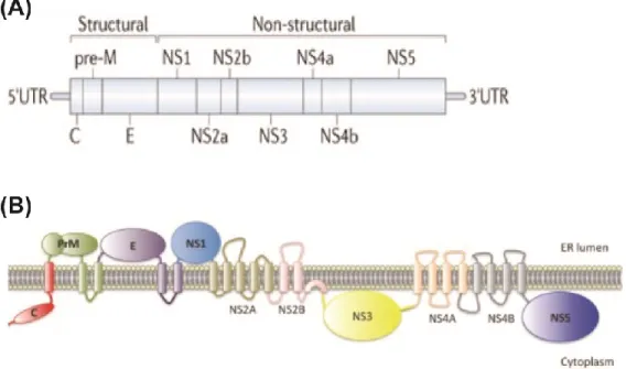
Figure 1.2 Dengue Virus Genome and Polyprotein ... 21
Figure 1.3 DENV Virion Structure ... 22
Figure 1.4 Antibody Dependent Enhancement of Dengue Virus ... 23
Figure 1.5 Temporal Regulation of Antibodies Following DENV Infection ... 24
Figure 2.1 Map and Location of Study Site (Ward 33) in Relation to Colombo Municipal Council and Colombo District in Sri Lanka ... 43
Figure 2.2 Representative Neutralization Cures for Confirmed Inapparent Dengue Cases ... 44
Figure 3.1 Comparison of Antibody Responses Following Primary Inapparent and Apparent Dengue Virus Infections ... 54
Figure 3.2 Temporal Analysis of Neutralizing Antibody Responses Following Primary Dengue Virus Infections ... 55
Figure 3.3 Temporal Analysis of Neutralizing Antibody Responses Following Repeat Dengue Virus Infections ... 57
Figure 4.1 Pre-existing Neutralizing Antibody Responses of Repeat Inapparent and Apparent Dengue Infections ... 70
xix
LIST OF ABBREVIATIONS
1°– primary 2° - secondary 3° - tertiary 4° - quaternary
Ae. aegypti – Aedes aegypti Ae. albopictus – Aedes albopictus
ADE – antibody dependent enhancement AG129 -
BALB/c - Bagg Albino B cell – B lymphocyte BoB – blockade of binding C – capsid
C57BL/6 – Black 6
CD14 – cluster of differentiation 14 c-type lection – calcium-dependent lectin DC – dendritic cell
DC-SIGN - dendritic cell-specific intercellular adhesion molecule-3-grabbing non-integrin DENV – dengue virus
DENV1 – dengue virus serotype 1 DENV2 – dengue virus serotype 2 DENV3 – dengue virus serotype 3 DENV4 – dengue virus serotype 4 DENV5 – dengue virus serotype 5 DF – dengue fever
xx DSS – dengue shock syndrome
E – envelope
EDI – envelope domain I EDII – envelope domain II EDIII – envelope domain III ER – endoplasmic reticulum FBS – fetal bovine serum
FcγR – fragment crystallizable gamma receptor FcγRI – fragment crystallizable gamma receptor 1 FcγRII – fragment crystallizable gamma receptor 2 FcγRIIa – fragment crystallizable gamma receptor 2a FcγRIIb – fragment crystallizable gamma receptor 2b FL – fusion loop
HAI - hemagglutination inhibition assay hsp70 – heat shock protein 70
hsp90 – heat shock protein 90
huMAb – human monoclonal antibody Gln - glutamine
IFN – interferon
xxi IP10 – interferon inducible protein
LAV – live attenuated vaccine M – membrane
mAb – monoclonal antibody
MCP-1 - monocyte chemotactic protein 1
MIP1β – macrophage inflammatory protein 1 beta mθ – macrophages
NEAA – non-essential amino acids neut50 – 50% neutralization
NHP – non-human primate NHS – normal human serum NK – natural killer
NS – nonstructural protein NS1 – nonstructural protein 1 NS2A – nonstructural protein 2A NS2B – nonstructural protein 2B NS3 – nonstructural protein 3 NS4A – nonstructural protein 4A NS4B – nonstructural protein 4B NS5 – nonstructural protein 5 Pen/Strep – penicillin/streptomycin
PDVI – Pediatric Dengue Vaccine Initiative PCR – polymerase chain reaction
xxii rE – recombinant envelope
RNA – ribonucleic acid SD – standard deviation
STAT2 - Signal transducer and activator of transcription 2 T cell – T lymphocyte
TGN – trans-Golgi network
1
CHAPTER ONE
Background and Significance
1.1 Dengue: the Virus
Dengue virus (DENV), the causative agent of dengue disease, is an enveloped positive-strand RNA Flavivirus, akin to other disease-causing viruses such as West Nile virus, Tick-borne Encephalitis, and Yellow Fever virus. DENVs have been thought to exist as four distinct but antigenically related serotypes, DENV1-4, but data describing a new fifth DENV serotype, DENV5, has recently arisen [1]. Further complicating the dengue virus species, DENV
serotypes can be additionally classified into regionally-specific genotypes. Infectious Life Cycle of Dengue Virus
DENV is transmitted to humans via the bite of infected Aedes aegypti and Aedes albopictus mosquitoes. Upon injection into the skin, DENV primarily targets mononuclear phagocytic cells, such as monocytes, macrophages, and dendritic cells [2]. To date, a specific human cellular receptor for DENV has not been identified. Several molecules have been
2
The methods by which DENV is internalized after attachment to appropriate receptors and/or co-receptors (figure 1.1A) are still active areas of research (reviewed in [10]). Clathrin-mediated endocytosis is largely accepted as the main method of internalization by DENV (figure 1.1B) [11], although a clathrin-independent, dynamin-dependent internalization method used by DENV2 was identified in Vero cells as well [12]. Upon internalization, DENV particles are trafficked in early endosomes (figure 1.1C). Early endosomes mature into acidic late endosomes; low pH of late endosomes triggers DENV envelope (E) conformational changes [13, 14]. E homodimers dissociate, exposing the fusion loop (FL), which is then inserted into the cellular membrane. E homotrimers form, and viral and host membranes bend toward each other and fuse together (figure 1.1D) [14, 15].
3 Dengue Virion Structure
Mature dengue virions have icosahedral enveloped structures with a round core, containing multiple copies of C encapsidating the DENV RNA genome. The core is surrounded by a host-cell lipid bilayer containing 180 copies of membrane (M) and E [17]. The structure of E is biologically important in regards to neutralizing antibody epitopes; briefly, E consists of three domains, EDI-III, and a fusion loop at the tip of EDII (figure 1.3A). On a mature DENV virion, E is organized in 90 homodimers, which lay in sets of triples along the viral surface, creating a smooth appearance (figure 1.3C). Oppositely, immature dengue particles have a rough appearance because trimers of prM/E heterodimers form spikes on the surface of the particle (figure 1.3B,D) [18]. Dengue maturation state, as well as maturation of other flaviviruses, has been shown to be relevant to pathogenesis and antibody responses [19, 20]. Dengue virion structure will be related to antibody responses later in this chapter.
1.2 Dengue: the Disease
Global Epidemiology of Dengue
Dengue virus infection is a widespread emerging global epidemic, with a projected two-thirds of the world’s population at risk of exposure to DENV [21, 22]. Of all Ae. aegypti-borne viruses, including yellow fever virus and chikungunya, DENV represents the greatest
epidemiological and economic burden [23, 24]. In the last 30 years, DENV has spread into over 100 countries, including the United States of America, leading to a four-fold increase of dengue disease [22]. Recent dengue outbreaks in Texas and Florida are true testaments to the
continuous spread of DENV into previously unaffected areas and the emergence of dengue as a serious international disease threat [25, 26].
There are several factors that are contributing to the global emergence of dengue virus infections [21]. For one, Ae. aegypti and Ae. albopictus,are populating new areas thus
4
years ago, estimates of the global dengue disease burden stated that 100 million total DENV infections occurred each year, 500,000 of which resulted in severe disease and over 20,000 that resulted in death [21, 22]. Since then, those estimates drastically increased as surveillance, detection, and records of dengue cases improved. In 2013, it was estimated that about 390 million dengue infections occur around the world each year, which is a staggering 300% increase over previously published estimates [22, 28].
Dengue in Sri Lanka
Risk of dengue virus infection is considered highest in tropical regions of Asia. Latest estimates from the global burden of dengue in 2010 state that Asia contributed to 67% of the world’s dengue infections, with 37% occurring in India alone [28]. Sri Lanka is an island country on the southern tip of the Indian subcontinent. Dengue virus has been present in Sri Lanka since at least 1966 [29, 30], though epidemiology of DENV infection in Sri Lanka has changed in recent years. There is documentation of a 10-fold increase in dengue fever (DF) and dengue hemorrhagic fever (DHF) cases in the last 10 years [30, 31]. Expansion of dengue across Sri Lanka coupled with introduction of new, possibly more virulent DENV genotypes have
contributed to an increase in Sri Lankan dengue cases [30-33]. Crude estimations, based on surveillance cases reported to the WHO and cartographic estimates of global burden, state that there are about 2 million dengue cases that occur in Sri Lanka per year. [28]. Until our studies were conducted, there was no data on the true burden of dengue virus infections in Sri Lanka, or even the entire Indian subcontinent.
Clinical Presentation of Dengue Disease
5
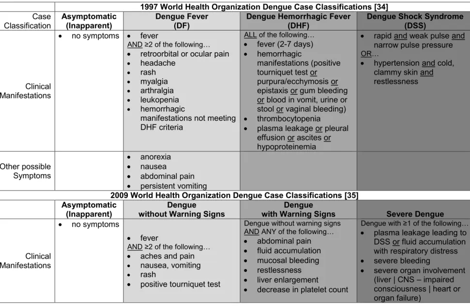
are thought to manifest clinically as dengue fever (DF), dengue hemorrhagic fever (DHF), or potentially-fatal dengue shock syndrome (DSS). DF consists of mild symptoms including, but not limited to, fever, joint pain, headache, and rash. DHF, the more severe form of dengue disease, includes symptoms of DF and is additionally characterized by plasma leakage. DSS, the most severe category of dengue disease, consists of signs of complete circulatory failure, manifesting as hypertension or irregular pulse [34]. In 2009, WHO modified its traditional classification system to broaden the complexity and applicability of apparent DENV case definitions. The newly revised system characterizes apparent dengue disease as dengue without warning signs, dengue with warning signs, and severe dengue, which are respectively similar to DF, DHF, and DSS, in regards to symptom descriptions [35]. Treatment of clinical dengue disease is simply a matter of close observation and symptom maintenance, mostly consisting of fluid replenishment [34]. (Refer to table 1.1 for a complete outline of WHO dengue case classification.)
1.3 Role of Viral Factors in Dengue Disease Severity
It is not fully understood why some individuals become more severely ill after dengue infections than others, although evidence shows viral factors play a role. Studies within the same cohort population have correlated specific DENV serotypes with severe disease outcomes. For example, a study in a Thai population showed DENV2 induced more severe disease than any of the other DENV serotypes [36, 37]. Introduction of new DENV clades into populations often have an effect on dengue disease severity, as well [32, 38-40]. DHF
6
immune system [43, 44]. NS5, for example, has the ability to inhibit type I interferon (IFN) signaling by binding to STAT2, which is a key component of the IFN pathway [43].
1.4 Role of Host Factors in Dengue Disease Severity
Immune Responses to Dengue Virus Infection
Although DENVs encode human immune system antagonists, as a whole, immune responses are typically efficient enough to clear DENV infection. Dendritic cells (DCs) residing in the skin are thought to be the first line of defense against invading DENVs [45]. DCs become activated and produce tumor necrosis factor alpha (TNFα) and IFNα [46]. Generally speaking, IFN responses to DENV infection are robust as evidenced by upregulation of IFN-related genes in DENV-infected cells [47-51]. In several cell types, type I IFNs, IFNα [52] and IFNβ [53], play a role in controlling DENV early in infection. Additionally, natural killer (NK) cells have been shown to induce proinflammatory type II IFN production and lyse DENV-infected cells [54].
Innate immune activation initiates an adaptive immune response to dengue infection. Antigen processing and presentation is an integral part of adaptive immunity. DCs are essential in trafficking DENVs to the lymphatic system, from where the virus then spreads to other cell types. Macrophages (mθ) process and present DENV antigen to T and B lymphocytes (T cells and B cells) [55-57]. Antigens stimulate CD4+ T cells to produce IFNγ [58]. Strong CD8+ T cell responses are stimulated by DENV infection as well [59-61]. Upon seeing antigen, B cells clonally expand to produce DENV-specific antibodies [55, 56], which are discussed later in this chapter.
Immunopathology of Dengue Virus Infection
7
dysregulation of cytokine responses, namely “cytokine storm”, correlates with severe dengue disease. Comprehensively, analysis of cytokine profiles in dengue patients reveals many cytokines, including IFNγ, TNFα, and several interleukins (ILs), are associated with dengue disease severity (reviewed in [62]). One of the more recent of these studies showed that MIP1β, IP-10, and MCP-1, which are all associated with inflammatory responses, are higher in patients with dengue with warning signs compared to patients with dengue without warning signs [63].
Cross-reactive CD8+ T cells are also correlated with DHF presence [59]. One
mechanism of T cell immunopathology is decreased degranulation [64], and another is cytokine induction [64]. The standing theory is that cross-reactive T cells produce elevated levels of cytokines, and subsequently, cytokine signaling contributes to vascular leakage, which is a classic symptom associated with DHF and DSS [64]. Complement, which is typically thought of as a viral clearance mechanism, is insinuated in inducing vascular leakage as well. While a helpful role for complement was shown in one study where DHF was associated with a reduction in complement components [65], other studies suggest an immunopathological role for complement. DENV NS1, an activator of complement signaling, and other key complement components are increased in fluids of DHF and DSS patients compared to DF patients [66, 67]. Also, in vitro, complement activation can enhance DENV uptake and viremia in myeloid cells [68].
1.5 Human Antibody Response to Dengue Virus
Role of Antibodies in Enhancement of Dengue Disease
8
of DENV antibodies induced after 1° infection are weakly neutralizing but cross-reactive [74-77]. Cross-reactive, sub-neutralizing DENV antibodies are thought to induce severe disease through a phenomenon known as antibody dependent enhancement (ADE) ([78] | reviewed in [79] and [80]) (figure 1.4).
Serotype cross-reactive antibodies have been mapped to NS1, prM, and E [81-84]. These weakly neutralizing cross-reactive antibodies bind to DENVs (figure 1.4A), and antibody-bound DENV particles then bind to Fc-gamma receptors, FcγRI and FcγRII, on appropriate moncocytic cells [85] (figure 1.4B). ADE via FcγRII can be tricky because isoforms FcγRIIa and FcγRIIb, which are found on most DENV target cells, have stimulatory and inhibitory effects on DENV ADE, respectively [86, 87]. Entry via ADE is thought to occur in one of two fashions: (1) endocytosis, similar to the classical DENV entry pathway described above, or (2)
phagocytosis, which occurs when aggregates of antibody-bound DENV particles sit atop a cell (figure 1.4C). In all, DENVs are engulfed by host cells thus increasing viral burden (extrinsic ADE) (figure 1.4E) and inducing cellular changes (figure 1.4D) that result in increased viral production (intrinsic ADE) (figure 1.4F).
In vitro studies suggest extrinsic ADE can result in upwards of a 1000-fold increase in virus infection [88]. Increased DENV infection by ADE has been proven repeatedly in multiple cell systems, including human-isolated mθ, monocytes, and DCs, using huMAbs [89], mouse MAbs [90], and serum from DENV-infected individuals [91]. ADE may be a more effective method of infection due to the ability of immature DENV particles, which are noninfectious via classical receptor-mediated pathways, to be bound by prM-specific antibodies and taken up by FcγRs [19, 81]. DENV infection via ADE pathways dramatically changes innate immune
9
pathogens and inducing cytokine and IFN production. DENV ADE may affect IFN production by reducing toll-like receptor (TLR) expression and signaling. [94]. Overall, the likely effect of intrinsic ADE is increased viral replication and progeny virus output per infected cell. Role of Antibodies in Protection of Dengue Disease
Similar to other DENV immune responses, antibody responses can be either pathogenic (enhancing), as discussed in detail above, or protective (neutralizing). The DENV neutralizing antibody response consists of type-specific antibodies and cross-neutralizing antibodies. Dogma would have it that these antibodies facilitate protection from severe disease upon 2° infection with a homotypic or heterotypic DENV serotype, respectively. Early in the quest to dissect DENV antibody epitopes, mouse MAbs revealed a large portion of the DENV antibody response was directed to EDIII [98]. However, more recent mapping studies, using huMAbs and
polyclonal human sera, show human antibody responses to DENV are different and more complex. In fact, human antibodies recognizing EDIII are relatively sparse and furthermore play only a minute role in DENV neutralization [99].
While strongly neutralizing mouse MAbs bind to recombinant E (rE) protein, strongly neutralizing huMAbs only recognize regions on intact DENV virions [77]. That finding was one of first to suggest a role for complex epitopes recognized by DENV neutralizing antibodies; since then more effort has been spent in understanding epitope specificity of DENV neutralizing antibodies. Strongly neutralizing huMAbs exerted antibody pressure on DENV in cell culture to render neutralization escape mutations around DENV EDI/II [77]. In studies of DENV1 vaccine recipients, it was found that amino acid mutations in EDI and EDII together were responsible for a type-specific antibody response [100, 101]. Structural analysis revealed a type-specific
10
Antibodies that confer potent neutralization across multiple DENV serotypes are less understood than type-specific antibodies. A few huMAbs that have potent broad neutralization capacity have been mapped to the FL region of E [104]. A potent broadly neutralizing huMAb that recognizes a region next to FL known as the “bc loop of EDII” was recently identified. However, FL-specific antibodies are typically weakly neutralizing, and, in fact, can confer enhancing activity [105]. The role for FL-specific antibodies in pathogenesis and/or protection is questionable, but a study using samples from a Nicaraguan cohort provides evidence that anti-FL antibodies generated from 1° DENV infections correlate with protection against 2°
heterologous infections [106].
Temporal Regulation of Dengue Antibodies
One might imagine that the balance between pathogenic and protective DENV antibody responses is largely dependent on temporal regulation of antibodies following DENV infections. The field’s current understanding of antibody timing following DENV infections is summarized in figure 1.5 [36, 107, 108]. Upon 1° DENV infection, a person may begin to show symptoms 4-10 days after inoculation. In the symptomatic or acute phase of illness, a person can exhibit viremia for up to 7 days; typical viremia lasts 5 days, peaking at day 3 of illness [36]. Length and
magnitude of viremia is shortened during 2° infections [36, 107]. NS1 antigen circulates in the blood stream during the viremic period and is often used as a quick and easy method for acute diagnosis of DENV infection [109-111].
IgG antibodies following 1° infection begin to appear after viremia wanes, at about day five post illness onset, and remain for a lifetime. Upon 2° infection, one is thought to experience a boost in IgG antibody levels that remains steady forever. IgM is more robust than IgG
immediately following 1° infection and quickly wane to undetectable levels by 3-6 months [112]. IgM levels following 2° infection are not as robust and are even shorter lived than those
11
primary and secondary DENV infections, but temporal regulation and biological significance of these antibody subtypes remain unclear [114].
More important than the timing of various types of antibodies is the timing of antibodies as they relate to pathogenic and protective disease outcomes. To that end, studies are being conducted to dissect temporal regulation of enhancing and protective antibody responses. Primary DENV infection with one serotype is dogmatically believed to lead to lifelong protective immunity to the infecting (homologous) serotype, but dengue-immune individuals remain susceptible to 2° infection with a different (heterologous) serotype [115]. It is thought that 2° infections induce antibody responses that are broadly-neutralizing and likely protective against post-secondary infections [116]. Even a 2° antibody response may not be completely protective because 3° and 4° infections, although rare, have been documented [70, 116-118].
Some of the more telling research on how long protective antibody responses last was conducted in the 1950s; Sabin infected humans with DENV and challenged them with either a homologous or heterologous serotype. That study pronounced the period of type-specific
protection to be at least 18 months while the period of cross-protection was only about 2 months [119]. Sabin’s last timepoint was 18 months, but studies have shown type-specific neutralization lasts up to 60 years post-infection [120]. Sequential infections in Thai and Nicaraguan cohorts reveal that 2° inapparent infections are more likely to occur within a shorter time span following 1° infection than 2° apparent infections. Together, these studies determined the period of cross-protection to be about 2 years [118, 121]. Perhaps, it is after 2 years that concentrations of weakly neutralizing cross-reactive antibodies decline and enhancement begins to occur [87].
1.6 Advantages of Studying Dengue Virus Infections in Human Cohorts
Mouse Models for Dengue Virus Pathogenesis
12
development, neurological effects and paralysis, which are not clinical symptoms of human dengue disease, were seen in DENV-infected animals [123, 124]. BALBc and C57BL/6 mice have shown human-like DHF symptoms, but only after being inoculated with high doses of DENV [125]. In studies of DENV pathogenesis, humanized mice have a stark advantage over other mouse models because their cell tropism is similar to that of an infected human [126], and their disease presentation is similar to that of human DF [126, 127]. ADE has been shown multiple times in IFNI/II receptor-deficient AG129 mice [128, 129]. In the AG129 model,
protection from ADE-induced disease is dependent on neutralizing antibodies [128]. In regards to antibody responses, it is important to restate that antibody responses in mice are drastically different than in humans [79, 99]. Furthermore, temporal regulation of DENV antibody
responses and sequential DENV infections cannot be studied in mice due to their short life spans.
Use of Non-human Primates in Vaccine-Related Studies
Despite aforementioned pitfalls, mice are still a telling model for preliminary vaccine studies, especially because many mouse models exhibit disease symptoms that can be used as suitable protection endpoints [123, 124]. For advanced vaccine-related studies, it is imperative that disease manifestation and antibody responses resemble those seen in humans’. DENV pathogenesis is less well-studied in non-human primates (NHP) because they do not develop human-like clinical symptoms. However, early on DENV-infected Rhesus macaques were shown to have similar viremia and antibody responses as humans [130]. Before introduction into humans, DENV vaccines are often tested in NHPs [122, 131-136], and NHPs have proven rather useful in predicting vaccine-induced immunogenicity.
Lessons Learned from Human Cohort Studies
13
completed (Table 1.2). The lessons that cohort studies have taught the dengue field are immense (reviewed in [138]). Using samples collected from cohort studies, it has been shown that severe dengue disease correlates with several biological factors, such as age, and clinical outcomes, such as platelet count [139-143]. Results from cohort studies [144] were even influential in WHO’s reevaluation of clinically apparent dengue symptomologies [145-148].
Additionally, researchers are making progress with understanding DENV antibody responses in human cohorts. Cohorts in Thailand were the first to show previous DENV infection is a risk factor for severe disease [69, 70]. Later, several cohorts followed suit with similar findings [78, 128, 149-152]. Also, maternal antibodies have been correlated with severe dengue disease in Vietnamese infants [153, 154]. Together, these findings justify the theory of ADE in naturally infected people. As far as protective antibody responses are concerned, determining the natural correlate of protection from DENV infections and/or disease is a continuing challenge for the vaccine field. As previously mentioned, cohort studies provided us with the first evidence of clade-induced shifts in DENV immunity [32, 38-40]. In Thai cohorts, pre-existing neutralization titers did not correlate with protection from DENV infection with the same serotype, as evidenced by clinical symptoms and viremia [152, 155], but Thai cluster studies show that neutralization titer does correlate with protection from DENV infection
[Buddhar, Darunee, et al. | unpublished data submitted to PLoS Neglected Tropical Diseases]. In a cohort in Peru, pre-existing neutralization breath correlated with protection from apparent post-secondary DENV infections [116]. Cross-protection following natural primary infections was determined to last about 2 years in Thai and Nicaraguan cohorts [118, 121].
1.7 Dengue Vaccine Development
In the wake of dengue emerging as a serious emerging global health problem,
14
susceptible to 3° and 4° DENV infections. Therefore, an ideal DENV vaccine should target a spectrum of age groups. Also, given the widespread circulation of DENV within poor nations, a DENV vaccine must be affordable. Most importantly, serotype-unbalanced vaccine responses run the risk of facilitating ADE upon natural challenge so an effective DENV vaccine must induce long-lived cross-protective responses against all DENV serotypes. Together these criteria present a vaccine development feat that, despite intense research efforts, has not been obtainable. To date, there is no DENV vaccine available to humans.
Prospective Dengue Virus Vaccine Candidates
Various approaches have been taken to develop a dengue vaccine. Subunit, subviral particles, or nonliving whole virus vaccines represent the safest class of DENV vaccines. There are a couple of nonliving DENV vaccines that stand out as potential vaccine candidates. The first is a tetravalent DNA vaccine that expresses DENV1-4 prM/E constructs [133], and the second is a tetravalent rE subunit vaccine [132]. These types of vaccines typically induce subpar immunogenicity and require combination with adjuvants and viral vectors to provide an immune boost [156, 157]. Live-attenuated or recombinant DENV vaccines have been tested and generally show stronger, longer-lived neutralizing antibody responses than nonliving
counterparts. Dengue virus can be attenuated via serial passage in cell culture so the virus no longer induces disease-causing infection but still induces an effective immune response. The efficacy of live attenuated DENV as a vaccine strategy was first tested by administering
attenuated DENV2 to naïve individuals. In that small trial, DENV2-specific neutralizing antibody responses lasted for up to 2 years post-vaccination [158]. Live-attenuated DENV vaccines have evolved since then into tetravalent recombinant formulas administered at various prime/boost timepoints [131, 134, 159-177].
15
attenuated DENV4 background. In human trial, vaccinees were challenged with DENV1 or DENV3 and were completely protected or partially protected, respectively [178]. DENVax is a LAV that has a DENV2 backbone with prM/E from the other serotypes inserted. In nonhuman primates, DENVax showed complete protection against DENV3 and DENV4 challenge, but only partial protection against DENV1 and DENV2 [135]. Recently completed human trials revealed DENVax induced broadly neutralizing antibody responses 30 days following inoculation [160]. Lastly is Sanofi Pasteur’s version of the DENV vaccine, CYD-TDV, which was thought to be the most promising candidate based on pre-phase IIB studies [179]. The chimeric tetravalent
formulation consists of each of DENV1-4 prM/E inserted into the yellow fever vaccine backbone. Unfortunately, this vaccine showed only showed ~30-70% protection against natural DENV challenge in clinical trials, depending on serotype [180-187], and was notably deficient in protecting against DENV2 infection in a Thai trial [186].
1.8 Outline of this Dissertation Research
Research Questions
With levels of protection being lower than predicted with the leading DENV vaccine [180-187], the field was reminded that there are fundamental aspects of protective responses that we do not understand. Natural DENV infection protects most people against severe secondary infection so using protective immune responses to natural infection as a gold-standard may prove beneficial to the vaccine field. Three overarching questions exist about human antibody responses to DENV infection:
(1) What are the differences between antibody responses induced by primary versus secondary infections?
(2) What role do enhancing antibodies play in disease severity?
16 Research Approach
To that end, the overall goal of this dissertation was to characterize the role of antibodies in natural inapparent and apparent dengue infections. Based on knowledge that a balance of pathogenic and protective antibody responses can influence the severity of dengue disease, we hypothesize that distinct properties of pre-existing antibodies correlate with inapparent
and apparent dengue disease outcome upon secondary infection. In collaboration with the
Pediatric Dengue Vaccine Initiative (PDVI) and investigators from the Ministry of Health in Sri Lanka, our group completed a prospective dengue study of 799 children, ages 0-12 years, who reside in Colombo, Sri Lanka. Using samples collected from the cohort, we addressed the following specific aims:
Specific Aim 1 (Chapter Two). Determine the incidence of DENV infection in cohort. We set out to characterize the true burden of DENV infection, including inapparent cases, on the Indian subcontinent for the first time.
Specific Aim 2 (Chapter Three). Investigate temporal regulation of antibodies following DENV infections.
2A. Compare quantity and neutralization capacity of antibodies acquired from 1°
inapparent and apparent DENV infections.
2B. Determine how time post-infection influences antibody neutralization capacity
following 1° infections.
2C. Characterize quantity and quality of antibodies following repeat DENV infections.
Specific Aim 3. Investigate qualities of pre-existing antibodies from repeat inapparent and apparent DENV infections.
3A. (Chapter Four) Compare quantity, neutralization capacity, and enhancement ability
17
3B. (Future Directions) Dissect infection history of children who acquired repeat DENV
infection. Innovation
Our study is innovative because most previous studies have only detailed differences between mild DF cases and severe DHF/DSS cases because they are hospital-based studies that enroll patients upon acquisition of symptoms [69, 139, 140]. Given the complexity and cost of cohort studies, very few groups have pre- and post-infection blood samples from both
18
Table 1.1. World Health Organization Dengue Case Classifications
1997 World Health Organization Dengue Case Classifications [34] Case Classification Asymptomatic (Inapparent) Dengue Fever (DF)
Dengue Hemorrhagic Fever (DHF)
Dengue Shock Syndrome (DSS)
Clinical Manifestations
no symptoms fever
AND ≥2 of the following… retroorbital or ocular pain headache rash myalgia arthralgia leukopenia hemorrhagic
manifestations not meeting DHF criteria
ALL of the following… fever (2-7 days) hemorrhagic
manifestations (positive tourniquet test or purpura/ecchymosis or epistaxis or gum bleeding or blood in vomit, urine or stool or vaginal bleeding) thrombocytopenia
plasma leakage or pleural effusion or ascites or hypoproteinemia
rapid and weak pulse and narrow pulse pressure OR…
hypertension and cold, clammy skin and restlessness
Other possible Symptoms
anorexia nausea
abdominal pain persistent vomiting
2009 World Health Organization Dengue Case Classifications [35] Asymptomatic
(Inapparent)
Dengue
without Warning Signs
Dengue
with Warning Signs Severe Dengue
Clinical Manifestations
no symptoms
fever
AND ≥2 of the following… aches and pain nausea, vomiting
rash
positive tourniquet test
Dengue without warning signs AND ANY of the following… abdominal pain fluid accumulation mucosal bleeding restlessness liver enlargement
decrease in platelet count
Dengue with ≥1 of the following… plasma leakage leading to
DSS or fluid accumulation with respiratory distress severe bleeding
19
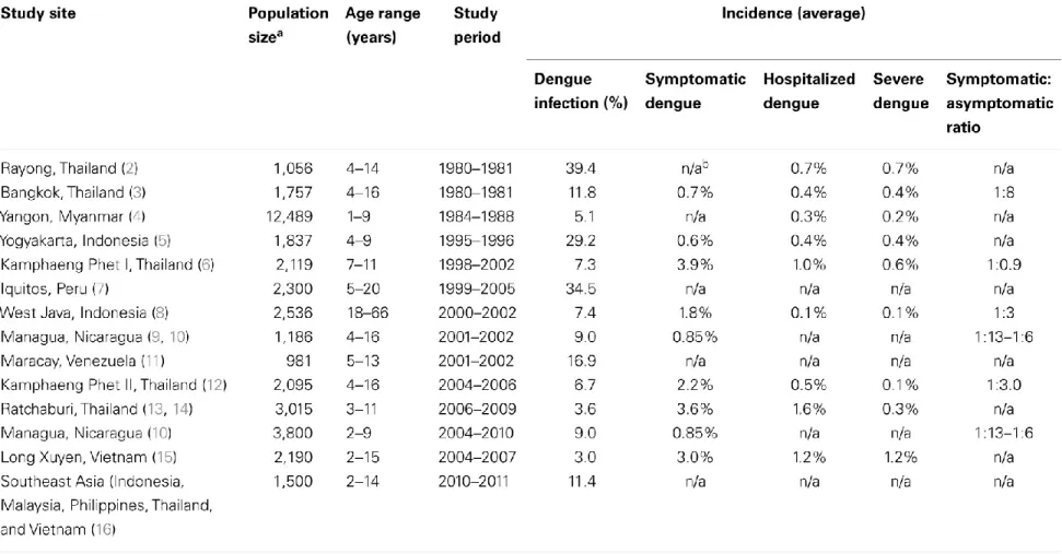
Table 1.2. List of Prospective Dengue Cohort Studies1
1This table was taken from 138. Endy, Timothy P, Human Immune Responses to Dengue Virus Infection: lessons learned from
20
21
22
23
24
Figure 1.5. Temporal regulation of antibodies following DENV infection. (This figure was
25
CHAPTER TWO
Burden of Dengue Infection and Disease in a Pediatric Cohort in Urban Sri Lanka
234
2.1 Introduction
Dengue is caused by four related mosquito-borne dengue viruses (DENV1-4), which are endemic in many tropical and subtropical regions of the world. Most individuals infected with DENV are asymptomatic or develop a febrile illness known as dengue fever (DF), but in a minority, disease can be more severe and progress to dengue hemorrhagic fever and dengue shock syndrome (DHF/DSS). Infection with one DENV serotype confers protective immunity to future infections with that serotype only and these individuals are susceptible to secondary infections with heterologous serotypes [79]. Individuals suffering from secondary DENV infections are at a greater risk of developing DHF/DSS compared to individuals experiencing their first infection [189, 193].
Globally, an estimated 2.5 billion people are at risk for DENV infection, with estimated 390 million annual DENV infections and 96 million dengue cases [28]. From prospective community and school based cohort studies, it has been possible to obtain estimates for the
2This chapter was previously published. | Tissera H, Amarasinghe A, de Silva A.D., Palihawadana P,
Kariyawasam P, Gunasena S, Corbett K.S., Katzelnick L, Tam C, Letson G.W., Margolis H.S., and de Silva A.M., Burden of dengue infection in a pediatric cohort in urban Sri Lanka. American Journal of Tropical Medicine and Hygiene,2014. 91(1): p. 132-7.
3Portions of this chapter are in press. | Corbett K.S., Katzelnick L, Tissera H, Amarasinghe A, de Silva
A.D., and de Silva A.M., Pre-existing Neutralizing Antibody Responses Distinguish Clinically Inapparent and Apparent Dengue Virus Infections in a Sri Lankan Pediatric Cohort. Journal of Infectious Diseases.
4This study was funded by the Pediatric Dengue Vaccine Initiative (Bill and Melinda Gates Foundation
26
incidence of DENV infection and disease for sites in southeast Asia [141, 194-198] and Latin America [199-201]. Although dengue is emerging in the Indian subcontinent and is considered a major health issue, we do not have estimates of the true burden of DENV infection and disease in this region.
Dengue was first reported in Sri Lanka in the 1960s [29]. Studies conducted from 1980-1984 showed a DENV seroprevalence of 50% among school children, with an annual
seroconversion rate of 10-15% among 5−7-year-old school children in Colombo, the capital of Sri Lanka [39, 202]. More recently, analysis of age-stratified seroprevalence data indicated that the annual seroconversion rate among children <12 years in Colombo is around 14% [202]. In the past, most dengue cases have been reported from the Colombo district and other
neighboring districts in the heavily urbanized southwestern region of the country. However, over the past 10-15 years dengue has been reported from nearly all districts of the island, and over the past two decades, the number of reported DF and DHF cases has increased by over 10-fold [31]. This increase in cases has been attributed to introduction of new genotypes of DENV as well the expansion of the range of the virus on the island [31-33]. In many ways, the changing epidemiology of dengue in Sri Lanka mirrors events in other parts of the subcontinent including India, which has also documented large increases in cases and appearance of new virus strains [203, 204]. We conducted a population-based study to determine the incidence of symptomatic and asymptomatic DENV infection among children living in Colombo, Sri Lanka, an urban setting of the Indian subcontinent.
2.2 Materials and Methods
Ethical Approval for Study
27
Study Site
The study was conducted in the city of Colombo, the commercial capital of Sri Lanka, which has a population of 647,100 and is the most densely populated area in the country with 17,353 persons per square kilometer [205]. The city is divided into 47 municipal wards and ward 33 was selected for the study because of its stable population, which reflects the
socio-economic status and demographics of the entire municipal area. The ward is endemic for dengue with the catchment population seeking healthcare in the tertiary care institution situated within its boundaries (Figure 2.1).
Study Population
A prospective cohort study was conducted between November 2008 and January 2010. The protocol for the study is described in detail elsewhere [206]. In brief, a house-to-house census was conducted by research assistants to determine the size, socio-demographic information and health-seeking patterns of the permanent resident childhood population <12 years of age. A representative sample of 800 children <12 years of age was recruited for the current study. The sample size calculation was based on the ability to detect an annual
incidence of dengue of 10% with an absolute precision of 3%. The estimated sample size was then inflated by 30% to allow for possible loss to follow-up during the study period. All children were enrolled in the study following written informed consent from parent/legal guardian and assent from children > 7 years of age. Each participating household was given a thermometer and each child was given an identification card with a unique study number. A febrile illness was defined as a temperature of ≥38°C lasting ≤7 days in any child in the study cohort documented by the parent, research assistant or health care professional. Following assessment and
28
Sample Collection
Blood samples were collected from all children at enrollment (Between November 2008- January 2009) and one year after enrollment (between November 2009 and January 2010) by finger prick and stored as blood spots on protein saver card (Whatman & ID Biological systems, USA) [207, 208]. From children with a documented fever, whole blood was collected by
venipuncture into EDTA containing tubes. Some of the whole blood was used to prepare dried blood spots for later serological testing. The remaining blood was centrifuged and the plasma used for molecular diagnostic testing. Ten or more days after recovery from fever, convalescent samples were collected by finger prick and stored as blood spots on protein saver cards. DENV Strains Used for Laboratory Assays
The WHO DENV reference strains, i.e. DENV1 West Pac 74, DENV2 S-16803, DENV3 CH54389 and DENV4 TVP-360 were used for preparing antigen and infectious stocks for serological assays. The WHO reference viruses were initially obtained from Dr. Robert Putnak (Walter Reed Army Institute of Research, Silver Spring, MD). Infectious stocks of virus were prepared using the C6/36 mosquito cell line and dengue antigens were harvested from Vero cells as previously described [209].
Molecular Detection of DENV
Plasma obtained during the acute phase of the febrile illness was tested by reverse transcriptase polymerase chain reaction (RT-PCR) to detect and serotype DENVs as previously described [31].
Detection of anti-DENV IgM and IgG Antibodies in Dried Blood Spots
29
We performed dengue IgM capture ELISA as described [211], except we used anti-flavivirus monoclonal antibody 4G2 followed by enzyme-conjugated goat anti-mouse IgG to detect captured DENV antigen. In brief, 96-well plates were coated (overnight, 4°C) with 100 μL/well (1 ng/μL) of goat anti-human IgM (Sigma, St. Louis, MO, USA) at a concentration of 0.1 mol/L in carbonate buffer (pH 9.6). Plates were washed 3× in Tris-buffered saline with 0.2% Tween 20 (TBST) and blocked with 200 μL/well of 1× Tris-buffered saline with 0.05% Tween 20 and 3% nonfat dry milk. Paired serum samples were tested on the same plate. Diluted serum (1:50) was loaded in duplicate and incubated (37°C, 1 h) to capture IgM antibody. Unbound antibody was washed, and wells were successively incubated with DENV antigen (mix of serotypes DEN1–4), mouse anti-flavivirus 4G2 mAb, and human-absorbed alkaline
phosphatase (AP)–conjugated goat anti-mouse IgG antibody (Sigma). Optical density (OD) was measured at 405 nm after final incubation with AP substrate.
Dengue IgG ELISA was performed as described [212]. Plates were coated overnight (4°C) with 100 μL/well of mouse anti-flavivirus 4G2 mAb at a concentration of 0.1 mol/L in carbonate buffer (pH 9.6) and then washed 3× in TBST. Plates were then blocked with standard diluents and successively incubated (37°C, 1 h) with DEN1–4 antigen, diluted serum (1:100) in duplicate wells, and AP-conjugated goat anti-human IgG (Fc portion), with 3 washings (TBST) between incubations. Plates were read at 405 nm after a final incubation with AP substrate (15 min, room temperature, in the dark).
Measurement of DENV Neutralizing Antibodies
30
were serially diluted (4 fold dilutions starting at 1:40 and ending at 1:10,240) and the serum dilution that neutralized 50% of the viruses was calculated by nonlinear, dose-response regression analysis with Prism 4.0 software (GraphPad Software, Inc., San Diego, CA). Determining Dengue Serostatus at Study Enrollment
Dengue virus serostatus (DENV naïve or immune) at enrollment was determined by dengue IgG immunoassay. An OD value > 0.3 were considered dengue antibody positive. During the study period, primary infections were defined as dengue naïve children at enrollment who experienced an infection. Secondary infections were defined as dengue immune children at enrollment who experienced an infection.
Criteria for Laboratory Diagnosis of DENV infections
A laboratory-confirmed clinically apparent dengue case was a child with a febrile illness who tested positive in at least two out of the three diagnostic assays (PCR, rising levels of IgM and/or IgG antibodies in paired acute and convalescent blood samples). In the few cases where there was only an acute blood sample, the diagnosis was based on RT-PCR testing only. Apparent dengue cases were further classified as primary or secondary based on baseline DENV naïve or immune status, respectively.
31
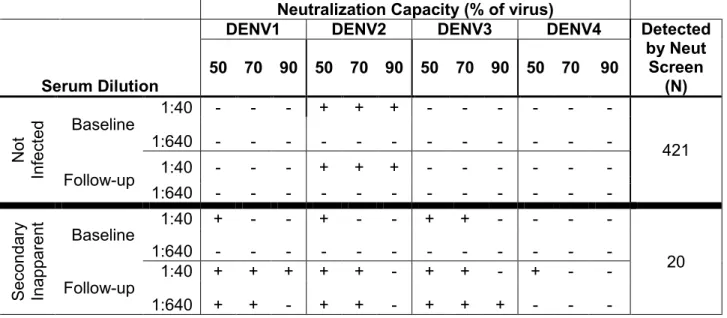
Therefore, all children were screened for increased neutralization capacity over the study year by subjecting paired baseline and 12-month follow up samples from all children (at dilutions of 1:40 and 1:640) to a FACS-based neutralization assay against each of the four dengue serotypes (Table 2.1). Those children showing an increase in neutralization capacity by 2-dilution screen were then confirmed by neutralization assay [213, 214] with full serum dilution series (Figure 2.2). To establish criteria for defining secondary infections using paired samples collected 12 months apart, we used a test set of 8 symptomatic secondary cases (detected by PCR and serology performed on samples collected within 1 month of acute infection). Only half the cases (4/8) cases displayed a 4-fold or greater increase whereas all cases (8/8) displayed a 2-fold or greater increase in neutralizing antibody levels when paired baseline and end-of-year samples from these children were tested. Therefore, children who had >2 fold increases in levels of neutralizing antibodies to one or more serotypes by the end of the year were designated as secondary infections. Samples were tested at least twice and only paired samples that displayed a reproducible 2 fold-increase were included as new secondary infections. Although it is possible that some inapparent infections were actually dengue fever cases that were missed, efforts were made to ascertain case detection by educating parents and having a study team visit each house weekly [215]. Loss to follow-up was minimal; of 800 children enrolled, only 1 child dropped out during the 12 months [215, 216].
2.3 Results
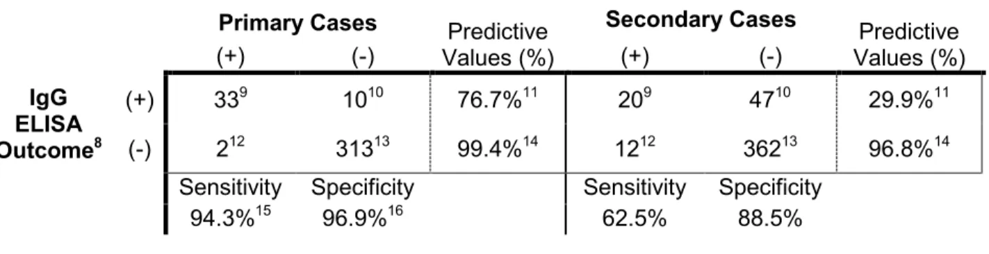
32
predictive value (PPV) of 76.7%), the assay performed poorly for secondary infections (sensitivity of 62.5% and a PPV of only 29.9%) These results indicate that IgG ELISA is an unreliable method for identifying true DENV infections, particularly 2° infections, when utilizing paired samples collected 12 months apart.
Between November 2008 and February 2009 a total of 800 children ages 0-12 years were enrolled in the study from ward 33 (Figure 2.1), these being representative of the age and demographic distribution of the 2527 children known to be permanent resident of the ward. Only 1 child was lost to follow up during the study period. The dengue seroprevalence at enrollment was determined by testing all children by ELISA for the presence of DENV binding IgG antibodies (Table 2.3). To confirm that IgG seropositivity was due to dengue infection and not exposure to related Japanese encephalitis virus (JEV), which is also present in Sri Lanka, all IgG positive samples were tested for the presence of DENV neutralizing antibodies. The vast majority (96%) of IgG positive sera also neutralized DENV indicating that in this population the results of the IgG ELISA reflected dengue seroprevalence. The overall dengue seroprevalence was 53% at enrollment (Table 2.3). The age specific seroprevalence steadily increased with advancing age from a low of 22% in the youngest (<1 year) age group to a high of 74.26 % in the oldest (10-12 year) age group (Table 2.3).
33
Out of the 65 new apparent and inapparent DENV infections, 34 were primary infections and 31 were secondary infections (Table 2.5). As might be expected, the ratio of primary to secondary infections decreased with age from 3 in the youngest (< 1 year) age group to 0.75 in the oldest (10-12) age group (data not shown). The ratios of inapparent to apparent infections were not statistically significantly different (P>0.1) between primary and secondary infections (Table 2.5).
The prevalence of each DENV serotype during the study year was estimated by identifying the serotypes responsible for primary infections (Table 2.6). DENV2 was the most common serotype (49%) followed by DENV3 (27%), DENV1 (23%) and DENV4 (3%). The number of people with monospecific neutralizing Abs to each serotype at enrollment into the study was used to estimate the prevalence of each serotype in this population before the start of our study (Table S1). This analysis demonstrated that DENV2 (43%) and DENV3 (29%) were more common than DENV1 (9%) and DENV4 (12%) in the period preceding the study.
2.4 Discussion
Over the past two decades dengue has emerged as a major health problem in the Indian subcontinent [203, 204]. Dengue epidemics in the region have been linked to more intense transmission, expansion of the range of the virus and introduction of new strains [31-33, 203, 204]. Most estimates of dengue incidence in the region are based on hospital-based studies and nationally reported cases, which grossly underestimate the true burden of disease and infection [217]. In this study, we followed a cohort of 799 children in Colombo, Sri Lanka to gauge the true burden of infection in this region.
34
children at 2 dilutions for the presence of neutralizing antibody before selecting samples for more comprehensive neutralization testing. Our study demonstrates that while simple to
perform, IgG ELISA and related assays are unreliable methods for detection of DENV infections when samples are collected 12 months apart, particularly for repeat infections.
Having fully characterized primary and secondary DENV infections in the cohort using neutralization assay, we estimated the incidence of infection and disease to be 8.39 and 3.38 per 100 children respectively. The ratio of clinically inapparent to apparent infections was 1.48 indicating that for every apparent infection there were approximately 1.5 inapparent infections in children. This high intensity of transmission was also supported by the seroprevalence data that demonstrated a gradually rising prevalence with age that ranged from 22% in the youngest age group to 74% in the oldest age group. The 4 DENV serotypes were circulating in this population both before and during our study, with serotypes 2 and 3 being more prevalent than 1 and 4 (Table 2.6). Our study establishes a rigorously measured, accurate burden of dengue infection and disease among children living in an urban area of Sri Lanka.
Recently, we used dengue seroprevalence data from this cohort to model the rate of primary infections in dengue naïve children [202]. Using a catalytic model, we estimated the incidence of primary infection to be 14.1% per year (95% CI: 12.7%– 15.6%), which is higher than the incidence 8.39 % (95% CI: 6.56-10.53) observed here for total infections (primary and secondary infections). A more legitimate comparison would be to compare the model based estimate for primary infections with the incidence of primary infections in our cohort. The incidence of primary infection noted in the current study was 9.8% (95% CI: 6.80-12.80), which is closer to the model-based estimate of 14.1% per year (95% CI: 12.7%– 15.6%). A previous study of school children in Colombo over a 5-year period from 1980-1985 estimated the


![Figure 1.5. Temporal regulation of antibodies following DENV infection. (This figure was adapted from [192].) A person can exhibit viremia for up to 7 days [36], although length and magnitude of viremia is shortened during 2° infections [36, 107]](https://thumb-us.123doks.com/thumbv2/123dok_us/8288299.2194809/46.918.126.783.105.548/temporal-regulation-antibodies-following-infection-magnitude-shortened-infections.webp)