Received 13 Nov 2015|Accepted 16 Aug 2016|Published 7 Oct 2016
Structural characterization of antibiotic
self-immunity tRNA synthetase in plant
tumour biocontrol agent
Shaileja Chopra
1,*, Andre
´s Palencia
2,*
,w, Cornelia Virus
1, Sarah Schulwitz
1, Brenda R. Temple
3,
Stephen Cusack
2& John Reader
1,wAntibiotic-producing microbes evolved self-resistance mechanisms to avoid suicide. The biocontrolAgrobacterium radiobacterK84 secretes the Trojan Horse antibiotic agrocin 84 that is selectively transported into the plant pathogenA. tumefaciensand processed into the toxin TM84. We previously showed that TM84 employs a unique tRNA-dependent mechanism to inhibit leucyl-tRNA synthetase (LeuRS), while the TM84-producer prevents self-poisoning by expressing a resistant LeuRS AgnB2. We now identify a mechanism by which the antibiotic-producing microbe resists its own toxin. Using a combination of structural, biochemical and biophysical approaches, we show that AgnB2 evolved structural changes so as to resist the antibiotic by eliminating the tRNA-dependence of TM84 binding. Mutagenesis of key resistance determinants results in mutants adopting an antibiotic-sensitive phenotype. This study illuminates the evolution of resistance in self-immunity genes and provides mechanistic insights into a fascinating tRNA-dependent antibiotic with applications for the development of anti-infectives and the prevention of biocontrol emasculation.
DOI: 10.1038/ncomms12928 OPEN
1Department of Cell Biology and Physiology, The University of North Carolina at Chapel Hill, Chapel Hill, North Carolina 27599-7090, USA.2European
M
icrobes that produce antimicrobial agents often prevent cell suicide by co-expressing self-immunity proteins that employ a variety of different resistance mechanisms1. Genes encoding self-immunity proteins can also act as a source of antibiotic resistance in the environment when they are horizontally transferred to microbial pathogens2. This threatens not only the successful treatment of human pathogens but also prevention of important animal and crop diseases3. One mechanism of self-immunity is to encode resistant variants of the enzyme targeted by the antibiotic. A fascinating example of such an antibiotic-producing organism is Agrobacterium radiobacter strain K84, which is used as a biocontrol agent of the plant pathogen A. tumefaciens that causes plant tumours in a number of agriculturally important species4–6. The biocontrol produces the ‘Trojan Horse’ antibiotic agrocin 84 (refs 7–9) that mimics a plant tumour-derived substrate to gain entry by subterfuge into the pathogen10,11, where it is processed into a toxin termed TM84 (refs 10,12) (Figs 1 and 2a). We recently identified the molecular target of TM84 in A. tumefaciens as leucyl-tRNA synthetase (LeuRSAt) which catalyses an essentialfirst step in protein synthesis12. To prevent cell suicide, A. radiobacter K84 contains a second, non-essential LeuRS, termed AgnB2, which is resistant to TM84 (refs 12–14) (Fig. 1). The agnB2 gene is encoded on the 44 kb plasmid pAgK84, along with all the genes required to biosynthesize agrocin 84 (refs 14,15). Importantly, horizontal gene transfer of the pAgK84 plasmid from A. radiobacter K84 to the pathogen has been previously reported to occur in the field, resulting in agrocin 84-resistant strains of A. tumefaciens encoding the agnB2 gene5,16,17. Thus, understanding the molecular mechanisms by which AgnB2 provides immunity to agrocin 84 is crucial for combating antibiotic resistance.
The molecular target of TM84, LeuRSAt, is an essential
enzyme that specifically ligates leucine to its cognate tRNALeu isoacceptors, a critical step before protein synthesis at the ribosome. The aminoacylation of tRNALeu is catalysed in a two-step mechanism in all LeuRSs: in the tRNA-independent first
step, leucine is activated by ATP to form the essential obligate intermediate leucyl-adenylate (Leu-AMP) that is stably bound to the enzyme. In the second step, a bound tRNALeurepositions its CCA 30-end into the LeuRS active site where activated-leucine is transferred to the tRNA. The essential catalytic nature of LeuRSs, and the 19 other canonical aminoacyl-tRNA synthetases (AARSs), has led to these enzymes emerging as prominent targets for anti-pathogens18,19, ranging in the prevention of fungal diseases in humans20 to bacterial plant pathogens12. A substantial amount of work has been performed on stable analogues of aminoacyl-adenylates that bind tightly to an AARS active site, and preventing activation of the amino acid with ATP21,22. However, these molecules often cannot be transported efficiently into cells and can lack discrimination between microorganism and the intended host18.
We recently showed that TM84 does not behave like other stable aminoacyl-adenylate analogues but, instead, employs a unique tRNA-dependent inhibition mechanism23,24. TM84 binds only weakly to the LeuRS-active site on its own and requires the presence of tRNALeuto form a tight binding ternary inhibition complex23. The X-ray crystal structure of the Escherichia coli LeuRSEctRNALeuTM84 ternary complex shows TM84 bound in
the Leu-AMP-binding site of the catalytic domain. The tRNALeu is bound to LeuRS in an ‘aminoacylation-like’ conformation with the CCA 30-end of the tRNALeuentering into the active site with the terminal adenosine hydrogen bonding (H-bonding) directly with TM84 (ref. 23). The positioning of the tRNALeu acceptor stem is accompanied with a substantial movement of the catalytic K619MSK622S motif. The K619 sidechain bonds to the penultimate tRNALeu cytidine base, while K622 extends across the adenylate analogue and interacts with the phosphate of TM84 via a non-bridging oxygen. The repositioning of the Lys-Met-Ser-Lys-Ser (KMSKS) loop and tRNALeucombine to effectively bury the bound TM84, thus explaining how the presence of tRNALeu induces the tight binding of LeuRSs to TM84.
AgnB2 exhibits a high degree of resistance to TM84 (ref. 12) despite the close similarity of TM84 to Leu-AMP (Fig. 2a) and the
TM84
Agrocin 84 Agrocinopine Agrocin 84
tRNA
Leucine
ATP AgnB2 LeuRS
tRNA
Leucine
ATP LeuRSAt
Sucrose
Uptake moiety TM84 pTiC58
pAgK84
PLANT TUMOR
Biocontrol Pathogen
Figure 1 | Biology of agrocin 84 produced by biocontrolA. radiobacterstrain K84.PathogenicA. tumefacienspossesses a TiC58 plasmid that upon infection of the host plant makes the plant produce a carbon and phosphate source agrocinopine. The pathogen takes up agrocinopine via an agrocinopine permease encoded on pTiC58. This transporter also recognizes the uptake moiety on agrocin 84, an antibiotic produced by the plant tumour biocontrol,
need for AgnB2 to adopt a true aminoacylation complex as exhibited in a recent LeuRSEctRNALeuLeu-AMS crystal
structure25. These observations raise the question of how does AgnB2 achieve resistance to TM84 without dramatically interfering with the catalysis of the aminoacylation reaction. This is particularly intriguing as primary sequence alignments of AgnB2 and TM84-sensitive LeuRSs show a high similarity of residues, including catalytic motifs (except a single Q instead of M at the KMSKS motif). Here, we examine the TM84 resistance mechanisms employed by AgnB2 using both thermodynamics and steady-state enzyme kinetics. We also present the X-ray crystal structure of AgnB2 in complex with tRNALeuand a stable leucyl-adenylate analogue and reveal tertiary interactions unique to AgnB2. We finally identify key TM84 immunity determinants in AgnB2 and the TM84-sensitive LeuRSAtand show that it is
possible to partially inter-convert the TM84 sensitivity properties of these enzymes.
Results
Analysis of AgnB2 steady state enzyme kinetics. The ability of AgnB2 to discriminate between TM84 and the obligate reaction intermediate Leu-AMP prompted us to examine differences between the two molecules. As shown in Fig. 2a, TM84 contains a stable N-acyl phosphoamidate bond, instead of the reactive phosphoanhydride linkage in Leu-AMP. In addition, TM84 has a deoxy-arabinose sugar group, instead of a ribose with subsequent loss of a 30-hydroxyl (OH) and a repositioned 20-OH above the sugar ring, and replacement of the primary amine with another OH group. For TM84-sensitive LeuRSs, these subtle differences between toxin and reaction intermediate allow TM84 to employ a tRNA-dependent inhibition mechanism where the CCA 30-end of the tRNALeubinds in an ‘aminoacylation-like conformation’ into the active site of the enzyme and directly hydrogen bonds with the antibiotic23. The ‘aminoacylation-like’ conformation of tRNA in the LeuRSEctRNALeuTM84 ternary inhibition complex
exhibits a high degree of similarity with the tRNALeu in the true LeuRS aminoacylation conformation25. Since the tRNALeu bound to AgnB2 still has to reposition its CCA 30-end into the active site during catalysis of the aminoacyl transfer reaction, we questioned whether destabilization of the TM84 ternary complex in AgnB2 might be reflected in the kinetic properties of AgnB2. Our steady-state kinetic analysis of AgnB2 examined both the overall aminoacylation reaction and the first step, the amino acid activation reaction, using the ATP/PPi exchange reaction. We firstly determined the apparent TM84 inhibition constant
(Ki) for AgnB2 in the aminoacylation reaction by altering the
ATP concentrations of the substrate and pre-incubating the AgnB2tRNALeucomplex before initiating the reaction. The data revealed an apparentKiof 4.72±0.75mM (Supplementary Fig. 1). We then determined the apparent TM84Kifor AgnB2 by altering the leucine concentration and keeping the ATP fixed giving a value of 1.38±0.28mM (Supplementary Fig. 1a,b). Both values are more than three orders of magnitude weaker than theKiapp determined previously for the natural target LeuRSAt(ref. 23).
Importantly, TM84 acted as a weak competitive inhibitor of AgnB2 with regards to both ATP and leucine (Supplementary Fig. 1a,b, respectively) and therefore still binds to the synthetic active site and not at an alternative site. We also found that AgnB2 is resistant to TM84, but is still potently inhibited by Leu-AMS (a non-hydrolysable analogue of the Leu-AMP reaction intermediate), for both the amino acid activation (Supplementary Fig. 2) and overall aminoacylation reactions (Fig. 2b). Substantial weakening of the binding of the aminoacyl adenylate to AgnB2 did not evolve as a solution to generating resistance to TM84, as this would severely affect the catalysis of amino acid activation. As shown in Table 1, AgnB2 and LeuRSAtshare similar kcat
values for all substrates. The higher Kmfor tRNALeu in AgnB2
may reflect an alteration in the interactions of AgnB2 with TM84
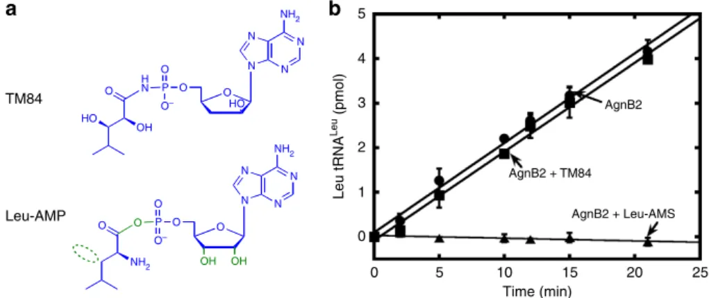
a
Leu-AMP
N
N N N
NH2
O O P O
O– O
O
NH2 OH OH
N
N N N
NH2
O HO O P H N
O– O
O
OH HO
b 5
4
3
2
1
0
0 5 10 15 20 25
Time (min)
Leu tRNA
Leu
(pmol) AgnB2
AgnB2 + Leu-AMS AgnB2 + TM84
Figure 2 | AgnB2 discriminates between a stable Leu-AMP analogue and TM84.(a) Chemical structures of Leu-AMP and TM84 with differences highlighted in green. A non-hydrolysable adenylate analogue called Leu-AMS, which contains a N-sulfamoyl linkage in place of the phosphoanhydride of Leu-AMP, was used in biophysical and crystallography experiments. (b) Effect of TM84 and Leu-AMS on the aminoacylation reaction of wt AgnB2. Aminoacylation reactions were carried out using 2 nM wt AgnB2 only (K) and wt AgnB2 in the presence of 1mM TM84 (’) and 20 nM Leu-AMS (m) at 28°C, pH 7.4 and initiated using 1 mM ATP. Error bars represent s.d. (n¼3).
Table 1 | Kinetic parameters obtained for the aminoacylation and ATP-PPi exchange reactions catalysed by LeuRSAtand AgnB2 at 28°C at pH 7.4.
Enzyme Substrate Km(lM) kcat(s1)
Aminoacylation
AgnB2 ATP 95.4±18.3 0.43±0.02
Leucine 28.7±7.3 0.35±0.03 tRNA 1.48±0.25 0.43±0.02 LeuRSAT* ATP 396±83 0.33±0.02
Leucine 38±3.2 0.38±0.01 tRNA 0.94±0.19 0.36±0.02
ATP-PPiExchange
AgnB2 ATP 1047±140 67±8.9
Leucine 12±2 73±3.6
LeuRSAT ATP 357±22 87±1.5
Leucine 9.7±0.7 85±1.6
All data in this table were average values obtained from three independent experiments with error±values representing s.d.
tRNALeu that is relevant to resistance to the tRNA-dependent TM84 inhibitor. The Km for ATP is a small 3.8-fold higher
affinity value for AgnB2 than for LeuRSAt, while the leucine
affinities show minimal differences. In the case of ATP/PPi exchange reaction, AgnB2 showed a higher Km for ATP
compared with the aminoacylation reaction (10-fold increase). Interestingly, these kinetic comparisons represent only small energetic differences between the two enzymes, yet still lead to enzymes with pronounced differences in TM84 sensitivity. In addition, the steady-state kinetic properties shows that the ATP saturation plot for the overall aminoacylation reaction shows a degree of substrate inhibition, whereas the ATP/PPi exchange shows no substrate inhibition for ATP (data not shown) when tRNALeuis absent. One possibility is the population of a second unproductive ATP-binding site in the AgnB2 active site that may in part explain the differences between the apparent TM84 Ki’s determined for AgnB2 when varying leucine and ATP (Supplementary Fig. 1). Interestingly, we do not observe any ATP substrate inhibition for LeuRSAt. The data suggest that
different substrate-dependent conformations populated by AgnB2 may be important for TM84 resistance.
tRNALeuhas reduced effect on the affinity of TM84 to AgnB2. If resistance of AgnB2 is based on abolishing of the normal tRNA-dependent inhibition mechanism employed by TM84 (ref. 23) then one would expect to see a decrease in the affinity for TM84, tRNALeuand/or for TM84 in the presence of tRNA or vice versa. To test these possibilities, we used isothermal calorimetry (ITC) (Supplementary Table 1) to investigate the ligand binding modes to AgnB2 (ref. 23).Kdvalues of 1052 nM for TM84 and
310 nM for tRNA were obtained, indicating reductions in AgnB2-binding affinities for both TM84 (6.9-fold) and tRNA (3.7-fold), compared with LeuRSAt (ref. 23). Further examination of
TM84 binding to AgnB2 (with pre-bound tRNA) showed a Kdof 332 nM, which suggests a substantial destabilization of
the ternary complex (AgnB2tRNATM84) (B330-fold decrease) as compared with the LeuRSAttRNATM84 complex
(Supplementary Table 2). Interestingly, we also see no evidence of a binary isotherm for TM84 binding to AgnB2tRNALeu species, unlike LeuRSAt. Thus, AgnB2 has a significantly
wea-kened affinity for TM84 as compared with LeuRSAt. Since TM84
and the Leu-AMP analogue bind to the same active site but potentially to different conformations, we examined if AgnB2-binding affinity for Leu-AMS had been substantially weakened. Our results show that Leu-AMS binds to AgnB2 with low nanomolar binding affinity (Kd¼4.4 nM), which is slightly
weaker than to LeuRSAt (1.6 nM). The fact that AgnB2 binds
tightly to Leu-AMS is not so surprising since, firstly, AgnB2 has highly conserved residues at the synthetic site compared with other LeuRSs and, secondly, the enzyme must bind tightly to the Leu-AMP intermediate to catalyze the aminoacylation reaction. This result was supported by our enzyme kinetic observations above which showed that Leu-AMS is a low nanomolar inhibitor of both the AgnB2-catalyzed aminoacylation and amino acid activa-tion reacactiva-tions, demonstrating that the analogue efficiently com-petes with ATP and leucine substrates (Supplementary Fig. 1).
Our thermodynamic results suggest that AgnB2 may achieve resistance to TM84 by not simply weakening the affinity for TM84 (and/or tRNALeu) but most significantly by destabilizing the formation of the AgnB2tRNALeuTM84 complex. Interestingly, this is accomplished with only minimal change to affinity for the adenylate, allowing the enzyme to still efficiently carry out aminoacylation.
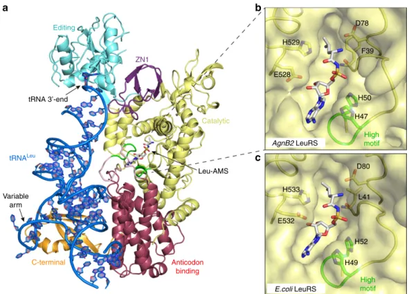
Structure of AgnB2tRNALeuLeu-AMS ternary complex. To identify structural interactions in AgnB2 that may contribute to
resistance, we performed extensive crystallizations trials with AgnB2, agrobacterium tRNALeu and multiple combinations of substrates or inhibitors. We obtained an X-ray crystal structure of AgnB2 in complex with tRNALeuand Leu-AMS; the final model was refined to a final R-factor of 19.4% (R-free¼23.8%) and a final resolution of 2.1 Å (Table 2, Fig. 3a and Supplementary Fig. 3).
The overall AgnB2tRNALeuLeu-AMS structure (Fig. 3a, and see domain architecture in Fig. 4) presents a similar conformation to a number of bacterial LeuRS structures obtained in complex with tRNAsLeu, in which the CCA 30-end is positioned into the editing active site and represents what appears to be the preferred LeuRS conformation20,25,26. This editing conformation of the tRNA in the AgnB2 complex is in contrast to the structures of the TM84 ternary inhibition complex of LeuRSEctRNALeuTM84 and the LeuRSEctRNALeuLeu-AMS
aminoacylation complex which have the CCA 30-end penetrating into the catalytic active site of the enzyme positioned next to the bound inhibitors23,25. Another important interaction observed in the structure is the C-terminal domain interaction with the variable loop ofAgrobacteriumtRNALeu(Supplementary Fig. 4a). A comparison of the aminoacylation and editing states in LeuRSEc, shows the C-term domain coupled to the variable arm
of E. coli tRNALeu rotates significantly upwards towards the catalytic domain in the aminoacylation state. It has been hypothesized that this C-term domain movement propagates towards the main body of the tRNALeuand towards the acceptor stem, and together with rotations of other external domains, or residues, forces the 30-end of the tRNA into the catalytic site.
Table 2 | Data collection and refinement statistics
Agrobacterium radiobacter strain K84
AgnB2 LeuRSþtRNALeuþLeu-AMS
Data collection
Space group P21
Cell dimensions
a,b,c(Å) 169.91, 50.32, 170.52
a,b,g(°) 90.00, 93.48, 90.00 Resolution (Å)* 50-2.1 (2.10-2.20)
Rsym 7.5 (65.2)
I/sI 10.6 (2.0)
Completeness (%) 99.5 (99.8)
Redundancy 3.7 (3.7)
Refinement
Resolution (Å) 48.6-2.1 (2.15-2.10) No. of reflections work/free 160,330/8,487
Rwork/Rfree 0.194 (0.309)/0.237 (0.331)
No. of atoms
Protein [6,307]A, [6,141]B tRNALeu [1,702]C, [1,721]D
Ligand 62 [2Leu-AMS]
Water/SO42 871/25 [5SO42]
Mn2þ 2
B-factors
Protein [52.3]A, [48.6]B
tRNALeu [57.7]C, [54.9]D
Ligand [27.8]E, [26.6]F
Water/SO42 44.4/86.2
Mn2þ 87.3
RMS deviations
Bond lengths (Å) 0.016
Bond angles (°) 1.736
Interestingly, the tRNALeu from Agrobacterium has a shorter variable stem than the E. coli tRNA, therefore questioning whether this propagation force due to coupling of tRNA with the
C-term domain is also required in AgnB2 for aminoacylation. In fact, a comparison of the C-terminal domain from AgnB2 compared with theE. coliediting structure (LeuRSEctRNALeu)25
b a
c Editing
H529
H50 D78
F39
D80
L41
H52
H49 E532
H533
H47
High motif
High motif E528
Leu-AMS
Variable arm
C-terminal Anticodon
binding Catalytic ZN1
tRNA 3’-end
tRNALeu
AgnB2 LeuRS
E.coli LeuRS
Figure 3 | Structure of the AgnB2tRNALeuLeu-AMS complex.(a) X-ray structure of wild-type AgnB2tRNALeuLeu-AMS complex with 30-tRNALeu
(blue) bound in the editing site (PDB Code—5AH5). The catalytic, editing, anticodon binding and C-terminal domains are displayed in yellow, cyan blue, red and gold, respectively. Leu-AMS bound to the synthetic active site is represented in a stick structure and the catalytic KQSKS and HIGH loops are highlighted in green. (b,c) Comparison of wt AgnB2tRNALeu-AMS complex and LeuRSEctRNALeuLeu-AMS complex binding site residues. The surface
of the catalytic active site of the protein is depicted in yellow with Leu-AMS bound (stick structure).
High motif
Catalytic LS Anticodon C-terminal
High KMSKS
Leucine-specific domain (LS) KMSKS motif
Tt K Y L M P Y S G H G H V P L M L K Q V F D 3 4 - 5 4 Ec K Y L M P Y S G H G H P L M V R Y C S L R 3 4 - 5 4 At K Y L M P Y S G H G H V P I M V R Y E F R 3 5 - 5 5 AgnB2 K Y L M P Y S G H G H V L I V R T F E F H 3 2 - 5 2 Hp K Y L M P Y S G H G H I P I M V R K S L E 3 0 - 5 0
Tt T I G Y N Q A K S L M A R E R L 1 0 1 - 1 1 6 Ec T I G Y N M K Q L L D A Y N K M 1 0 1 - 1 1 6 At T I G Y N T M R Q L M Q A G K S 1 0 2 - 1 1 7 AgnB2 T I G S M K S M L A Q D D R M Q 9 9 - 1 1 4 Hp T I G Y N N M L E E Q K E F E A 9 7 - 1 1 2
Tt Q G M V S K S N G A D L M K V V A W T D F G P V E V E G S V V R L P E P T R I R L E I P E S A L S L E D V R K M G A E L R P H E D G T L H L W K P A V G G M V G P F K E Q 5 7 4 - 6 5 8 Ec Q G M V S K S N G A D L K M K I P M V Y A D A F Y . . . Y V G E N G E R N W V S P V D A I V E R D E K G R I V K A K D A A G H E L V Y T G M S N G D Q V E R 5 6 6 - 6 4 0 At Q G M V S K S N G A D V K M K V P I I Y H E T Y S . . . R G E G L T R E W V P P A E L R I E E N D G T R R A F L L S S G E E V K I G S I E K V D D D A S 5 8 1 - 6 5 3 AgnB2 Q G M V S K S N G A D K P V V F Q T N D V H G . . . R L G V D S V Q E 5 6 2 - 5 9 3 Hp Q G M V S K S N G A D L K M K V P I L Y K N G . . . A G V S K E K K 5 6 3 - 5 9 3
Editing
Tt R L W S R Q R Y W G V T Y D I R R L 4 1 4 - 4 3 1 Ec R L W S R Q R Y W G V N Y D V K R G 4 1 2 - 4 2 9 At R L W S R Q R Y W G V N F D I K R G 4 2 6 - 4 4 3 AgnB2 R L W S R Q R Y W G Q Y I Q S T N S 4 0 7 - 4 2 4 Hp R L W S R Q R Y W G I N Y D V V Q G 4 0 7 - 4 2 4 A102
T42 Q413
Q573 T581
Figure 4 | Domain structure and primary sequence alignment of LeuRSs and AgnB2.Domain structure of LeuRS and primary sequence alignment of the catalytic domain and LS domains ofThermus thermophilusLeuRS (LeuRSTt), LeuRSEc, LeuRSAt, plasmid encoded AgnB2 (fromA. radiobacterstrain
K84) and LeuRSHp. The highly conserved regions are depicted in grey and the residues highlighted in bold (blue) represent the non-conserved residues in
shows there is a rotation of about 18.5°and the shorter variable loop of agrobacterium only makes one contact with the phosphate of nucleotide 46f (P46f) vs the E. coli tRNA which makes multiple contacts with the variable domain25 (Supplementary Fig. 4a,b). This structure may give new insights into the function of this stem-loop.
Unique amino acid substitutions in the AgnB2 catalytic site. Close inspection of the Leu-AMS-binding site in the AgnB2 structure reveals that the residues at the catalytic site are very similar compared with those found in LeuRSEc Fig. 3b,c. There are small differences including F39 in AgnB2, forming part of the hydrophobic pocket for the leucine, vs the corresponding L41 found in the LeuRSEc. Additionally, E528 in AgnB2 exhibits a
different rotamer away from ribose hydroxyl vs the correspond-ing E532 residue in the LeuRSEcaminoacylation complex, which is situated more closely to the ribose hydroxyl. The LeuRSEcE532 plays an important role in catalysis of the aminoacylation reaction, as suggested by the LeuRSEctRNALeuLeu-AMS
com-plex structure and confirmed by biochemical experiments25. The observation that TM84-sensitive LeuRSEc and resistant
AgnB2 showed no substantial differences between active site residues directly interacting with Leu-AMS might explain how the two enzymes can bind equally well to Leu-AMS and carry out aminoacylation with close kinetic parameters (see above). It therefore follows that no AgnB2 residues are directly in a position to interrogate the 50 N-acyl phosphoramidate bond of TM84 when it is bound to the enzyme alone. The weaker binding of TM84 to AgnB2, with or without tRNALeu, compared with LeuRSAtmight be explained by residues or domains that do not
directly interact with the adenylate analogue, but which could promote long range effects or conformational changes affecting the catalytic site or perturbing the interaction with tRNALeu.
The KMSKS catalytic peptide motif, along with the HIGH loop, is present in all Class I aaRSs, including LeuRSs, and has been shown to play a critical role in the both the amino acid activation reaction and the aminoacyl transfer reactions27,28. Biochemical and biophysical studies on class I synthetases, including LeuRS, shows that ATP binding to the catalytic active site leads to a conformational change in the mobile KMSKS loop shifting it from an ‘open’ to a ‘closed’ conformation with the two lysine residues of the KMSKS loop interacting with the a and b
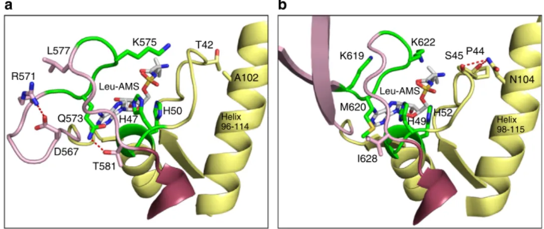
phosphates of the ATP27–29. Once the aminoacyl-adenylate is formed, the KMSKS loop moves into a ‘semi-open’ conformation to allow for the entry of the CCA 30-end of the tRNA into the active site. Finally, once the CCA 30-end of the tRNA is in the active site, the KMSKS loop once again adopts the closed conformation25,27,30. In the case of AgnB2, the highly conserved methionine of the KMSKS motif is interestingly replaced by a glutamine residue (Q573). Our structure shows that Leu-AMS is bound in the active site with the mobile KQSKS loop present in a semi-open conformation (Supplementary Fig. 5). This loop position is in contrast to the fully closed position exhibited by the KMSKS loop in the LeuRSEctRNATM84 ternary complex
where the second lysine of the KMSKS loop (K622) directly interacts with the non-bridging phosphate of the nucleotide analogue and the first lysine of the KMSKS loop (K619) interacts with cytosine 75 of the tRNA 30-end23. In Class I aaRSs, this conserved methionine forms a number of hydrophobic packing interactions and is thought to play an important role in aminoacylation31–34. In LeuRS structures it also forms a main chain interaction with the adenine ring of the bound adenylate25,35. The equivalent position in AgnB2, Q573, forms a hydrogen bond side-chain interaction with the side chain of T581 (Fig. 5a), which is found to be a conserved hydrophobic residue in TM84 sensitive LeuRSs (I628 in LeuRSEc) (Fig. 5b). It is
interesting to note that the AgnB2 KQSKS loop exhibits a distinctly shifted position relative to the Leu-AMP binding pocket (Supplementary Fig. 5), perhaps due to these changes. The fact that a glutamine residue is found in this critical catalytic motif but not in any other LeuRS in nature, may indicate an important role for Q573 in TM84 resistance. It maybe that a substantial number of compensatory mutations are required to accommodate Q573 in AgnB2.
T42 and A102 are two other residues that are specific to AgnB2. T42 is positioned upstream from the catalytic H47IGH50 motif, while the side chain of A102 is situated in close proximity to T42 on a neighbouring a-helix (Fig. 5). Importantly, the presence of A102 in AgnB2, instead of an asparagine as found in the LeuRSEc structure (N104), leads to loss of an H-bond
interaction between the a-helix (96–114) and a main-chain peptide bond located at T42 (P45 LeuRSEc). Both these
modifications could allow for subtle changes in the positioning of the neighbouring HIGH motif that binds TM84.
b a
Leu-AMS Leu-AMS
L577
R571
K575
H47 H50
T42
K619
K622
M620
I628
N104
Helix 98-115 Helix
96-114
Q573
D567 T581
S45 P44
H52 H49 A102
Figure 5 | Interactions of key residues in the catalytic active site of AgnB2.Ribbon structure of the catalytic active site of (a) AgnB2tRNALeuLeu-AMS complex and (b)E. colitRNALeuLeu-AMS complex. AgnB2 key residues interacting with Leu-AMS (shown as white sticks) or adjacent to the catalytic site
AgnB2 has modified tRNA elements compared with LeuRSEc. A prominent feature of the AgnB2 structure is the lack of an idiosyncratic leucyl-specific insertion domain (LS-domain), which is present in bacterial LeuRS, except in a few cases such, as Bacillus subtilis or Mycoplasma mobile25,36, and as seen in the Helicobacter pylori LeuRS (LeuRSHp) primary amino acid
sequence in Fig. 4. The LS-domain undergoes substantial movement between the LeuRSEc editing, and the TM84 and
Leu-AMS bound ‘aminoacylation-like’ conformations, buttressing the highly mobile KMSKS loop as well as guiding the tRNA into the catalytic site, as found in the aminoacylation conformation25. The missing LS-domain in AgnB2 is replaced by a small connecting loop that contains a salt bridge between residues D567 and R571. While the LS domain was shown not to be absolutely required for the aminoacylation or editing reactions of LeuRS per se37, it has been shown to be important for tRNA binding and is able to distinguish between the isoacceptors of tRNALeu in E. coli38. Our thermodynamic studies showed a
B3.5-fold reduction in tRNALeu affinity between LeuRSAt and
AgnB2 (Supplementary Tables 1 and 2), raising the possibility that the modification of tRNA interacting elements in AgnB2 may play a role in TM84 resistance (tested below).
It has been reported that residues distant from the active site can also be crucial in forming interactions with the negatively charged backbone of the tRNALeu CCA 30-end and its repositioning in catalysis38. Mutation of the residue R418 to a neutral amino acid, such as glutamine, decreased the aminoacylation activity in LeuRSEcby reduction of the residence
of the 30-end in the enzyme active site37,38. Importantly, the
AgnB2 structure shows the equivalent residue to R418 in LeuRSEc
is the neutrally charged Q413 (Fig. 4). Thus, it is possible that Q413 in AgnB2 has a negative effect on binding of the tRNA-dependent inhibitor TM84.
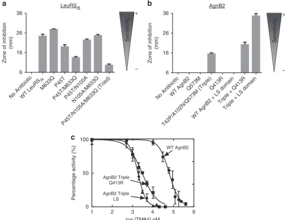
Identification of permissive mutations in the catalytic core. Having identified possible TM84 sensitivity/resistance determi-nants by comparison of the AgnB2 structure with other LeuRSs known to be sensitive to the toxin, we made mutants of AgnB2 and also LeuRSAtto test the contribution of these sites to
anti-biotic resistance. Because of the limited number of possible mutants and permutations that could be constructed using this approach, not all potential TM84 resistance/sensitivity determinants could be tested. We initially assayed candidate mutants using a biological assay (see Methods), followed by in vitro enzyme inhibition experiments combined with assessment of direct binding by calorimetry on promising constructs.
Three of the putative resistance determinants in AgnB2, Q573, A102 and T42 described above (Fig. 5a), reside in the catalytic domain and differ from the highly conserved residues in TM84-sensitive LeuRSs. We introduced these mutations at correspond-ing positions (as determined by homology modellcorrespond-ing) in the TM84-sensitive enzyme LeuRSAt(P45T, N105A and M633Q) and
tested whether these mutations imparted resistance using our in vivoassay (Fig. 6a). The single M633Q mutation in the K(M/ Q)SKS loop is restrictive in nature, as it was not tolerated in the LeuRSAtenzyme, most likely resulting in a functionally inactive
enzyme. In contrast, the P45T (Fig. 6a) or N105A (data not
a
c
LeuRSAt b AgnB2
Sensitivity Sensitivity
No AntibioticWT LeuRS
At
M633QP45T
P45T/M633QP45T/N105A N105A/M633Q
P45T/N105A/M633Q (Triad)
6 16 26 36
Zone of inhibition
(mm)
No AntibioticWT AgnB2 Q573M
T42P/A102N/Q573M (Triple) Q413R
WT AgnB2 + LS domain Triple + Q413R
Triple + LS domain 6
16 26 36
Zone of inhibition
(mm)
1 2 3 4 5 6
0 50 100
log [TM84] nM
Percentage activity (%)
WT AgnB2
AgnB2 Triple Q413R
AgnB2 Triple LS
– +
– +
Figure 6 | Comparison of antibiotic sensitivity of wt and mutant forms of LeuRSAtand AgnB2.A. tumefaciensNTL4 T7pol-Gm-FRT(pYW15d-acsDaccR)
strain transformed with pBBR1MCS-3 plasmids containing genes encoding wt or mutant LeuRSAtor AgnB2 constructs were tested for their sensitivity
towards 50ml of agrocin 84 (60mM) placed in central well of a bioassay plate. For negative control, sterile ddH2O was used instead of the antibiotic.
(a) Bioassay results in graphical form indicating the sensitivity of wt LeuRSAtand LeuRSAtmutants; and (b) the resistance properties of wt AgnB2 and
selected AgnB2 mutants; (c) Comparison of the TM84 inhibition of the aminoacylation reactions of wt AgnB2 (K), AgnB2 Triple Q413R mutant (’) and AgnB2 Triple LS domain mutant (m). Dose–response curves and the resulting IC50values were obtained at saturating leucine and (0.675 mM) ATP
shown) are permissive mutations and had a small effect on enzyme function or TM84 sensitivity. A P45T/N105A double mutant showed no increase in resistance for LeuRSAt. When the
P45T was introduced with M633Q in a double mutant, the P45T acted as a permissive mutation, increasing the resistance of the mutant enzyme. Finally, when all three mutations were introduced into LeuRSAt(P45T/N105A/M633Q, termed LeuRSAt
Triad) there was a significant increase in resistance to the antibiotic (Fig. 6a) compared with wild type (wt).
Kinetic inhibition experiments on LeuRSAtTriad showed the
enzyme was resistant to to concentrations of TM84 up to 500 nM (Supplementary Fig. 6a). This is in contrast to wt LeuRSAt, which
is sensitive to very low concentrations of TM84 (Kiapp¼0.3±0.1
nM)23. The introduction of the Triad mutations also negatively affected the percentage of active enzyme, presumably because these modified residues not only alter engagement of the enzyme with the antibiotic but also modify the interaction of the active site with its substrates. Subsequent thermodynamic analysis of the binding of TM84 to the LeuRSAt Triad alone and the LeuRSAt
Triad pre-incubated with tRNALeushowed respective 10-fold and 28-fold decreases in binding affinity, as compared with LeuRSAt
(Supplementary Table 2).
To see if the three reverse mutations in AgnB2 could make the enzyme more sensitive to TM84, a variant containing all three mutations (T42P/A102N/Q573M termed AgnB2 Triple) was constructed. The AgnB2 Triple showed a distinct increase in TM84 sensitivity (larger clearance zone) as detected by bioassay (Fig. 6b). An individual, Q573M mutant did not increase TM84 sensitivity of AgnB2 (Fig. 6b). Kinetic analysis of the AgnB2 Triple mutant showed a 1.8-fold increase in resistance using a fixed concentration of ligand (Supplementary Fig. 6b). Thermodynamic analysis of the binding of TM84 to the AgnB2 Triple alone showed no significant effect on TM84 binding, while there was a 3.5-fold increase in binding of TM84 to the AgnB2tRNALeucomplex (Supplementary Table 1).
We also tested whether disruption of the H-bond interaction between Q573 of the KQSKS loop and T581 side chain (Fig. 5a) could affect TM84 binding by mutating the threonine residue to a valine. This single T581V mutation, as well as the double mutation (Q573M/T581V) had no detectable effect on TM84 resistance in our bioassays (Supplementary Fig. 7a). However, addition of the T581V mutation to the AgnB2 Triple construct did produce an apparent increase in antibiotic sensitivity (Supplementary Fig. 7a).
A miniloop substitutes for the LS domain in AgnB2 with implications for TM84-resistance and aminoacylation. Since AgnB2 lacks the LS domain and possesses a short peptide with a unique salt bridge between residues D567 and R571 (Fig. 5a), we tested if removal of the salt bridge had any effect on TM84 sensitivity. The salt bridge was disrupted using a number of mutations (D567N, R571E, M and Q), and tested using the bioassays. No significant change in resistance was detected for these mutants compared with wt AgnB2 (Supplementary Fig. 7b). An L577K mutation designed to compete with R571 (Fig. 5) for the salt bridge to the residue D567 produced a clearing zone in our bioassays (Supplementary Fig. 7b) but that is likely due to loss of stability of the enzyme. Taken together, these results indicate that disruption of the D567-R571 salt bridge that replaces the LS domain is not sufficient to cause resistance to TM84, but it may play still play an important structural or canonical catalytic role in AgnB2.
Conversion of a TM84-resistant enzyme into a sensitive form by altering key tRNA interactions. To test whether an LS
domain can have a substantial effect on the TM84 sensitivity of a LeuRS, we examined recombinant LeuRS H. pylori (LeuRSHp),
which lacks an LS domain (Fig. 4). Interestingly, we found using in vitro aminoacylation assays that the LeuRSHp was
still significantly inhibited by TM84 (IC50¼34±1.14 nM;
Supplementary Fig. 8) although to a lesser degree than LeuRSAt
(ref. 23). Interestingly, this enzyme also contains a neutral Q413 residue in a homologous position to R418 from LeuRSEC
is thought to be involved in positioning of the tRNA CCA 30-end in the enzyme active site25,38. We took this one step further, by introducing all three core mutations implicated in TM84 resistance in AgnB2 into LeuRSHp (P40T, N100A and
M573Q). However, ourin vitroaminoacylation analysis revealed that this enzyme was inactive under the conditions used in our study (Supplementary Fig. 8).
To pursue further a possible role of the LS domain in TM84 sensitivity, we inserted the LS domain from LeuRSAtinto AgnB2
LeuRS and tested the chimeric enzyme’s sensitivity to TM84 in our bioassay. In addition, we also inserted the LS domain into the background of the AgnB2 Triple variant (T42P/A102N/Q573M). Our results show that insertion of the LS domain into AgnB2 wt had no effect on antibiotic sensitivity in our bioassays but when the LS domain was inserted into the AgnB2 triple background there was a substantial increase in clearing zone size (Fig. 6b). A comparison of the TM84 inhibition of aminoacylation activity of the AgnB2 Triple mutant þ LS domain construct against wt AgnB2, at a fixed ATP concentration of 0.675 mM ATP (where no substrate inhibition of either enzyme is detected) showed a 34-fold (IC50 of 1.8mM) decrease in the IC50 of the chimeric
mutant compared with wild-type (IC50 of 61.3mM) (Fig. 6c).
ITC analysis of this construct was used to deconvolute the contribution of tRNA to the binding of TM84 to the mutant. Our results showed that TM84 binding alone (Kd¼258 nM) increased
by fourfold (Fig. 7a) and TM84 binding in presence of tRNA (Kd ¼19 nM) increased by 17-fold respectively, compared with
wt AgnB2 (Fig. 7b).
The positively charged R418 residue in LeuRSEc has been
implicated in controlling the translocation of the negatively charged backbone of the tRNALeu CCA 30-end into the synthetic active site25,38. We tested whether modification of the homologous position in AgnB2, Q413, from a neutral to positively charged arginine might modulate TM84 resistance through stabilization of the tRNA-dependent inhibition complex. Introduction of a single-point Q413R mutation into AgnB2 showed no effect on the size of clearing zones in our bioassays compared with wt (Fig. 6b). However, when we placed the Q413R mutation in the background of the AgnB2 Triple mutant (T42P/ A102N/Q573M) there was a substantial increase in the clearing zone diameter suggesting an increase in TM84 sensitivity compared with the AgnB2 Triple (Fig. 6b). Analysis of the TM84 inhibition kinetics of the AgnB2 Triple þ Q413R mutant showed an IC50 of 2.9mM, 21-fold smaller than wt-AgnB2 at a
fixed ATP concentration (Fig. 6c) indicating this construct had a significant increase in TM84 sensitivity compared with wt. We then analyzed the thermodynamics of binding of TM84 to the AgnB2 Triple Q413RtRNALeucomplex and found it was sixfold tighter than wt AgnB2 (Fig. 7b). However, the binding of TM84 to the AgnB2 Triple Q413R enzyme alone was only 1.3-fold tighter than the wt enzyme (Fig. 7a). This further suggested the importance of tRNA interacting residues for imparting sensitivity towards TM84.
Discussion
TM84 (agrocin 84) is a highly potent LeuRS toxin that employs a unique tRNA-dependent inhibition mechanism. During production of agrocin 84, the biocontrol A. radiobacter strain 84 expresses the self-immunity LeuRS AgnB2 to prevent cell suicide. Our data show that AgnB2 achieves resistance to TM84 by minimizing the effect tRNALeuhas on the toxin’s affinity for the enzyme as well as weakening its affinity for tRNALeu and the inhibitor alone. Remarkably, this resistance phenotype is achieved without dramatically affecting the catalysis of the overall aminoacylation reaction (Table 1). Consistent with this observation, the X-ray crystal structure of the AgnB2tRNALeuLeu-AMS ternary complex reveals that residues in the synthetic active site that directly bind Leu-AMP are not substantially different from other bacterial LeuRSs.
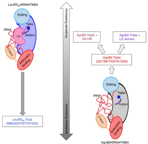
We identified key molecular determinants imparting TM84 resistance (or sensitivity) to LeuRSs by utilizing an approach based on the analysis of structures of the naturally resistant and sensitive enzymes AgnB2 and LeuRSEc (Fig. 8). LeuRS mutants
that affected TM84 sensitivity were then tested usingin vitroand in vivoapproaches. Our data indicate that the molecular elements in LeuRSs that contribute to TM84 sensitivity or resistance are much more complex than one would expecta priori. Surprisingly, some of these determinants were not located directly in the active site but on the periphery (with distances B10–15 Å for most residues and 25 Å for Q413), including some residues interacting with the tRNALeu. The core resistance mutations in the catalytic domain of TM84 resistant AgnB2 are located in KQSKS loop (Q573), just upstream of the HIGH loop (T42) or buttressing next to T42 in the case of A102. It is possible that the nature of the amino acids at these particular sites in LeuRSs may affect the molecular dynamics of these two critical catalytic motifs and ultimately altering the LeuRSs TM84 sensitivity phenotype. The exact contribution of other determinants to TM84 sensitivity at a greater distance from the active site is more difficult to discern. For AgnB2, alterations in tRNA-binding contacts, due to lack of an LS domain, C-term domain modifications, and the effect of Q413 on CCA 30-end localization in the active site, may all play a role in destabilizing a TM84 ternary complex leading to resistance. It maybe that evolution has traversed a narrow
energetic landscape towards a resistance phenotype by exploiting small energetic differences between mutants to produce larger scale changes in only certain kinetic and/or thermodynamic parameters. Presumably, there must be other additional unconserved residues that have played subtle compensatory roles on the pathway towards evolution of TM84 resistance.
Why did nature evolve TM84 to target LeuRSAt and not
another AARSs from A. tumefaciens, and in particular the other Class I AARSs that have similar active site architectures such as IleRS and ValRS. TM84 does appear to be LeuRS specific, as the antibiotic does not inhibit agrobacterial IleRS12, and the single LeuRSagnb2gene is capable of providing agrocin 84-immunity to the pathogen. In silico docking analysis of TM84, from the LeuRSEc structure (PDB: 3ZGZ), into the catalytic domain of
existing IleRS (PDB code: 1JZQ) and ValRS (PDB code: 1GAX) structures reveals steric clashes of the ‘leucine-like’ region of TM84 with hydrophobic side chains in the active site (Supplementary Fig. 9). This analysis may provide some understanding of the LeuRS specificity of TM84. One possible reason that agrocin 84/TM84 evolved to target a LeuRS maybe because leucine is one of the most abundant amino acids in proteins, and so translation could be particularly sensitive to interference of LeuRS activity. A more plausible reason is that agrocin 84/TM84, or its evolutionary ancestor, had other roles in Agrobacteriumbiology that predisposed the toxin to evolve into a LeuRS inhibitor. The recent finding that the uptake molecule of agrocin 84, when cleaved from TM84, can act as signalling molecule binding to a key agrobacterial transcriptional regulator that controls quorum-sensing signal synthesis9provides some support for this latter view.
Horizontal gene transfer of the agnB2 gene on the pAgK84 plasmid fromA. radiobacterstrain K84 to the pathogen threatens emasculation of the biocontrol5,16,17. Genetic engineering of pAgK84 to prevent conjugative transfer of the plasmid to the pathogen has resulted inA. radiobacter K1026 strain39, the first bioengineered microbe approved for release into the environment40. Yet despite not containing any foreign DNA, this genetically altered strain is still only registered for use as a biopesticide in a limited number of countries (Gary Bullard and Allen Kerr, personal communications). In addition, as with all
a b
WT AgnB2
AgnB2 Triple
AgnB2 Triple Q413R
AgnB2 Triple LS 0
250 500 750 1,000 1,250
Dissociation constant
(
Kd
) nM
Dissociation constant
(
Kd
) nM
WT AgnB2
AgnB2 Triple
AgnB2 Triple Q413R
AgnB2 Triple LS 0
100 200 300 400
AgnB2•tRNA AgnB2•tRNA•TM84
TM84 +
AgnB2•TM84 AgnB2
TM84 +
Figure 7 | The accumulative mutation of tRNA interacting elements with core Triple mutations in AgnB2 increases the enzyme’s affinity to TM84.
antibiotic interventions, continued success against resistance can never be completely assured. Thus, understanding the resistance mechanism of agrocin 84 immunity protein AgnB2 not only provides key insights into the mechanism of a fascinating tRNA-dependent toxin but also informs research into the continued crop protection provided by this biocontrol against the spectre of antibiotic resistance.
Methods
Generation of mutants of AgnB2 and LeuRSAt.Mutations in AgnB2 and LeuRSAtwere introduced using the Quikchange site-directed mutagenesis method
(Stratagene). Mutagenesis was carried out in theagnB2gene previously cloned12 into the pET-28a vector (Novagen) for use in recombinant protein expression, and also in theagnB2gene subcloned into the broad host range plasmid pBBR1MCS-3 (ref. 41) for use in bioassay studies. Similarly, LeuRSAt,mutants were generated in
theA. tumefaciens leuSgene cloned into the pET-21b vector (for recombinant protein expression) and pBBR1MCS-3 (bioassays). Multiple rounds of mutagenesis were performed to obtain more than one mutation in an enzyme. DNA sequence analysis was used to confirm the correct construction of the entire gene sequence for all single, and multiple site mutants, used in this study.
Insertion of the LS domain of LeuRSAtinto AgnB2.The LS domain of LeuRSAt
(141 bp) was inserted into AgnB2 (replacing the region between amino acid no. 566 and 572 with the LS domain)37To obtain this, the forward PCR primer was designed to contain the region of AgnB2 (B25 bp) preceding the LS domain insertion followed by the first 25 bases of the LS domain (50–30). Similarly, the reverse primer was designed so as to include the last 25 bp of the LS domain (30–50) and the gene sequence of AgnB2 after the LS domain insertion. An extension reaction was carried out to obtain mega primers, (forward and reverse) encoding the entire LS domain (of LeuRSAt) flanked by the sequence of AgnB2 on both sides
using the protocol by Wang and Malcolm42. DpnI digestion was then carried out to remove the wt template. The resultant mega primer was gel purified using the
Qiaex II gel extraction kit (Qiagen) to be used in the second step. The PCR amplified mega primers (generated in the first step) were then added to the extension reaction in presence of the pET-28a plasmid containing theagnB2gene (using the Quikchange site-directed mutagenesis protocol II). Positive clones encoding AgnB2 with the entire LS domain of LeuRSAtwere identified by colony
PCR and finally confirmed by DNA sequencing analysis. Using a similar strategy, LS domain was added to the AgnB2 Triple mutant as well as the AgnB2 Triple Q413R mutant.
Construction ofA. tumefaciensbioassay strains.A modifiedacsoperon encoding the agrocin 84 transporter system (agrocinopine permease)43–45was cloned into pYW15d plasmid containing a T5 promoter and an antibiotic marker (carbenicillin)46. Importantly, theaccRrepressor gene, responsible for the regulation of the agrocinopine permease47, had been removed from our construction in order to produceA. tumefaciensstrains with maximum agrocin 84 uptake. The pYW15d-acsDaccRplasmid encoding the agrocin 84 transporter and the pBBR1MCS-3 plasmid41containing theagnB2/leuSAtgene and a T7 promoter
and a tetracycline marker were then transformed into an electrocompetent A. tumefaciensNTL4 T7pol-Gm-FRTstrain containing the DE3 lysogen encoding a T7 RNA polymerase48. Briefly, electroporation was performed at 2400 V and 400O
and the culture was then grown in 3 ml LB broth for 2.5 h. TheA. tumefaciens culture was then streaked on LB agar plates containing 100mg ml1of carbenicillin, 15mg/ml of gentamycin and 10mg/ml of tetracycline. The final constructA. tumefaciensNTL4 T7pol-Gm-FRTcontaining both pYW15d-acs DaccR(encoding the agrocin 84 transporter) and a pBBR1MCS-3 plasmid (containing a gene encoding either wt or mutant AgnB2/LeuRSAtproteins was then
used for bioassay analysis. Individual colonies were picked and grown in 3 ml cultures to make glycerol stocks for storage at 80°C.
Protein expression and purification.All enzymes (mutants of AgnB2 and LeuRSAt) were overexpressed inE. coliBL21 (DE3) RIL codon plus cells (Agilent)
(containing pET-28a plasmid encoding C-terminal 6X His-tagged AgnB2 gene or pET-21b plasmid encoding C-terminal 6X histidine tagged LeuRSAtgene).
AgnB2 Triple + Q413R
AgnB2 Triple (Q573M/T42P/A102N)
AgnB2 Triple + LS domain
Editing Editing
TM84
KMSKS LS
tRNA tRNA
TM84
KQSKS C-ter
m
C-ter
m Anticodon
binding
Anticodon binding
Increasing sensitivity
Increasing resistance
LeuRSAt•tRNA•TM84
AgnB2•tRNA•TM84
LeuRSAt Triad
(M663Q/P45T/N105A)
Figure 8 | Model illustrating determinants of TM84 sensitivity and resistance in LeuRSAtand AgnB2.The ternary complex of LeuRSAtforms a tight
binding complex with the CCA 30-end of the tRNA and TM84. Upon mutation of three critical residues, P45, M633 and N105, a TM84 resistant mutant (LeuRSAtTriad) is obtained. AgnB2 on the other hand forms a weak ternary complex with tRNA and TM84, thereby explaining its high level of resistance
towards TM84. Mutagenesis of the corresponding residues in AgnB2 with the residues in LeuRSAtresults in the AgnB2 Triple mutant that has reduced
LeuRS enzymes were purified in a similar manner as detailed previously for LeuRSAt23. In brief, His-tagged proteins were purified by affinity chromatography
using Ni-NTA agarose beads (Qiagen), followed by purification using a strong anion-exchange Mono-Q column (GE Healthcare) by FPLC. All proteins were confirmed to beB95% pure using SDS–polyacrylamide gel electrophoresis analysis and the active concentration determined by active-site titration.
Purification of TM84.Agrocin 84 and TM84 were obtained fromA. tumefaciens NT1 (pAgK84::Tn5A1-B5) agrocin 84 secretion strain grown at 28°C in D-glucose minimal media supplemented with 50mg ml1kanamycin as described previously9,23. Briefly, agrocin 84 and TM84 was separated from other media components by adsorption on charcoal, and then eluted using 70–90% reagent alcohol, dried and both further purified by reverse-phase HPLC. The concentration of TM84 was determined using an extinction coefficient (e¼260 nm) of 0.0154 M1cm1.
Bioassays.In vivoassays to determine the agrocin 84 sensitivity of AgnB2/ LeuRSAtand their mutants were performed. Stationary phase cultures ofA.
tumefaciensNTL4 T7pol-Gm-FRTstrains transformed with both pYW15d-acs DaccR(encoding the agrocin 84 transporter) and the pBBR1MCS-3 plasmids (containing genes encoding the wt or mutant AgnB2/LeuRSAtproteins) were first
diluted 1:100 and then added to 20 ml LB agar plates containing 100mg/ml of carbenicillin, 15mg ml1of gentamycin, 10mg ml1of tetracycline and 1mM IPTG. A well was punched in the centre of the plate and 50ml of 60mM agrocin 84 was added to determine the sensitivity of agrocin 84 towards AgnB2 and LeuRSAt.
The plates were then incubated at 28°C for 48 h. The sensitivity of the strain towards agrocin 84 was determined by measuring the zone of clearance around the well containing agrocin 84. The control strain containing only theacsoperon, but no pBBR1MCS-3 plasmid showed a zone of clearance of around 4 cm. Therefore, any sample having a zone of clearance44.1 cm was considered inactive.
tRNALeutranscription.In this study, we used purifiedin vitrotranscribed tRNALeu(UAA) isoacceptor based on the nucleotide sequence fromA. tumefaciens (referred to in text as agrobacterium tRNALeu) and notA. radiobacter. This allowed a direct comparison ofin vitroaminoacylation kinetics and ITC analysis with our in vivostudies of AgnB2 mutants. Importantly, both tRNALeuisoacceptors from both species share a substantial similarity. Agrobacterium tRNALeuwith (UAA) isoacceptor was obtained by treating plasmids encoding the tRNALeugene with BstNI overnight at 60°C. The linearized DNA was then used as a template in the in vitrotranscription reaction containing 40 mM Tris HCl (pH 8.0), 25 mM MgCl2,
40 mM DTT, 0.1% Triton X-100, 1 mM spermidine and 2 mM rNTPs, 1 U ml1 RNase inhibitor and 9mM T7 RNA polymerase23. After DNase I treatment for removal of template DNA and quenching the reaction with EDTA, the tRNA was precipitated using 0.3 M sodium acetate and absolute ethanol. The tRNA obtained was further purified on a 12% denaturing PAGE gel (19:1) containing 8 M urea and 1X TBE buffer (pH 8.3). Gel extraction of the tRNA was then performed, followed by refolding of tRNA at room temperature using 1 mM MgCl2after incubating
1 min at 80°C (refs 23,49). The concentration of tRNA was determined and corrected by a factor of 1.34 to account for the hypochromic effect on absorbance23.
Active-site titration assay.Active enzyme concentrations of AgnB2, LeuRSAtand
their respective mutants was determined using the method described by Beebe et al.50and Ferst51. In brief, purified enzymes in the range of 1-2mM (as determined by the Bio-Rad Protein assay) were added to a reaction mix containing 20 mM KCl, 10 mM MgCl2, 10 mMb-mercaptoethanol (b-ME), 1 mM L-leucine,
5mg ml1yeast inorganic pyrophophatase (NEB), 5mM ATP and 4mCi [g-32P] ATP. The reaction was initiated at 28°C and 5ml aliquots of the reaction quenched using 7% perchloric acid and 1 M HCl over a time period of 5–30 min. The amplitude of the burst phase of Leu-AMP formation was used to determine the amount of Leu-AMP formed and subsequently the active concentration of the enzymes.
ATP/PPi exchange assay.The formation of Leu-AMP by ATP/PPi exchange assays was performed as described earlier23,50. Briefly, the reaction buffer contained 50 mM HEPES buffer (pH 7.4), 20 mM KCl, 10 mM MgCl2, 500mM L-leucine, 4 mM ATP, 1 mM tetrasodium pyrophosphate (NaPPi), 10mCi/mmol [32P] NaPPi and 1mM bovine serum albumin (BSA). The reaction was initiated by addition of 1–4 nM of active enzyme and incubated at 28°C. Aliquots (5ml each) were collected over 30 min and quenched in a 10% charcoal slurry containing 0.5% HCl and 50ml of 200 mM sodium pyrophosphate in 1 M HCl. Liquid scintillation counting was then used to measure the amount of [32P]-PPi incorporation into ATP that was adsorbed onto the charcoal.
Aminoacylation assay.Aminoacylation activities of AgnB2, LeuRSAtand their
mutants were measured in 50 mM HEPES buffer (pH 7.4) containing 20 mM KCl, 25 mM MgCl2, 25 mMb-ME, 4 mM ATP, 500mM [3,4,5-3H] L-leucine
(20mCi/mmole), 5mg/ml inorganic pyrophosphatase, 1mM BSA, 10mM of active in vitrotranscribed tRNALeuand enzymes in the range of 2–8 nM. Reactions were pre-incubated with or without substrate and/or inhibitor and then carried out at 28°C (37°C-LeuRSHp). Aliquots of the reaction (5–10ml) were quenched on 3 mM
filter pads (Whatman, GE Healthcare) presoaked with 5% trichloroacetic acid (TCA). The pads were washed thrice with 5% TCA and 90% ethanol at 4°C and then dried. The amount of Leu-tRNALeuwas then quantified using scintillation counting. Each reaction was performed in triplicate and analyzed using Kaleidagraph (forKmandkcatdeterminations) or GraphPad Prism software
(for IC50determination).
Isothermal titration calorimetry.The affinity of TM84 binding to AgnB2, LeuRSAtand their mutants was determined using ITC as described earlier23. Binary
interactions (TM84 binding to protein) and ternary interactions (TM84 binding to proteinþtRNA (1:1.2 molar ratio)) were tested using an Auto-ITC200 microcalorimeter (from MicroCal/GE Healthcare). Typically, about 4–8mM active protein solution and a 10-fold higher ligand concentration were prepared in buffer containing 50 mM HEPES (pH 7.4), 20 mM KCl 10 mM MgCl2and 1 mMb-ME.
The titrations were carried out in triplicate at 28°C. The data was then analyzed using Origin software (version 7) and fitted to a one-site or a two-site binding model to obtain the binding affinity (Kd), stoichiometry (N) and enthalpy (DH) for
each complex.
Crystallization.Crystallization was carried out at 20°C by the hanging drop vapor diffusion method. Crystals of the ternary complex AgnB2 LeuRStRNALeu Leu-AMS were obtained by mixing 1ml of a solution containing AgnB2 LeuRS at 43mM, tRNALeu(A. tumefaciens) at 34mM and Leu-AMS at 2 mM; with 1ml of reservoir solution containing 0.1 M MES (pH 6.5), 0.2 M ammonium sulfate and 27% (w/v) PEG 5000 MME. Quality and size of crystals were optimized by macro-seeding with protein at 2 mg ml1. The crystals were frozen in liquid nitrogen after transfer for a few seconds in the mother liquor plus 15% (v/v) ethylene glycol as cryoprotectant.
Structure determination and refinement.The diffraction data sets of the complexAgnB2LeuRS-tRNALeu-Leu-AMS were collected at the European Synchrotron Radiation Facility (ESRF, France). The data sets were integrated and scaled with the XDS suite52. Further data analysis was performed with the CCP4 suite53. The structure was initially solved by molecular replacement with PHASER54using as a model the catalytic and anticodon-binding domains of a model built with SWISS-Model55based on homology to LeuRS
Tt(PDB 2BTE)56
and LeuRSEc(PDB: 4AS1)25. The molecular replacement solution was used to
search with PHASER for AgnB2 tRNALeuusing as a model the core ofE. coli tRNALeu(bases 1–73, except the bases belonging to the long-variable arm). The obtained model was completed by manual placing of the ZN1 domain, leucine-specific domain, C-terminal domain and editing domain. Manual adjustments were done with COOT57and the final model was refined using REFMAC5 (ref. 58). Structure quality was analysed with MOLPROBITY (http://molprobity.biochem.duke.edu/) and showed all residues in allowed regions. Figures were drawn with PYMOL (http://www.pymol.org/).
Molecular modelling.A homology model of LeuRSAtwas built using the Insight II
Molecular Modeling System (www.accelrys.com) based upon the template structure of the LeuRSTt(PDB ID 1H3N)35. A normalized sequence-structure
compatibility score of 0.77 was calculated for the LeuRSAthomology model using
the Verify the three-dimensional module of Insight II59,60.
Data availability.Atomic coordinates and structure factors for the AgnB2tRNALeu-AMS ternary complex have been deposited in the RCSB Protein Data Bank with the accession code 5AH5. All other data associated with this manuscript are available from the corresponding author on reasonable request.
References
1. Walsh, C.Antibiotics : Actions, Origins, Resistance335 (ASM Press, 2003). 2. Wright, G. D. The antibiotic resistome: the nexus of chemical and genetic
diversity.Nat. Rev. Microbiol.5,175–186 (2007).
3. Levy, S. B.The Antibiotic Paradox: How the Misuse of Antibiotic Destroys Their Curative Powers(Perseus Publishing, 2002).
4. Kerr, A. & Htay, K. Biological control of Crown Gall through bacteriocin production.Physiol. Plant Pathol.4,37–44 (1974).
5. Kerr, A. Biological control of crown gall through production of agrocin 84. Plant Dis.64,25–30 (1980).
6. Kerr, A. & Tate, M. E. Agrocins and the biological control of crown gall. Microbiol. Sci.1,1–4 (1984).
7. Roberts, W. P., Tate, M. E. & Kerr, A. Agrocin 84 is a 6-N-phosphoramidate of an adenine nucleotide analogue.Nature265,379–381 (1977).
9. El Sahili, A.et al.A pyranose-2-phosphate motif is responsible for both antibiotic import and quorum-sensing regulation inAgrobacterium tumefaciens.PLoS Pathog.11,e1005071 (2015).
10. Murphy, P. J., Tate, M. E. & Kerr, A. Substituents at N6 and C-5’ control selective uptake and toxicity of the adenine-nucleotide bacteriocin, agrocin 84, in Agrobacteria.Eur. J. Biochem.115,539–543 (1981).
11. Ryder, M. H., Tate, M. E. & Jones, G. P. Agrocinopine A, a tumor-inducing plasmid-coded enzyme product, is a phosphodiester of sucrose and L-arabinose.J. Biol. Chem.259,9704–9710 (1984).
12. Reader, J. S.et al.Major biocontrol of plant tumors targets tRNA synthetase. Science309,1533 (2005).
13. Ryder, M. H., Slota, J. E., Scarim, A. & Farrand, S. K. Genetic analysis of agrocin 84 production and immunity inAgrobacteriumspp.J. Bacteriol.169, 4184–4189 (1987).
14. Kim, J. G.et al.Bases of biocontrol: sequence predicts synthesis and mode of action of agrocin 84, the Trojan horse antibiotic that controls crown gall.Proc. Natl Acad. Sci. USA103,8846–8851 (2006).
15. Farrand, S. K., Slota, J. E., Shim, J. S. & Kerr, A. Tn5 insertions in the agrocin 84 plasmid: the conjugal nature of pAgK84 and the locations of determinants for transfer and agrocin 84 production.Plasmid13,106–117 (1985).
16. Panagopoulos, C. G., Psallidas, P. G. & Alivizatos, A. S. inSoil-Borne Plant Pathogens. (ed. Schiffers, B.a.G.,W.) 569–578 (Academic Press, 1979). 17. Ellis, J. G. & Kerr, A. inSoil-Borne Plant Pathogens. (ed. Schiffers, B.a.G.,W.)
579–583 (Academic Press, 1979).
18. Schimmel, P., Tao, J. & Hill, J. Aminoacyl tRNA synthetases as targets for new anti-infectives.FASEB J.12,1599–1609 (1998).
19. Dewan, V., Reader, J. & Forsyth, K. M. Role of aminoacyl-tRNA synthetases in infectious diseases and targets for therapeutic development.Top. Curr. Chem. 344,293–329 (2014).
20. Hernandez, V.et al.Discovery of a novel class of boron-based antibacterials with activity against gram-negative bacteria.Antimicrob. Agents Chemother.57, 1394–1403 (2013).
21. Hughes, J. & Mellows, G. Inhibition of isoleucyl-transfer ribonucleic acid synthetase inEscherichia coliby pseudomonic acid.Biochem. J.176,305–318 (1978).
22. Hughes, J. & Mellows, G. On the mode of action of pseudomonic acid: inhibition of protein synthesis inStaphylococcus aureus.J. Antibiot. (Tokyo)31, 330–335 (1978).
23. Chopra, S.et al.Plant tumour biocontrol agent employs a tRNA-dependent mechanism to inhibit leucyl-tRNA synthetase.Nat. Commun.4,1417 (2013). 24. Chopra, S. & Reader, J. tRNAs as antibiotic targets.Int. J. Mol. Sci.16,321–349
(2014).
25. Palencia, A.et al.Structural dynamics of the aminoacylation and proofreading functional cycle of bacterial leucyl-tRNA synthetase.Nat. Struct. Mol. Biol.19, 677–684 (2012).
26. Rock, F. L.et al.An antifungal agent inhibits an aminoacyl-tRNA synthetase by trapping tRNA in the editing site.Science316,1759–1761 (2007).
27. Kobayashi, T.et al.Structural snapshots of the KMSKS loop rearrangement for amino acid activation by bacterial tyrosyl-tRNA synthetase.J. Mol. Biol.346, 105–117 (2005).
28. Kuratani, M.et al.Crystal structures of tyrosyl-tRNA synthetases from Archaea.J. Mol. Biol.355,395–408 (2006).
29. Xin, Y., Li, W. & First, E. A. The ’KMSKS’ motif in tyrosyl-tRNA synthetase participates in the initial binding of tRNA(Tyr).Biochemistry39,340–347 (2000). 30. Yaremchuk, A., Kriklivyi, I., Tukalo, M. & Cusack, S. Class I tyrosyl-tRNA
synthetase has a class II mode of cognate tRNA recognition.EMBO J.21, 3829–3840 (2002).
31. Datt, M. & Sharma, A. Conformational landscapes for KMSKS loop in tyrosyl-tRNA synthetases.J. Struct. Funct. Genomics15,45–61 (2014). 32. First, E. A. & Fersht, A. R. Analysis of the role of the KMSKS loop in the
catalytic mechanism of the tyrosyl-tRNA synthetase using multimutant cycles. Biochemistry34,5030–5043 (1995).
33. Hountondji, C., Dessen, P. & Blanquet, S. The SKS of the KMSKS signature of class I aminoacyl-tRNA synthetases corresponds to the GKT/S sequence characteristic of the ATP-binding site of many proteins.Biochimie75, 1137–1142 (1993).
34. Hountondji, C.et al.Crucial role of conserved lysine 277 in the fidelity of tRNA aminoacylation byEscherichia colivalyl-tRNA synthetase.Biochemistry41, 14856–14865 (2002).
35. Cusack, S., Yaremchuk, A. & Tukalo, M. The 2 A crystal structure of leucyl-tRNA synthetase and its complex with a leucyl-adenylate analogue. EMBO J.19,2351–2361 (2000).
36. Li, L.et al.Naturally occurring aminoacyl-tRNA synthetases editing-domain mutations that cause mistranslation in Mycoplasma parasites.Proc. Natl Acad. Sci. USA108,9378–9383 (2011).
37. Yan, W., Tan, M., Eriani, G. & Wang, E. D. Leucine-specific domain modulates the aminoacylation and proofreading functional cycle of bacterial leucyl-tRNA synthetase.Nucleic Acids Res.41,4988–4998 (2013).
38. Tan, M.et al.The Yin and Yang of tRNA: proper binding of acceptor end determines the catalytic balance of editing and aminoacylation.Nucleic Acids Res.41,5513–5523 (2013).
39. Jones, D. A., Ryder, M. H., Clare, B. G., Farrand, S. K. & Kerr, A. Construction of the Tra- deletion mutant of pAgK84 to safeguard the biological control of crown gall.Mol. Gen. Genet.212,207–214 (1988).
40. Clare, B. G., Kerr, A. & Jones, D. A. Characteristics of the nopaline catabolic plasmid inAgrobacteriumstrains K84 and K1026 used for biological control of crown gall disease.Plasmid23,126–137 (1990).
41. Kovach, M. E.et al.Four new derivatives of the broad-host-range cloning vector pBBR1MCS, carrying different antibiotic-resistance cassettes.Gene166, 175–176 (1995).
42. Wang, W. & Malcolm, B. A. Two-stage PCR protocol allowing introduction of multiple mutations, deletions and insertions using QuikChange site-directed mutagenesis.Biotechniques26,680–682 (1999).
43. Kim, H. & Farrand, S. K. Characterization of the acc operon from the nopaline-type Ti plasmid pTiC58, which encodes utilization of agrocinopines A and B and susceptibility to agrocin 84.J. Bacteriol.179,7559–7572 (1997). 44. Hayman, G. T. & Farrand, S. K. Characterization and mapping of the
agrocinopine-agrocin 84 locus on the nopaline Ti plasmid pTiC58.J. Bacteriol. 170,1759–1767 (1988).
45. Hayman, G. T., Beck von Bodman, S., Kim, H., Jiang, P. & Farrand, S. K. Genetic analysis of the agrocinopine catabolic region ofAgrobacterium tumefaciensTi plasmid pTiC58, which encodes genes required for opine and agrocin 84 transport.J. Bacteriol.175,5575–5584 (1993).
46. Wang, Y., Mukhopadhyay, A., Howitz, V. R., Binns, A. N. & Lynn, D. G. Construction of an efficient expression system forAgrobacterium tumefaciens based on the coliphage T5 promoter.Gene242,105–114 (2000).
47. Beck von Bodman, S., Hayman, G. T. & Farrand, S. K. Opine catabolism and conjugal transfer of the nopaline Ti plasmid pTiC58 are coordinately regulated by a single repressor.Proc. Natl Acad. Sci. USA89,643–647 (1992). 48. Kang, Y., Son, M. S. & Hoang, T. T. One step engineering of T7-expression
strains for protein production: increasing the host-range of the T7-expression system.Protein Expr. Purif.55,325–333 (2007).
49. Vu, M. T. & Martinis, S. A. A unique insert of leucyl-tRNA synthetase is required for aminoacylation and not amino acid editing.Biochemistry46, 5170–5176 (2007).
50. Beebe, K., Waas, W., Druzina, Z., Guo, M. & Schimmel, P. A universal plate format for increased throughput of assays that monitor multiple aminoacyl transfer RNA synthetase activities.Anal. Biochem.368,111–121 (2007). 51. Fersht, A. R.et al.Active site titration and aminoacyl adenylate binding
stoichiometry of aminoacyl-tRNA synthetases.Biochemistry14,1–4 (1975). 52. Kabsch, W. Xds.Acta Crystallogr. D Biol. Crystallogr.66,125–132 (2010). 53. Collaborative Computational Project, N. The CCP4 suite: programs for protein
crystallography.Acta Crystallogr. D Biol. Crystallogr.50,760–763 (1994). 54. McCoy, A. J., Grosse-Kunstleve, R. W., Storoni, L. C. & Read, R. J.
Likelihood-enhanced fast translation functions.Acta Crystallogr. D Biol. Crystallogr.61,458–464 (2005).
55. Arnold, K., Bordoli, L., Kopp, J. & Schwede, T. The SWISS-MODEL workspace: a web-based environment for protein structure homology modelling. Bioinformatics22,195–201 (2006).
56. Tukalo, M., Yaremchuk, A., Fukunaga, R., Yokoyama, S. & Cusack, S. The crystal structure of leucyl-tRNA synthetase complexed with tRNALeu in the post-transfer-editing conformation.Nat. Struct. Mol. Biol.12,923–930 (2005). 57. Emsley, P. & Cowtan, K. Coot: model-building tools for molecular graphics.
Acta Crystallogr. D Biol. Crystallogr.60,2126–2132 (2004).
58. Murshudov, G. N., Vagin, A. A. & Dodson, E. J. Refinement of macromolecular structures by the maximum-likelihood method.Acta Crystallogr. D Biol. Crystallogr.53,240–255 (1997).
59. Ginalski, K., Elofsson, A., Fischer, D. & Rychlewski, L. 3D-Jury: a simple approach to improve protein structure predictions.Bioinformatics19, 1015–1018 (2003).
60. Luthy, R., Bowie, J. U. & Eisenberg, D. Assessment of protein models with three-dimensional profiles.Nature356,83–85 (1992).
Acknowledgements
Author contributions
S.Ch. and J.S.R performed project design and the data interpretation, and prepared the manuscript. S.Ch. also conducted the biochemical and biophysical experiments,in vivo mutant constructions and bioassays as well as data analyses. A.P. and S.Cu. solved the structure of the AgnB2 protein ternary complex, provided experimental suggestions and assisted in editing and figures to the final manuscript. C.V. developed the agrobacterial in vivoscreen and constructed and assayed initial mutants for this project. S.S. developed AgnB2 and LeuRSHpprotein expression protocols and performed initial biochemical
experiments for project. B.T. assisted with structural interpretation of the data and sug-gestion of mutants to be tested, preparation of figures and assistance in editing manuscript.
Additional information
Supplementary Informationaccompanies this paper at http://www.nature.com/ naturecommunications
Competing financial interests:The authors declare no competing financial interests.
Reprints and permissioninformation is available online at http://npg.nature.com/ reprintsandpermissions/
How to cite this article:Chopra, S.et al.Structural characterization of antibiotic self-immunity tRNA synthetase in plant tumour biocontrol agent.Nat. Commun.
7,12928 doi: 10.1038/ncomms12928 (2016).
This work is licensed under a Creative Commons Attribution 4.0 International License. The images or other third party material in this article are included in the article’s Creative Commons license, unless indicated otherwise in the credit line; if the material is not included under the Creative Commons license, users will need to obtain permission from the license holder to reproduce the material. To view a copy of this license, visit http://creativecommons.org/licenses/by/4.0/