Stem Cell Reports
Article
Long Noncoding RNA Moderates MicroRNA Activity to Maintain
Self-Renewal in Embryonic Stem Cells
Keriayn N. Smith,1Joshua Starmer,1Sarah C. Miller,1Praveen Sethupathy,1and Terry Magnuson1,* 1Department of Genetics, University of North Carolina, Chapel Hill, NC 27599, USA
*Correspondence:[email protected] http://dx.doi.org/10.1016/j.stemcr.2017.05.005
SUMMARY
Of the thousands of long noncoding RNAs expressed in embryonic stem cells (ESCs), few have known roles and fewer have been func-tionally implicated in the regulation of self-renewal and pluripotency, or the reprogramming of somatic cells to the pluripotent state. In ESCs,Cyranois a stably expressed long intergenic noncoding RNA with no previously assigned role. We demonstrate thatCyrano contrib-utes to ESC maintenance, as its depletion results in the loss of hallmarks of self-renewal. Delineation ofCyrano’s network through tran-scriptomics revealed widespread effects on signaling pathways and gene expression networks that contribute to ESC maintenance.Cyrano shares unique sequence complementarity with the differentiation-associated microRNA,mir-7, andmir-7overexpression reduces expres-sion of a key self-renewal factor to a similar extent asCyranoknockdown. This suggests thatCyranofunctions to restrain the action of mir-7. Altogether, we provide a view into the multifaceted function ofCyranoin ESC maintenance.
INTRODUCTION
Pluripotent stem cells hold significant therapeutic poten-tial in the context of degenerative disease. To use pluripo-tent cells in transplantation therapies, a thorough under-standing of the molecular mechanisms that regulate immortality through self-renewal becomes a key require-ment. The specialized cells that arise from pluripotent cells during development do so through temporal restrictions in their cellular plasticity. The blueprint behind this cell-fate determination is the transcriptome, whose status is based upon regulatory networks consisting of epigenetic machin-ery, transcription factors, and noncoding RNAs (ncRNAs). Although well studied, protein-coding sequences ac-count for only approximately 2% of the genome and 28%–40% of the transcriptome in humans (Alexander et al., 2010; Harrow et al., 2012). This suggests that non-protein-coding RNAs may have heretofore unidentified functions. Indeed, it has been demonstrated that ncRNAs are abundant regulatory components of vertebrate tran-scriptomes. This is particularly evident for long noncoding RNAs (lncRNAs; >200 nt long) and the noncoding class of small RNAs termed microRNAs (miRNAs), which influence numerous biological processes including proliferation, apoptosis, and differentiation.
Mechanistically, lncRNAs have emerged as multifaceted regulators of various cellular processes, with roles that include influencing epigenetic landscapes, transcriptional circuitry, and post-transcriptional regulatory processes (Rinn and Chang, 2012; Wang and Chang, 2011). While lncRNAs generally have no significant open reading frame, many share characteristics of mRNAs such as 50capping, splicing, and polyadenylation (Cabili et al., 2011; Guttman et al., 2010). Typically, tissue-specific expression of
lncRNAs is more demarcated than that of mRNAs, and it follows that several lncRNAs have been implicated in organ development and cell-fate specification (Fatica and Boz-zoni, 2014). These data point to the need for the elucida-tion of lncRNA funcelucida-tion in both specialized and unspecial-ized cell types.
To date, thousands of lncRNAs have been identified through transcriptomics, particularly RNA sequencing (RNA-seq), in embryonic stem cells (ESCs) (Cabili et al., 2011; Guttman et al., 2010), yet well-defined biological functions are known for very few. However, loss-of-func-tion approaches can provide insight into roles for lncRNAs in the maintenance of self-renewal and pluripotency, re-programming, and differentiation (Guttman et al., 2011; Kelley and Rinn, 2012; Kim et al., 2015; Lin et al., 2014; Loewer et al., 2010). Such functional characterization would address precise roles for individual lncRNAs in pluripotent stem cell maintenance.
Cyrano (linc-oip5, 1700020I14Rik) is a long intergenic ncRNA (lincRNA) transcribed in mouse ESCs (Chew et al., 2013; Guttman et al., 2010; Ulitsky et al., 2011) that was first characterized in zebrafish (Ulitsky et al., 2011). In zebrafish, it is a key regulator that functions in brain, eye, and nasal development (Ulitsky et al., 2011). This is at least in part mediated by a short region of high sequence conser-vation among vertebrate genomes that is critical for func-tion. Rescue experiments in zebrafish utilizing higher-order orthologs provided the first insight thatCyranomay have a functional role in mice (Ulitsky et al., 2011).
repress gene expression at the post-transcriptional level (Bartel, 2009). More recently, broader miRNA functionality has been recognized. This includes noncanonical binding to non-30 UTR regions including the coding sequence of target genes, as well as cross-regulatory interactions that exist between miRNAs and lncRNAs to affect either miRNA or lncRNA stability and/or function, and the regulation of downstream targets (Jeggari et al., 2012; Paraskevopoulou et al., 2013).
One such lncRNA/miRNA interaction has been postu-lated betweenCyrano andmir-7 (Ulitsky et al., 2011). At the cellular level,mir-7 is associated with differentiation (Cui et al., 2013; Kong et al., 2012; Nguyen et al., 2010), with its levels increasing during neural specification from neural stem cells (Cui et al., 2013). In various cellular con-texts, it acts by inhibiting receptor-mediated signaling pathways, including EGFR and STAT3 signaling, to pro-mote differentiation and modulate cellular adhesion (Kefas et al., 2008; Nguyen et al., 2010; Tazawa et al., 2012; Zhang et al., 2014). Antagonism ofmir-7function, mediated by sequestration and inactivation via molecular sponges or decoy RNAs, is a well-known strategy for moderating its ac-tivity on target transcripts. One of the best-studied exam-ples is the circular RNACD1Ras/CiRS-7, which possesses multiple seed matches tomiR-7(Hansen et al., 2013; Mem-czak et al., 2013). Sponge-based regulation of miRNA activ-ity is also employed in the ESC regulatory landscape to pre-vent post-transcriptional degradation of key pluripotency factors includingOct4,Sox2, andNanog(Wang et al., 2013). Here, we demonstrate thatCyranois essential for mainte-nance of self-renewing ESCs. Our studies revealed that interplay betweenCyranoandmir-7affects key properties including cell adhesion in colony maintenance to support ESC immortality. Importantly, Cyranodepletion disrupts
self-renewal signaling and gene expression regulatory net-works, particularly the expression of Nanog. Aberrations in these properties including the loss ofNanogexpression, cell adhesion, and colony survival to maintain self-renewal capacity are recapitulated inmir-7gain-of-function experi-ments. This supports the existence of a competing relation-ship betweenmir-7andCyranoin ESCs.
RESULTS
lncRNACyranoExhibits Stability and Is Broadly
Localized in ESCs
While lncRNAs exhibit a range of localization patterns (Cabili et al., 2015), their basic localization provides pre-liminary insight into their cellular functions. For instance, nuclear-domain localized lncRNAs, including Xist and
Kcnq1ot1, function to silence vast chromatin domains, while the cytoplasmic lncRNAH19is a primary miRNA pre-cursor (Cai and Cullen, 2007; Keniry et al., 2012).
To characterize the function ofCyranoin ESCs, we first used single-molecule fluorescence in situ hybridization (smFISH) and fractionation methods to examineCyrano’s subcellular localization. Through z-stack imaging of multi-ple ESC lines, smFISH revealed distinct signals in the nucleus and cytoplasm throughout ESC colonies (Figures 1A andS1A–S1D), averaging40 molecules per cell ( Fig-ure 1B). This distribution was confirmed by subcellular fractionation comparing the localization of splicedCyrano
to the unspliced nuclear form, cytoplasmic H19 (Keniry et al., 2012) and nuclear speckle-localized Malat1 ( Miya-gawa et al., 2012) (Figure 1C). Furthermore, ENCODE data from human ESCs (ENCODE Project Consortium, 2012; Yue et al., 2014) showed similar subcellular
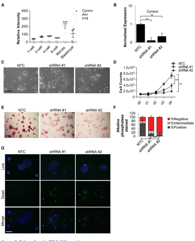
Figure 1. CyranoDisplays Dispersed Sub-cellular Localization and Exhibits Stabil-ity in ESCs
(A) smFISH analysis of a representative ESC colony shows Cyrano localization in the nucleus and cytoplasm of ESCs. Nuclei, blue (DAPI). Scale bar, 10mm.
(B) Quantitation ofCyranomolecules/ESC. (C) Subcellular fractionation and qRT-PCR confirms Cyrano’s presence in the nucleus and cytoplasm.
(D) Assessment of the stability ofCyrano. Data are from three independent experi-ments. Error bars represent SEM. See also
localization of the unspliced and spliced human ortholog,
OIP5-AS1(Figure S1E). Consistent with the lack of enrich-ment in either cellular compartenrich-ment (Clark et al., 2012; Tani et al., 2012), we found thatCyranodisplayed moderate stability of t1/26 hr in ESCs (Figure 1D).Cyrano’s distribu-tion in the cell could indicate that nuclear and cytoplasmic
Cyrano pools have distinct functions or that it interacts with proteins that shuttle from the nucleus to the cytoplasm.
CyranoIs Required for Maintenance of ESC Self-Renewal
In addition to consistent localization and expression among ESC lines, microarray analysis of early embryonic developmental stages (Xie et al., 2010) revealed an increase inCyrano expression in morulae and blastocysts relative to two well-studied lncRNAs, H19 and Airn (Figure 2A). Similar results were obtained upon examination of single-cell RNA-seq data (Deng et al., 2014) from ESCs ( Fig-ure S2A). ESCs are derived from blastocysts and are an excellent model system for early developmental processes. We used short hairpin RNA (shRNA) knockdown (KD) to examine the effect of loss ofCyranoexpression in ESCs. Independent shRNAs reproducibly achieved greater than 60%–85% reduction ofCyranolevels (Figures 2B,S2B, and S2C) compared with a nontargeting control (NTC). Within 2–3 days of expression of shRNAs, we found thatCyrano -depleted cells were unable to robustly maintain a typical ESC phenotype of tightly packed cells assembled in a dome-shaped colony in leukemia inhibitory factor (LIF)-containing ESC medium (Figure 2C). Because of possible shortcomings in KD efficiency due to multiple lncRNA splice variants, differential subcellular localization, and possible nontargeting of shRNAs, the loss-of-self-renewal phenotype was confirmed using additional shRNAs ( Fig-ures S2C and S2D) and in an independent ESC line ( Fig-ure S2E). Specifically, we observed increased numbers of cells floating in the medium and prominent partitioning of cell-cell contacts. This is unlike the standard pluripotent ESC state in which cell-cell boundaries within a colony are difficult to define. Along with this breakdown in the colony maintenance of self-renewing ESCs, we observed a sharp reduction in cell numbers in Cyrano-depleted cells ( Fig-ure 2D). Loss of self-renewal was further underscored by a qualitative and quantitative loss of alkaline phosphatase activity (Figures 2E, 2F, S2D, and S2E) and increases in cell death (Figure 2G). Altogether, these data indicate that
Cyranois required for maintenance of ESC self-renewal.
The ESC Gene Expression Signature Is Disrupted by CyranoDeficiency
Pluripotent ESCs are characterized by a well-documented gene expression profile that supports their ability to
self-renew while maintaining the capacity for differentiation into embryonic germ layer derivatives (Boyer et al., 2005). We next examined modifications in gene expres-sion profiles uponCyranoKD to determine whether cells assumed a particular cellular identity upon Cyrano loss. Total read number in RNA-seq experiments for samples ranged from 23 to 29 million reads, with mapped reads ranging from 88% to 90% of total read number. Concom-itant with morphological anomalies, RNA-seq uncovered significant differential gene expression due to shRNA-mediated KD (Figure 3A). Sample comparisons revealed 489 and 380 up- and downregulated genes, respectively, between control and KD samples (Figure 3A). Validation of differentially expressed non-pluripotency-related genes was carried out by qRT-PCR (Figure S3A). Ingenuity Pathway Analysis (IPA) classification revealed that the top dysregulated pathways post KD were primarily related to cell adhesion, signaling, and motility (Figure 3B), obser-vations consistent with the loss-of-function phenotype. Based on these results and phenotypic observations ( Fig-ure 2C), we further assessed anomalies in cell adhesion by examining localization of E-cadherin (Figure 3C) and F-actin (Figure S3B) in immunofluorescence assays. E-cad-herin is a key pluripotency cell-signaling modulator ( Red-mer et al., 2011) that typically mediates cell-cell adhesion and is normally found at cell-cell boundaries in ESC colonies. We observed aberrant localization of cell membranous E-cadherin onCyranodepletion (Figure 3C). Furthermore, phalloidin staining (Figure S3B) revealed a loss of F-actin localization as is typically seen at the cell cortex in ESCs (Schratt et al., 2002). In addition to anomalous cell and colony morphology, this unchar-acteristic localization of qualitative markers, whose local-ization is normally indicative of self-renewing ESC col-onies, further indicates atypical cell adhesion in colony maintenance.
CyranoDepletion DisruptsNanogExpression
We next rank-ordered genes that showed a decrease in expression (false discovery rate [FDR] < 0.05). As expected, the reduction in Cyrano expression was most significant using this threshold (Figure 4A). Interestingly, the master pluripotency regulatorNanogwas one of the more signifi-cantly downregulated genes (Figure 4A). Further examina-tion of expression of other key factors in self-renewal main-tenance (Xu et al., 2014) includingOct4, revealed that they remained mostly unchanged while there was a decrease in
Nanoglevels (Figures 4B,S4A–S4D). The specific downregu-lation ofNanogand not other master regulators, including
Figure 2. CyranoDeficiency Impairs ESC Self-Renewal
(A) Expression analysis (GEO: GSE18290) ofCyrano,Airn, andH19lncRNAs in early development.
(B) qRT-PCR shows significant reduction inCyranoexpression upon KD using independent shRNAs, compared with a nontargeting control. Experiments were performed in triplicate, normalized toGAPDH, with error bars representing 95% CI.
(C)CyranoKD results in loss of the ESC characteristic colony morphology. Scale bar, 100mm.
(D) KD ofCyranoresults in a decrease in cell numbers as determined by cell counts beginning with plating on day 1 post transfection. (E and F) Significant reduction in alkaline phosphatase staining of ESC colonies afterCyranoKD. n > 200. Scale bar, 100mm.
(G) Increases in cell death observed uponCyranoKD 2 days post transfection. Nuclei (blue, live; green, dead). Scale bar, 200mm. Counts were performed in triplicate with error bars representing SD. Data are from three independent experiments. **p < 0.01. See also
Aberrant Expression of Anti-Self-Renewal Factors Is
Associated withCyranoDepletion
The phenotype uponCyrano loss led us to more closely examine genes that showed significant increases in expres-sion. This revealed key anti-pluripotency genes, including developmental regulators not associated with the ESC state. These include lineage specification markers (Otx2,
Nestin, Gata4, Pdgfra, and Sox11; Figures 4C–4G) and the epithelial-mesenchymal transition factor,Snai1 ( Fig-ure 4H). Furthermore, we observed increases in Apela
expression (Figure 4I), a recently described regulatory RNA with the ability to encode a small peptide hormone (Chng et al., 2013; Li et al., 2015b; Pauli et al., 2014). The upregulation ofApelais consistent with it being a genomic target of Nanog, whose expression increases upon RNAi-mediated depletion of Nanog (Loh et al., 2006), and its role in mesendoderm specification in zebrafish (Chng et al., 2013).Apelaexpression abruptly increases upon LIF withdrawal in mouse ESCs (Figure S4E), consistent with the reduced capacity to differentiate into mesoderm and
Figure 3. Cyrano Depletion Results in Aberrant Gene Expression in ESCs
(A) Smear plot comparing gene expression in NTC and KD samples reveals significant gene expression changes 3 days post KD. Differentially expressed genes are indicated in red, compared with insignificant genes in black (FDR < 0.05).
(B) IPA analysis of differentially expressed genes shows an enrichment of pathways afterCyranoKD.
(C) Immunofluorescence examination of E-cadherin in control and KD cells, showing aberrant localization. Nuclei, blue (DAPI); Scale bar, 100mm.
Figure 4. Depletion ofCyranoResults in Altered Expression ofNanogand Lineage-Related Genes
(A) Ranking of the top-10 decreased genes in RNA-seq based on FDR (black bars) shows significant decreases inNanoglevels withCyrano
KD. Fold change is also indicated (white bars).
(B) Assessment of pluripotency regulators onCyranoKD reveals thatNanogdisplays the most significant differential regulation.
endoderm in embryoid bodies with Apela depletion (Li et al., 2015b). As Apela has been recently shown to be important for self-renewal maintenance in human ESCs (Ho et al., 2015), the rapid upregulation may represent a transition from naive to primed pluripotency prior to spon-taneous differentiation. Consistent with this, subsequent downregulation ofApelais seen later in mouse ESC differ-entiation (Figure S4E), similar to the decrease observed in human ESC differentiation (Chng et al., 2013). These re-sults suggest that deviations from idealApelalevels antago-nize maintenance of the ESC state.
The constitutive cell cycle of ESCs, which lacks the peri-odicity of differentiated cell types, is associated with a lack of cyclin D regulation (White and Dalton, 2005). Consis-tent with the phenotypic change and decreased prolifera-tive capacity, we observed an increase in levels of the cyclin D-Cdk inhibitor,Cdkn2a, inCyrano-deficient cells relative to controls (Figure 4J).
Taken together, these gene expression alterations support the failure to retain self-renewal capacity upon Cyrano
depletion. The function ofCyranodoes not appear to pri-marily be via proximalcismechanisms, as no significant gene expression changes were observed in RNA-seq (FDR < 0.05) for neighboring genes within a 100-kb window (Figure S4F), and consistent changes were not observed for both shRNAs in qRT-PCR analyses (Figure S4G).
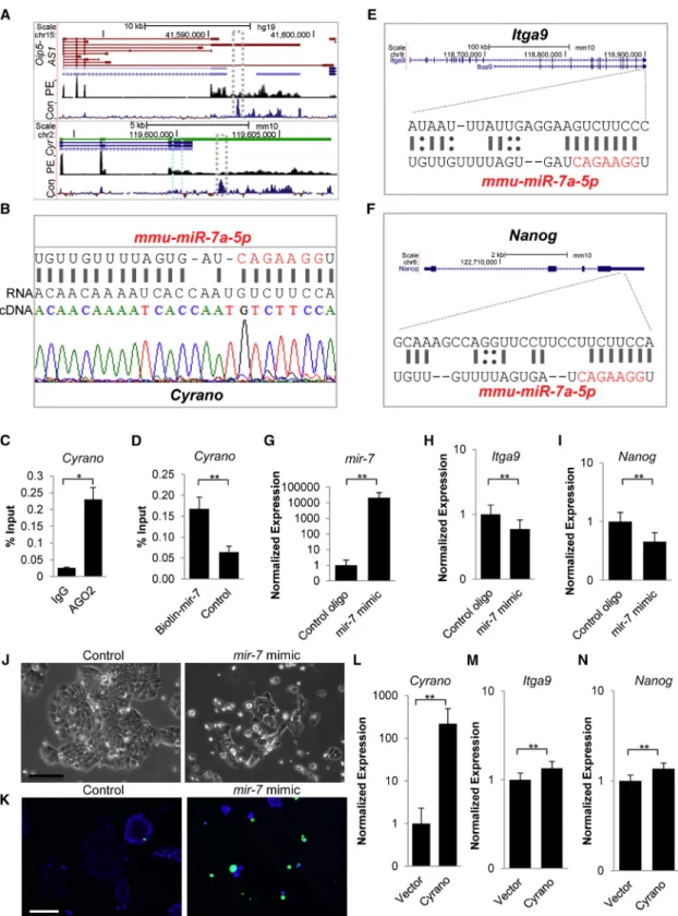
CyranoCounteractsmir-7Action in ESCs to Support Self-Renewal Maintenance
We sought to determine howCyranomediates these survival, adhesion, and anti-differentiation roles in ESCs, and hy-pothesized thatCyranofunctions were at least partially deter-mined through its unique relationship withmir-7(Ulitsky et al., 2011). Several lines of evidence support this hypothe-sis. (1) In addition to a conventional seed match (Figure S5A andTable S1), the prominently expressed long splice variant (Figure 5A) ofOip5-AS1/Cyrano(Sigova et al., 2013) possesses an almost complete binding site tomir-7in mouse ESCs ( Fig-ure 5B) and this binding site is conserved across vertebrates (Figure 5A andUlitsky et al., 2011). (2) Similar to previous ob-servations in mouse brain (Zhang and Darnell, 2011) and HEK293 (Kishore et al., 2011),Cyranoor its human ortholog is bound by Argonaute in ESCs (Figure 5C), similar to the known mir-7 target gene Igf1R (Figure S5B) (Jiang et al., 2010). (3)mir-7is associated with differentiation in various cell types (Cui et al., 2013; Kong et al., 2012; Nguyen et al., 2010). (4)mir-7is a documented Stat3 pathway antagonist
(Zhang et al., 2014) and the LIF-Stat3-Nanog axis is required for mouse ESC self-renewal. (5) Similar to previous observa-tions from Bartel and colleagues, we found thatmir-7and
Cyranophysically interact (Figure 5D), as observed for the
mir-7targetIgf1R(Jiang et al., 2010) (Figure S5C), suggesting that this molecular interaction could be a mechanism for
Cyranoregulation ofmir-7function. Indeed, small RNA-seq data from ENCODE indicated thatmir-7is expressed and localized similarly toCyranoin ESCs (Figure S5D). Further-more, nuclear enrichment ofmir-7has been found in addi-tional contexts including HEK293 and human carcinoma cell lines (Liao et al., 2010), similar to that observed for
Cyrano(Figures 1A, 1C, andS1A–S1E).
We then determined whether mir-7 could affect ESC maintenance by downregulating key adhesion and self-renewal regulators. First, we used mirWalk (Dweep and Gretz, 2015) (Figure S5E andTable S2), TargetScan (Agarwal et al., 2015; Lewis et al., 2003, 2005), and RNA22 (Miranda et al., 2006), and found that the 30UTR of the ESC-enriched integrinItga9(Nagano et al., 2008; Rugg-Gunn et al., 2012) (Figure 5E) contains a strong 8mer mir-7 seed sequence. Interestingly, examination ofNanog’s 30UTR also revealed a 7mer-A1mir-7seed match (Figure 5F). Second, we trans-fected ESCs with amir-7mimic (Figures 5G,S5F, and S5G) and observed a decrease in Itga9 (Figure 5H) andNanog
levels (Figure 5I). Third, as the mir-7-Nanog interaction was based on a weaker prediction, we introduced the
mir-7 mimic along with a luciferase reporter plasmid constituting of theNanog30UTR together with a transfec-tion control and observed inhibitransfec-tion of luciferase expres-sion (Figure S5H). Also, inhibition of mir-7 resulted in increased Nanog levels (Figure S5I). We also found that
Cyrano levels decreased upon introduction of the mir-7
mimic, suggesting that mir-7 also regulates Cyrano in ESCs (Figure S5J). Importantly, when we transfected ESCs with themir-7mimic and monitored the ESC phenotype in LIF-containing medium, we observed a loss of ESC col-ony maintenance within 2 days ofmir-7introduction ( Fig-ure 5J). This was accompanied by anomalies in cell and col-ony adhesion with numerous detached cells (Figure 5J) and increases in cell death (Figure 5K), similar to observations made uponCyranodepletion. Finally, asItga9andNanog
levels decreased upon mir-7 overexpression as well as uponCyranodepletion (Figures S5K andS4B–S4D), we hy-pothesized thatItga9andNanoglevels would increase with modulation of Cyrano levels and found that increased expression of Cyrano (Figure 5L) augmented their
(C–J) UCSC genome browser plots shows RNA-seq reads mapped to mm9 and normalized to remove sequencing depth biases, and inde-pendent qRT-PCR examination uponCyranoKD indicates increased expression of factors that antagonize self-renewal in mouse ESCs. Data are from three independent experiments with error bars representing 95% CI. *p < 0.05, **p < 0.01.
Figure 5. CyranoRestrainsmir-7Activity to Support ESC Self-Renewal
(A) Genome browser blots showingOip5-AS1/Cyranosplice variants. Despite the presence of multiple splice variants ofOip5-AS1, the long variant containing the conservedmir-7binding site (gray box; segment of this region with themir-7interaction site is expanded in B) is prominently expressed in human ESCs (top panel, GEO: GSE41009) and mouse ESCs (bottom panel, GEO: GSE36799). Blue box marks
expression (Figures 5M and 5N). This suggests thatCyrano
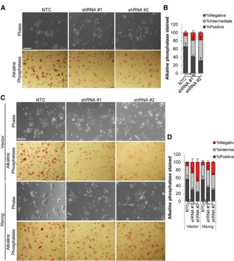
andmir-7act in opposition to one another in ESCs. It is now well established that compared with culture in serum + LIF, mouse ESCs can be cultured more homoge-nously in the presence of GSK3 and MEK inhibitors (2i,
Ying et al., 2008). Due to the heterogeneity observed in serum + LIF, we also tested the effect ofCyrano deple-tion under condideple-tions of more homogeneity. Under 2i conditions, Nanog expression is somewhat higher than that of growth conditions in serum-containing medium (Abranches et al., 2014), whereasCyrano’s levels remain un-changed (Figure S6). To further delineate whetherCyrano
functions upstream ofNanog, we examined the effect of
Cyrano KD in ESCs under conditions of higher Nanog
expression: (1) cells cultured in 2i and (2)Nanog overex-pression. Similar to observations in serum-containing LIF medium, KD ofCyranoin 2i resulted primarily in a loss of self-renewal maintenance, and increasedNanogexpression resulted in partial recovery of self-renewal capacity (Figures 6A–6D). This suggests that additional mechanisms of
Cyranoaction are functional in ESC maintenance. Delinea-tion ofCyranofunction can be further enhanced through single-cell analyses of ESC lines harboring endogenously encoded reporters and mutant alleles ofCyranoand puta-tive direct and indirect targets.
Altogether, we provide evidence forCyrano’s role in cell survival and colony maintenance in self-renewing ESCs. Furthermore, our results provide evidence for one direct mechanism by whichCyranofunctions, namely the exis-tence of a negative-feedback loop between Cyrano and
mir-7(see Graphical Abstract), to support the maintenance of ESC self-renewal through factors includingNanogand
Itga9that sustain key properties of pluripotent cells.
DISCUSSION
Of the nearly 9,000 known lncRNAs (Derrien et al., 2012; Harrow et al., 2006, 2012), the mechanism of action is
understood for only a small fraction. We have described a previously undefined role for Cyrano in the mainte-nance of the self-renewing state of mammalian pluripo-tent cells, a model cell type for establishing mechanisms of action relevant to early development and regenerative medicine.
In our characterization ofCyranofunction, we show that depletion of Cyranoresults in disarray in the pluripotent gene expression signature and defects in self-renewal maintenance. Driving this is aberrations in colony survival and preservation, which requires maintenance of cellular adhesion and signaling, as well as the decrease inNanog
expression, which itself is required for cell growth and apoptosis avoidance in ESCs (Chen et al., 2012). Further-more, it is well established that Nanog function has far-reaching implications in the repression of negative regula-tors of the ESC state, such asDkk1andGata6(Loh et al., 2006; Singh et al., 2007). Mining gene expression data pro-duced in a recent shRNA screen for lncRNAs that regulate pluripotency further supportsCyrano’s key role (Lin et al., 2014).
Misregulation and aberrant expression of lncRNAs are increasingly associated with disease states (Wapinski and Chang, 2011; Batista and Chang, 2013; Fatica and Bozzoni, 2014). Similar signaling pathways (e.g., Jak-Stat, PI3K/AKT) and transcription factors (e.g., Myc, Stat3) activities sup-port stem cell growth and survival as well as tumorigenesis (Kim et al., 2010). As Cyrano displays particularly high expression in ENCODE cancer cell lines, a priority will be to determine the requirement ofCyranoin tumor cell sur-vival and cellular reprogramming to a malignant state. Indeed, similar to our findings in ESCs, Cyrano supports glioma cell proliferation in addition to cell migration and tumorigenesis (Hu et al., 2017).
Complex relationships exist between miRNAs and lncRNAs in transcriptional, post-transcriptional, and trans-lational regulatory processes. Previous studies have shown that lncRNAs can function as miRNA precursors and miRNA targets, or compete as decoys/sponges/competing
additional conventionalmir-7seed sequence. Con denotes conservation: Vertebrate Multiz Alignment & Conservation; PE, paired-end RNA-seq; Cyr,Cyrano.
(B) Near-completemir-7sequence complementarity is observed inCyranosequenced from ESCs.
(C) RNA immunoprecipitation indicates thatCyranois bound by Ago2 in ESCs. Experiments were performed in triplicate with error bars representing SEM.
(D) miRNA pull-down and qPCR indicates a physical interaction betweenCyranoandmir-7. Experiments were performed in triplicate with error bars representing SEM.
(E and F) The 30UTRs ofItga9andNanogcontainmir-7target sites (seeFigure S5;Tables S1andS2for further information on target sites).
(G–N) Overexpression ofmir-7(G) results in a decrease inItga9(H) andNanoglevels (I), loss of ESC self-renewal maintenance (J), and increased cell death (K) in ESCs 2 days post transfection. Nuclei (blue, live; green, dead); Scale bar, 100mm. qRT-PCR monitoring ofCyrano
endogenous RNAs to prevent inhibition of mRNA targets (Jeggari et al., 2012; Paraskevopoulou et al., 2013). In the context of skeletal muscle differentiation,H19antagonizes
let-7, releasing the repression oflet-7targets such asHMGA2
(Kallen et al., 2013). Similarly, if Cyrano has a negative influence on mir-7 function, it would be expected that depletion ofCyranowould freemir-7to repress its targets, while overexpression ofCyranowould boostmir-7 target-transcript levels, consistent with our observations.
Moreover, the combination of multiple sites for bind-ing to Cyrano, including the unique ultra-conserved binding site, along with the intermediate expression of
mir-7 in ESCs (Tang et al., 2006), suggests that it is feasible forCyrano to attenuate mir-7 activity to sustain self-renewal. This is particularly relevant for supporting the maintenance of expression of targets such asNanog, which display some heterogeneity in expression in ESCs (Abranches et al., 2014; Singh et al., 2007). While
Nanogtranscripts range from 0 to 500 mRNA molecules per ESC in normal culture conditions, a fraction of pluripotent cells exhibit low Nanog expression in the range of <25 molecules/cell (Abranches et al., 2014).
One hypothesis is thatCyranois one factor that presents a barrier to lineage commitment in conditions of subop-timal fluctuations in Nanog levels. Additionally or alter-natively,Cyrano may also directly repress lineage specifi-cation factors. Further studies may reveal mechanisms behind the molecular interplay between Cyrano and its interacting RNAs at the single-molecule and single-cell levels.
Based on our and previously available data, we postulate that regulating RNAs and proteins through interactions is a prominent role of the 8.2-kbCyrano molecule. Indeed, the ortholog OIP5-AS1 has been found to bind 37 RNA binding proteins (RBPs) (Li et al., 2015a), and a recent report provided further evidence for Cyrano’s interac-tion with other RNAs and highlighted the capacity for
Cyrano to function as a sponge of RBPs from mRNAs (Kim et al., 2016). Overall, our observations along with other emerging data onCyrano, which in various systems include anti-proliferative functions and roles in organo-genesis/embryonic development, are strongly indicative of complex biological function, which clearly warrants further study.
Figure 6. Ability of Increased Nanog Expression to Rescue the Cyrano KD Phenotype
(A and B) A reduction in alkaline phos-phatase staining is observed in ESC colonies afterCyranoKD in 2i medium.
(C and D) Nanog overexpression results in qualitative/partial rescue ofCyrano KD phenotype. n > 120.
Scale bar, 100 mm. Data are from three independent experiments and error bars represent SD. *p < 0.05, **p < 0.01. See also
EXPERIMENTAL PROCEDURES
Cell Culture and RNAi
Mouse R1 (XY,Nagy et al., 1993) ESCs were maintained in com-plete medium supplemented with LIF on gelatin-coated dishes. Mouse ES2-1 (XX,Royce-Tolland et al., 2010) were maintained in complete medium with LIF on feeders or gelatin-coated dishes. Embryoid body differentiation was carried out on low adherent dishes under LIF withdrawal conditions.
RNA Extraction and qRT-PCR
Total RNA was isolated using the RNeasy Mini Kit (Qiagen) or the Quick-RNA MiniPrep Kit (Zymo), followed by DNase treatment (Ambion) and reverse transcription using the iScript reagent (Bio-Rad). qRT-PCR for lncRNA and mRNA transcripts was performed using primers listed inTable S3with the SsoFast Evagreen Super-mix (Bio-Rad) or Taqman Assays (Applied Biosystems).
miRNAs were extracted with the Quick-RNA MiniPrep Kit (Zymo) and reverse transcription carried out with the TaqMan MicroRNA Reverse Transcription Kit.miR-7and theU6control were amplified using the TaqMan Universal Master Mix II, no UNG and TaqMan microRNA assays formmu-miR-7a-5pandU6, respectively.
Data were analyzed based on the 2DDCt, after log transforma-tion, mean centering, and autoscaling (Willems et al., 2008). For these and other quantitative data, experiments were carried out in at least three independent replicates and unpaired, two-tailed Student’s t-tests used to determine statistical significance (**p < 0.01, *p < 0.05,yp < 0.1 in figures).
Alkaline Phosphatase Assay
Alkaline phosphatase activity was detected using a leukocyte alka-line phosphatase staining kit (Sigma), and the effect ofCyranoKD assessed on day 3 post-transfection.
smFISH
A pool of 36 FISH probes forCyranowas used for hybridization (Biosearch Technologies) for cells cultured on coverslips for approximately 24–30 hr. After fixing with 4% paraformaldehyde (PFA) and permeabilization, cells were hybridized in 100 mg/mL dextran sulfate, 1% formamide, and 23saline sodium citrate over-night at 37C.
Transcriptomics
Libraries for RNA-seq were prepared from total RNA with a modi-fied dUTP strand-specific method (Zhong et al., 2011). RNA-seq reads were aligned to the mm9 mouse genome using TopHat (Trap-nell et al., 2009) and reads per gene were counted using HTSeq-count (Anders et al., 2015). Normalized bedgraphs were generated for the UCSC genome browser using bedtools (Quinlan and Hall, 2010). Differential expression between NTC and shRNA KD was performed using edgeR (Robinson et al., 2010) after removing genes with fewer than 20 reads, mapping to them across all repli-cates. Expression changes were visualized using ggplot2 (Wick-ham, 2009).
Publicly available microarray data (GEO: GSE18290) (Xie et al., 2010) of 1-cell, 2-cell, 4-cell, 8-cell, morula, and blastocyst embryos
were used to examine the expression ofCyrano, Airn, and H19 lncRNAs. Publicly available data (GEO: GSE45719, Deng et al., 2014) were used for single-cell RNA-seq analysis of Cyrano (1700020I14Rik) expression in preimplantation embryos.
Publicly available paired-end RNA-seq data (GEO: GSE36799, GSE41009;Sigova et al., 2013) were used to examine expression ofCyranosplice variants in mouse and human ESCs.
RNA Immunoprecipitation and miRNA Target Isolation
Coimmunoprecipitation of Argonaute-bound RNAs was per-formed generally as previously described (Moran et al., 2012). Ago2 antibody (04-642, Millipore) or immunoglobulin G control was coupled to protein A/G magnetic beads (Thermo Fisher) and incubated with cellular lysate prepared from approximately 1.03107cells previously crosslinked with 0.3% formaldehyde. Immunoprecipitation was carried out overnight at 4C with gentle rotation followed by extensive washes. RNA was eluted in the presence of proteinase K and incubated at 65C for 2 hr to reverse crosslinks. RNA was purified using the RNA Clean & Concentrator Kit (Zymo) followed by reverse transcription (iScript, Bio-Rad) for use in qPCR.
Cells for miRNA pull-down were transfected with biotin-tagged mir-7at a concentration of 50 nM and harvested approximately 30 hr post transfection. Cleared cellular lysate was incubated with streptavidin-magnetic beads preblocked with RNase-free BSA and yeast tRNA for 1 hr at 4C. After washing, RNA was isolated with TRIzol, followed by reverse transcription (iScript) for use in qPCR.
Western Blot Analysis and Immunofluorescence Antibodies were used for lamin A/C (E-1, sc-376248, Santa Cruz Biotechnology), Oct4 (C-10, sc-5279, Santa Cruz), Nanog (A300-397A, Bethyl Laboratories; #8822, Cell Signaling Technologies), E-cadherin (13-1900, Zymed), and b-actin (ab8226, Abcam) on cell extracts prepared using a modified RIPA buffer or fixed for immunofluorescence experiments with 4% PFA.
miRNA Binding Site Prediction and Luciferase Assays miRNA binding sites for Itga9 andNanog were predicted using miRWalk (Dweep and Gretz, 2015), TargetScan (Agarwal et al., 2015; Lewis et al., 2003, 2005), and RNA22 (Miranda et al., 2006). Cells were cotransfected in 24-well plates using Lipofectamine 3000 (Thermo Fisher) for Dual-Luciferase Reporter Assays (Prom-ega) according to the manufacturer’s protocol. miRNA mimics (50 nM, Dharmacon) were introduced along with 50 ng of the firefly luciferase vector pGL3 containing theNanog30UTR (Luc-Nanog-30UTR was a gift from Lin He, Addgene plasmid #63893) (Choi et al., 2011) and 5 ng of the control Renilla luciferase vector, pRL-TK (Promega). Firefly luciferase and Renilla luciferase activities were consecutively measured 48 hr post transfection.
ACCESSION NUMBERS
SUPPLEMENTAL INFORMATION
Supplemental Information includes Supplemental Experimental Procedures, six figures, and three tables and can be found with this article online at http://dx.doi.org/10.1016/j.stemcr.2017. 05.005.
AUTHOR CONTRIBUTIONS
Conceptualization, K.N.S. and T.M.; Methodology, K.N.S. and J.S.; Investigation, K.N.S. and S.C.M.; Formal Analysis, K.N.S, J.S. and T.M.; Data Curation, J.S.; Writing – Original Draft, K.N.S.; Writing – Review & Editing, K.N.S., J.S., S.C.M., P.S., and T.M.; Funding Acquisition, T.M.; Resources, P.S. and T.M.; Supervision, K.N.S and T.M.
ACKNOWLEDGMENTS
We thank Dr. Barbara Panning for the ES2-1 line, Dr. Mauro Calabr-ese for helpful discussions and critical comments on the manu-script, and the members of the T.M. laboratory for comments. Grant support: NIH R01 GM101974 to T.M.
Received: August 14, 2016 Revised: May 2, 2017 Accepted: May 3, 2017 Published: June 1, 2017
REFERENCES
Abranches, E., Guedes, A.M., Moravec, M., Maamar, H., Svoboda, P., Raj, A., and Henrique, D. (2014). Stochastic NANOG fluctua-tions allow mouse embryonic stem cells to explore pluripotency. Development141, 2770–2779.
Agarwal, V., Bell, G.W., Nam, J., and Bartel, D.P. (2015). Predicting effective microRNA target sites in mammalian mRNAs. Elife4, e05005.
Alexander, R.P., Fang, G., Rozowsky, J., Snyder, M., and Gerstein, M.B. (2010). Annotating non-coding regions of the genome. Nat. Rev. Genet.11, 559–571.
Anders, S., Pyl, P.T., and Huber, W. (2015). HTSeq—a Python frame-work to frame-work with high-throughput sequencing data. Bioinformat-ics31, 166–169.
Bartel, D.P. (2009). MicroRNAs: target recognition and regulatory functions. Cell136, 215–233.
Batista, P.J., and Chang, H.Y. (2013). Long noncoding RNAs: cellular address codes in development and disease. Cell 152, 1298–1307.
Boyer, L.A., Lee, T.I., Cole, M.F., Johnstone, S.E., Levine, S.S., Zucker, J.P., Guenther, M.G., Kumar, R.M., Murray, H.L., Jenner, R.G., et al. (2005). Core transcriptional regulatory circuitry in human embryonic stem cells. Cell122, 947–956.
Cabili, M.N., Trapnell, C., Goff, L., Koziol, M., Tazon-Vega, B., Regev, A., and Rinn, J.L. (2011). Integrative annotation of human large intergenic noncoding RNAs reveals global properties and specific subclasses. Genes Dev.25, 1915–1927.
Cabili, M.N., Dunagin, M.C., McClanahan, P.D., Biaesch, A., Pado-van-Merhar, O., Regev, A., Rinn, J.L., and Raj, A. (2015).
Localiza-tion and abundance analysis of human lncRNAs at single-cell and single-molecule resolution. Genome Biol.16, 20.
Cai, X., and Cullen, B.R. (2007). The imprinted H19 noncoding RNA is a primary microRNA precursor. RNA13, 313–316.
Chen, T., Du, J., and Lu, G. (2012). Cell growth arrest and apoptosis induced by Oct4 or Nanog knockdown in mouse embryonic stem cells: a possible role of Trp53. Mol. Biol. Rep.39, 1855–1861.
Chew, G.L., Pauli, A., Rinn, J.L., Regev, A., Schier, A.F., and Valen, E. (2013). Ribosome profiling reveals resemblance between long non-coding RNAs and 5’ leaders of coding RNAs. Development
140, 2828–2834.
Chng, S.C., Ho, L., Tian, J., and Reversade, B. (2013). ELABELA: a hormone essential for heart development signals via the apelin receptor. Dev. Cell27, 672–680.
Choi, Y.J., Lin, C., Ho, J.J., He, X., Okada, N., Bu, P., Zhong, Y., Kim, S.Y., Bennett, M.J., Chen, C., et al. (2011). miR-34 miRNAs provide a barrier for somatic cell reprogramming. Nat. Cell Biol.13, 1353– 1360.
Clark, M.B., Johnston, R.L., Inostroza-Ponta, M., Fox, A.H., Fortini, E., Moscato, P., Dinger, M.E., and Mattick, J.S. (2012). Genome-wide analysis of long noncoding RNA stability. Genome Res.22, 885–898.
Cui, Y., Xiao, Z., Chen, T., Wei, J., Chen, L., Liu, L., Chen, B., Wang, X., Li, X., and Dai, J. (2013). The miR-7 identified from collagen biomaterial-based three-dimensional cultured cells regulates neu-ral stem cell differentiation. Stem Cells Dev.23, 393–405.
Deng, Q., Ramsko¨ld, D., Reinius, B., and Sandberg, R. (2014). Single-cell RNA-seq reveals dynamic, random monoallelic gene expression in mammalian cells. Science343, 193–196.
Derrien, T., Johnson, R., Bussotti, G., Tanzer, A., Djebali, S., Tilgner, H., Guernec, G., Martin, D., Merkel, A., Knowles, D.G., et al. (2012). The GENCODE v7 catalog of human long noncoding RNAs: analysis of their gene structure, evolution, and expression. Genome Res.22, 1775–1789.
Dweep, H., and Gretz, N. (2015). miRWalk2. 0: a comprehensive atlas of microRNA-target interactions. Nat. Methods12, 697.
ENCODE Project Consortium (2012). An integrated encyclopedia of DNA elements in the human genome. Nature489, 57–74.
Fatica, A., and Bozzoni, I. (2014). Long non-coding RNAs: new players in cell differentiation and development. Nat. Rev. Genet.
15, 7–21.
Guttman, M., Garber, M., Levin, J.Z., Donaghey, J., Robinson, J., Adiconis, X., Fan, L., Koziol, M.J., Gnirke, A., Nusbaum, C., et al. (2010). Ab initio reconstruction of cell type-specific transcrip-tomes in mouse reveals the conserved multi-exonic structure of lincRNAs. Nat. Biotechnol.28, 503–510.
Guttman, M., Donaghey, J., Carey, B.W., Garber, M., Grenier, J.K., Munson, G., Young, G., Lucas, A.B., Ach, R., Bruhn, L., et al. (2011). lincRNAs act in the circuitry controlling pluripotency and differen-tiation. Nature477, 295–300.
Harrow, J., Denoeud, F., Frankish, A., Reymond, A., Chen, C.K., Chrast, J., Lagarde, J., Gilbert, J.G., Storey, R., Swarbreck, D., et al. (2006). GENCODE: producing a reference annotation for ENCODE. Genome Biol.7(Suppl 1), S4.1–S4.9.
Harrow, J., Frankish, A., Gonzalez, J.M., Tapanari, E., Diekhans, M., Kokocinski, F., Aken, B.L., Barrell, D., Zadissa, A., Searle, S., et al. (2012). GENCODE: the reference human genome annotation for the ENCODE Project. Genome Res.22, 1760–1774.
Ho, L., Tan, S.Y., Wee, S., Wu, Y., Tan, S.J., Ramakrishna, N.B., Chng, S.C., Nama, S., Sczerbinska, I., Chan, Y., et al. (2015). ELABELA is an endogenous growth factor that sustains hESC self-renewal via the PI3K/AKT pathway. Cell Stem Cell17, 435–447.
Hu, G., Wu, L., Kuang, W., Chen, Y., Zhu, X., Guo, H., and Lang, H. (2017). Knockdown of linc-OIP5 inhibits proliferation and migra-tion of glioma cells through down-regulamigra-tion of YAP-NOTCH signaling pathway. Gene610, 24–31.
Jeggari, A., Marks, D.S., and Larsson, E. (2012). miRcode: a map of putative microRNA target sites in the long non-coding transcrip-tome. Bioinformatics28, 2062–2063.
Jiang, L., Liu, X., Chen, Z., Jin, Y., Heidbreder, C.E., Kolokythas, A., Wang, A., Dai, Y., and Zhou, X. (2010). MicroRNA-7 targets IGF1R (insulin-like growth factor 1 receptor) in tongue squamous cell car-cinoma cells. Biochem. J.432, 199–205.
Kallen, A.N., Zhou, X., Xu, J., Qiao, C., Ma, J., Yan, L., Lu, L., Liu, C., Yi, J., Zhang, H., et al. (2013). The imprinted H19 lncRNA antago-nizes let-7 microRNAs. Mol. Cell52, 101–112.
Kefas, B., Godlewski, J., Comeau, L., Li, Y., Abounader, R., Hawkin-son, M., Lee, J., Fine, H., Chiocca, E.A., Lawler, S., and Purow, B. (2008). microRNA-7 inhibits the epidermal growth factor receptor and the Akt pathway and is down-regulated in glioblastoma. Can-cer Res.68, 3566–3572.
Kelley, D., and Rinn, J. (2012). Transposable elements reveal a stem cell-specific class of long noncoding RNAs. Genome Biol.13, R107.
Keniry, A., Oxley, D., Monnier, P., Kyba, M., Dandolo, L., Smits, G., and Reik, W. (2012). The H19 lincRNA is a developmental reservoir of miR-675 that suppresses growth and Igf1r. Nat. Cell Biol.14, 659–665.
Kim, J., Woo, A.J., Chu, J., Snow, J.W., Fujiwara, Y., Kim, C.G., Cantor, A.B., and Orkin, S.H. (2010). A Myc network accounts for similarities between embryonic stem and cancer cell transcription programs. Cell143, 313–324.
Kim, D.H., Marinov, G.K., Pepke, S., Singer, Z.S., He, P., Williams, B., Schroth, G.P., Elowitz, M.B., and Wold, B.J. (2015). Single-cell transcriptome analysis reveals dynamic changes in lncRNA expres-sion during reprogramming. Cell Stem Cell16, 88–101.
Kim, J., Abdelmohsen, K., Yang, X., De, S., Grammatikakis, I., Noh, J.H., and Gorospe, M. (2016). LncRNA OIP5-AS1/cyrano sponges RNA-binding protein HuR. Nucleic Acids Res.44, 2378–2392.
Kishore, S., Jaskiewicz, L., Burger, L., Hausser, J., Khorshid, M., and Zavolan, M. (2011). A quantitative analysis of CLIP methods for identifying binding sites of RNA-binding proteins. Nat. Methods
8, 559–564.
Kong, D., Piao, Y., Yamashita, S., Oshima, H., Oguma, K., Fushida, S., Fujimura, T., Minamoto, T., Seno, H., Yamada, Y., et al. (2012).
Inflammation-induced repression of tumor suppressor miR-7 in gastric tumor cells. Oncogene31, 3949–3960.
Lewis, B.P., Shih, I., Jones-Rhoades, M.W., Bartel, D.P., and Burge, C.B. (2003). Prediction of mammalian microRNA targets. Cell
115, 787–798.
Lewis, B.P., Burge, C.B., and Bartel, D.P. (2005). Conserved seed pairing, often flanked by adenosines, indicates that thousands of human genes are microRNA targets. Cell120, 15–20.
Li, J., Liu, S., Zheng, L., Wu, J., Sun, W., Wang, Z., Zhou, H., Qu, L., and Yang, J. (2015a). Discovery of protein-lncRNA interactions by integrating large-scale CLIP-seq and RNA-seq datasets. Front. Bioeng. Biotechnol.2, 88.
Li, M., Gou, H., Tripathi, B.K., Huang, J., Jiang, S., Dubois, W., Way-bright, T., Lei, M., Shi, J., Zhou, M., et al. (2015b). An apela RNA-containing negative feedback loop regulates p53-mediated apoptosis in embryonic stem cells. Cell Stem Cell16, 669–683. Liao, J., Ma, L., Guo, Y., Zhang, Y., Zhou, H., Shao, P., Chen, Y., and Qu, L. (2010). Deep sequencing of human nuclear and cytoplasmic small RNAs reveals an unexpectedly complex subcellular distribu-tion of miRNAs and tRNA 30trailers. PLoS One5, e10563. Lin, N., Chang, K., Li, Z., Gates, K., Rana, Z.A., Dang, J., Zhang, D., Han, T., Yang, C., and Cunningham, T.J. (2014). An evolutionarily conserved long noncoding RNA TUNA controls pluripotency and neural lineage commitment. Mol. Cell53, 1005–1019.
Loewer, S., Cabili, M.N., Guttman, M., Loh, Y., Thomas, K., Park, I.H., Garber, M., Curran, M., Onder, T., Agarwal, S., et al. (2010). Large intergenic non-coding RNA-RoR modulates reprogramming of human induced pluripotent stem cells. Nat. Genet.42, 1113– 1117.
Loh, Y., Wu, Q., Chew, J., Vega, V.B., Zhang, W., Chen, X., Bourque, G., George, J., Leong, B., Liu, J., et al. (2006). The Oct4 and Nanog transcription network regulates pluripotency in mouse embryonic stem cells. Nat. Genet.38, 431–440.
Memczak, S., Jens, M., Elefsinioti, A., Torti, F., Krueger, J., Rybak, A., Maier, L., Mackowiak, S.D., Gregersen, L.H., Munschauer, M., et al. (2013). Circular RNAs are a large class of animal RNAs with regula-tory potency. Nature495, 333–338.
Miranda, K.C., Huynh, T., Tay, Y., Ang, Y., Tam, W., Thomson, A.M., Lim, B., and Rigoutsos, I. (2006). A pattern-based method for the identification of MicroRNA binding sites and their corre-sponding heteroduplexes. Cell126, 1203–1217.
Miyagawa, R., Tano, K., Mizuno, R., Nakamura, Y., Ijiri, K., Rakwal, R., Shibato, J., Masuo, Y., Mayeda, A., Hirose, T., and Akimitsu, N. (2012). Identification of cis- and trans-acting factors involved in the localization of MALAT-1 noncoding RNA to nuclear speckles. RNA18, 738–751.
Moran, V.A., Niland, C.N., and Khalil, A.M. (2012). Co-immuno-precipitation of long noncoding RNAs. Methods Mol. Biol.925, 219–228.
Nguyen, H.T., Dalmasso, G., Yan, Y., Laroui, H., Dahan, S., Mayer, L., Sitaraman, S.V., and Merlin, D. (2010). MicroRNA-7 modulates CD98 expression during intestinal epithelial cell differentiation. J. Biol. Chem.285, 1479–1489.
Paraskevopoulou, M.D., Georgakilas, G., Kostoulas, N., Reczko, M., Maragkakis, M., Dalamagas, T.M., and Hatzigeorgiou, A.G. (2013). DIANA-LncBase: experimentally verified and computationally pre-dicted microRNA targets on long non-coding RNAs. Nucleic Acids Res.41, D239–D245.
Pauli, A., Norris, M.L., Valen, E., Chew, G.L., Gagnon, J.A., Zimmer-man, S., Mitchell, A., Ma, J., Dubrulle, J., Reyon, D., et al. (2014). Toddler: an embryonic signal that promotes cell movement via Apelin receptors. Science343, 1248636.
Quinlan, A.R., and Hall, I.M. (2010). BEDTools: a flexible suite of utilities for comparing genomic features. Bioinformatics26, 841–842.
Redmer, T., Diecke, S., Grigoryan, T., Quiroga-Negreira, A., Birch-meier, W., and Besser, D. (2011). E-cadherin is crucial for embry-onic stem cell pluripotency and can replace OCT4 during somatic cell reprogramming. EMBO Rep.12, 720–726.
Rinn, J.L., and Chang, H.Y. (2012). Genome regulation by long noncoding RNAs. Annu. Rev. Biochem.81, 145–166.
Robinson, M.D., McCarthy, D.J., and Smyth, G.K. (2010). edgeR: a Bioconductor package for differential expression analysis of digital gene expression data. Bioinformatics26, 139–140.
Royce-Tolland, M.E., Andersen, A.A., Koyfman, H.R., Talbot, D.J., Wutz, A., Tonks, I.D., Kay, G.F., and Panning, B. (2010). The A-repeat links ASF/SF2-dependent Xist RNA processing with random choice during X inactivation. Nat. Struct. Mol. Biol.17, 948–954.
Rugg-Gunn, P.J., Cox, B.J., Lanner, F., Sharma, P., Ignatchenko, V., McDonald, A.C., Garner, J., Gramolini, A.O., Rossant, J., and Ki-slinger, T. (2012). Cell-surface proteomics identifies lineage-spe-cific markers of embryo-derived stem cells. Dev. Cell22, 887–901. Schratt, G., Philippar, U., Berger, J., Schwarz, H., Heidenreich, O., and Nordheim, A. (2002). Serum response factor is crucial for actin cytoskeletal organization and focal adhesion assembly in embry-onic stem cells. J. Cell Biol.156, 737–750.
Singh, A.M., Hamazaki, T., Hankowski, K.E., and Terada, N. (2007). A heterogeneous expression pattern for Nanog in embryonic stem cells. Stem Cells25, 2534–2542.
Sigova, A.A., Mullen, A.C., Molinie, B., Gupta, S., Orlando, D.A., Guenther, M.G., Almada, A.E., Lin, C., Sharp, P.A., Giallourakis, C.C., et al. (2013). Divergent transcription of noncoding RNA/ mRNA gene pairs in embryonic stem cells. Proc. Natl. Acad. Sci. USA110, 2876–2881.
Tang, F., Hajkova, P., Barton, S.C., Lao, K., and Surani, M.A. (2006). MicroRNA expression profiling of single whole embryonic stem cells. Nucleic Acids Res.34, e9.
Tani, H., Mizutani, R., Salam, K.A., Tano, K., Ijiri, K., Wakamatsu, A., Isogai, T., Suzuki, Y., and Akimitsu, N. (2012). Genome-wide determination of RNA stability reveals hundreds of short-lived noncoding transcripts in mammals. Genome Res.22, 947–956. Tazawa, H., Yano, S., Yoshida, R., Yamasaki, Y., Sasaki, T., Hashi-moto, Y., Kuroda, S., Ouchi, M., Onishi, T., Uno, F., et al. (2012).
Genetically engineered oncolytic adenovirus induces autophagic cell death through an E2F1-microRNA-7-epidermal growth factor receptor axis. Int. J. Cancer131, 2939–2950.
Trapnell, C., Pachter, L., and Salzberg, S.L. (2009). TopHat: discov-ering splice junctions with RNA-Seq. Bioinformatics 25, 1105– 1111.
Ulitsky, I., Shkumatava, A., Jan, C.H., Sive, H., and Bartel, D.P. (2011). Conserved function of lincRNAs in vertebrate embryonic development despite rapid sequence evolution. Cell147, 1537– 1550.
Wang, K.C., and Chang, H.Y. (2011). Molecular mechanisms of long noncoding RNAs. Mol. Cell43, 904–914.
Wang, Y., Xu, Z., Jiang, J., Xu, C., Kang, J., Xiao, L., Wu, M., Xiong, J., Guo, X., and Liu, H. (2013). Endogenous miRNA sponge lincRNA-RoR regulates Oct4, Nanog, and Sox2 in human embry-onic stem cell self-renewal. Dev. Cell25, 69–80.
Wapinski, O., and Chang, H.Y. (2011). Long noncoding RNAs and human disease. Trends Cell Biol.21, 354–361.
White, J., and Dalton, S. (2005). Cell cycle control of embryonic stem cells. Stem Cell Rev.1, 131–138.
Wickham, H. (2009). ggplot2: Elegant Graphics for Data Analysis (Springer Science & Business Media).
Willems, E., Leyns, L., and Vandesompele, J. (2008). Standardiza-tion of real-time PCR gene expression data from independent bio-logical replicates. Anal. Biochem.379, 127–129.
Xie, D., Chen, C.C., Ptaszek, L.M., Xiao, S., Cao, X., Fang, F., Ng, H.H., Lewin, H.A., Cowan, C., and Zhong, S. (2010). Rewirable gene regulatory networks in the preimplantation embryonic devel-opment of three mammalian species. Genome Res.20, 804–815.
Xu, H., Ang, Y., Sevilla, A., Lemischka, I.R., and Ma’ayan, A. (2014). Construction and validation of a regulatory network for pluripo-tency and self-renewal of mouse embryonic stem cells. PLoS Com-put. Biol.10, e1003777.
Ying, Q.L., Wray, J., Nichols, J., Batlle-Morera, L., Doble, B., Wood-gett, J., Cohen, P., and Smith, A. (2008). The ground state of embry-onic stem cell self-renewal. Nature453, 519–523.
Yue, F., Cheng, Y., Breschi, A., Vierstra, J., Wu, W., Ryba, T., Sand-strom, R., Ma, Z., Davis, C., Pope, B.D., et al. (2014). A comparative encyclopedia of DNA elements in the mouse genome. Nature515, 355–364.
Zhang, C., and Darnell, R.B. (2011). Mapping in vivo protein-RNA interactions at single-nucleotide resolution from HITS-CLIP data. Nat. Biotechnol.29, 607–614.
Zhang, H., Cai, K., Wang, J., Wang, X., Cheng, K., Shi, F., Jiang, L., Zhang, Y., and Dou, J. (2014). MiR-7, inhibited indirectly by LincRNA HOTAIR, directly inhibits SETDB1 and reverses the EMT of breast cancer stem cells by downregulating the STAT3 pathway. Stem Cells32, 2858–2868.