Mechanical Ventilation and Clinical
Outcomes in Patients with Acute Myocardial
Infarction: A Retrospective Observational
Study
Antonio Eduardo P. Pesaro1*, Marcelo Katz1, Jason N. Katz2, Carmen Sílvia Valente Barbas1,3, Marcia R. Makdisse1, Alessandra G. Correa1, Marcelo Franken1,
Carolina Pereira1, Carlos V. Serrano, Jr.1,4, Renato D. Lopes5,6
1Hospital Israelita Albert Einstein, São Paulo, SP, Brazil,2Division of Cardiology, University of North Carolina at Chapel Hill, Chapel Hill, North Carolina, United States of America,3Pulmonary Department, Heart Institute (INCOR), University of São Paulo Medical School, São Paulo, SP, Brazil,4Heart Institute (INCOR), University of Sao Paulo Medical School, São Paulo, SP, Brazil,5Duke University Medical Center, Duke Clinical Research Institute, Durham, North Carolina, United States of America,6Federal University of São Paulo, Paulista School of Medicine, São Paulo, SP, Brazil
Abstract
Purpose
Patients with acute myocardial infarction (AMI) and respiratory impairment may be treated with either invasive or non-invasive mechanical ventilation (MV). However, there has been little testing of non-invasive MV in the setting of AMI. Our objective was to evaluate the inci-dence and associated clinical outcomes of patients with AMI who were treated with non-invasive or non-invasive MV.
Methods
This was a retrospective observational study in which consecutive patients with AMI (n = 1610) were enrolled. The association between exclusively non-invasive MV, invasive MV and outcomes was assessed by multivariable models.
Results
Mechanical ventilation was used in 293 patients (54% invasive and 46% exclusively non-invasive). In-hospital mortality rates for patients without MV, with exclusively non-invasive MV, and with invasive MV were 4.0%, 8.8%, and 39.5%, respectively (P<0.001). The median lengths of hospital stay were 6 (5.8–6.2), 13 (11.2–4.7), and 28 (18.0–37.9) days, respectively (P<0.001). Exclusively non-invasive MV was not associated with in-hospital death (adjusted HR = 0.90, 95% CI 0.40–1.99,P= 0.79). Invasive MV was strongly associ-ated with a higher risk of in-hospital death (adjusted HR = 3.07, 95% CI 1.79–5.26,
P<0.001). OPEN ACCESS
Citation:Pesaro AEP, Katz M, Katz JN, Barbas CSV, Makdisse MR, Correa AG, et al. (2016) Mechanical Ventilation and Clinical Outcomes in Patients with Acute Myocardial Infarction: A Retrospective Observational Study. PLoS ONE 11(3): e0151302. doi:10.1371/journal.pone.0151302
Editor:Yiru Guo, University of Louisville, UNITED STATES
Received:August 13, 2015
Accepted:February 25, 2016
Published:March 15, 2016
Copyright:© 2016 Pesaro et al. This is an open access article distributed under the terms of the
Creative Commons Attribution License, which permits unrestricted use, distribution, and reproduction in any medium, provided the original author and source are credited.
Data Availability Statement:All relevant data are within the paper and its Supporting Information files.
Funding:This research received no specific grant from any funding agency in the public, commercial, or not-for-profit sectors.
Competing Interests:The authors have declared
that no competing interests exist.
Conclusions
In AMI setting, 18% of the patients required MV. Almost half of these patients were treated with exclusively non-invasive strategies with a favorable prognosis, while patients who needed to be treated invasively had a three-fold increase in the risk of death. Future pro-spective randomized trials are needed to compare the effectiveness of invasive and non-invasive MV for the initial approach of respiratory failure in AMI patients.
Introduction
Approximately 17% of patients with acute myocardial infarction (AMI) typically experience respiratory impairment due to decompensated heart failure [1]. Depending on the severity of the respiratory impairment, patients may be treated with either invasive or non-invasive mechanical ventilation (MV) [2,3]. Prior studies have shown that nearly 8% of patients with AMI are treated with invasive MV during their hospital stay [3], forming a very high-risk sub-group with almost 50% short-term mortality rates [4].
On the other hand, several trials and meta-analyses have demonstrated that non-invasive MV may be useful for the treatment of cardiogenic pulmonary edema [5,6]. Both continuous positive airway pressure and bi-level positive airway pressure ventilation, by improving gas exchange and hemodynamics, have been proven effective and safe in reducing the need for endotracheal intubation [7]. Unfortunately, most of these studies were not performed in acutely ischemic patients, and only small trials have tested non-invasive MV in the setting of AMI [8,9].
Given the limitations of the contemporary literature, the objective of our study was to assess the incidence and associated clinical outcomes of unselected AMI patients with respiratory fail-ure requiring non-invasive or invasive MV.
Materials and Methods
Population
This was a retrospective observational study in which 1610 consecutive patients with ST-seg-ment elevation myocardial infarction (STEMI) and non-STEMI were enrolled between 2004 and 2012.
AMI was defined according to international guideline criteria [10]: typical rise and gradual fall of biochemical markers of myocardial necrosis (troponin or creatine kinase-MB) with at least one of the following: 1) ischemic symptoms, 2) development of pathologic Q waves on the electrocardiogram (ECG), 3) ECG changes indicative of ischemia (ST-segment elevation or depression), or 4) coronary artery intervention (e.g., coronary angioplasty). The registry design, methods, and main results have been reported previously [11].
Clinical characteristics and in-hospital outcomes were compared among three groups of patients: (1) those who did not receive MV, (2) those treated exclusively with non-invasive MV, and (3) those that required invasive MV. Patients who used both types of MV could have used non-invasive MV as part of the initial treatment, before intubation, or as part of the MV weaning protocol after endotracheal extubation. Data on the chronology of the use of invasive and non-invasive MV in these patients was unavailable. Thus, patients treated initially with non-invasive MV prior to intubation, or during respiratory weaning, were considered part of the invasive MV group for the purposes of this analysis.
The diagnosis of respiratory failure, the indication for MV, the timing of support initiation, MV weaning strategies and all medical treatment decisions were determined by the medical staff in charge. Non-invasive MV was performed routinely by a respiratory physiotherapist with bi-level positive airway pressure according to standardized procedures. Noninvasive ven-tilation was delivered by a total face mask, secured with head straps, coupled to a BIPAP Vision™(Respironics INC1, Pennsylvania, USA). For patients with a nasogastric tube, a seal connector in the dome of the mask was used to minimize air leakage. After the mask was attached to the patient, pressure support was set at 5 cmH20 to obtain an exhaled tidal volume of 6 mL/kg of predicted body weight, a respiratory rate lower than 30 breaths per minute, atten-uation of respiratory accessory muscle activity and achievement of patient’s comfort. Positive end-expiratory pressure (PEEP) was initiated at 10 cm H2O and increased in steps of 2 to 3 cm
H2O up to 15 cm H2O until the FiO2requirement was 60% or less. All ventilator settings could
be re-adjusted by the attending physician and by a chest physiotherapist, based on the results of continuous oximetry, measurements of arterial blood gases (specially PaCO2and pH) and
ventilator parameters (expiratory tidal volume, respiratory rate, and mask leakage) as well as on patients’comfort. Criteria for endotracheal intubation included failure to maintain an arte-rial oxygen partial pressure (PaO2)>60 mmHg or SpO2>90% with an FiO2equal to or
greater than 60%, PaCO2higher than 60 mmHg with pH lower than 7.25, inability to protect
the airways or to manage copious tracheal secretions, hemodynamic or electrocardiographic instability, inability to tolerate the face mask, inability to correct dyspnea and progression of respiratory failure. Non-invasive MV success patients were maintained coupled to a BIPAP vision continuously during a 24-hour period. Afterwards, parameters were re-adjusted based on SpO2, arterial blood gas analysis (specially PaCO2levels), ventilator parameters (expiratory
tidal volume, respiratory rate and mask leakage) and patient’s comfort. When FiO2was lower
than 50%, respiratory rate lower than 30 breaths per minute, expiratory tidal volume higher than 5 mL/kg of predicted body weight with a pressure support lower than 10 cm H2O and
PEEP lower than 8 cm H2O, non-invasive MV was discontinued and oxygen ventury mask of
50% initiated. If an oxygen ventury mask of 50% was well tolerated during a one-hour period, a ventury mask of 50% was alternated with non-invasive MV (1 hour in ventury mask of 50% and 3 hours in NIV) until the patient could stay spontaneously breathing. The maximal time allowed on full non-invasive MV support was 24 hours. After 24 hours, patients who could not stay for at least one hour on oxygen ventury mask was defined dependent on non-invasive MV and were intubated and mechanically ventilated.
A research nurse team was specifically designated to measure all variables in this registry, including daily reevaluation of data on MV.
The study protocol was conducted in accordance with the Declaration of Helsinki and was approved by the Institutional Review Board of the Hospital Israelita Albert Einstein. The study was granted a waiver for informed consent.
Statistical analysis
Baseline patient characteristics were summarized by ventilation modality. Continuous vari-ables were expressed as mean ± standard deviation (SD) or medians with interquartile range. Categorical variables were described as absolute and relative frequencies. Analysis of variance or the Kruskal-Wallis test was used to compare numerical variables among ventilation modali-ties. The chi-square test was used for categorical variable comparisons.
considered to select variables for adjustment. Consequently, fourteen variables were selected by this method: age, gender, diabetes, troponin peak levels, Killip classification on admission, left ventricular ejection fraction (LVEF), body mass index (BMI), STEMI, previous stroke, use of aspirin, thienopyridines, beta-blockers, angiotensin converting enzyme (ACE) inhibitors/ angiotensin II receptor blockers (ARBs) and coronary reperfusion for STEMI patients. All sta-tistical tests were two-sided, and the criterion for stasta-tistical significance wasP<0.05. All statis-tical analyses were performed using SPSS statisstatis-tical software version 20.0.
Results
Mechanical ventilation was used in 293 patients (18.2% of the overall population; 54% invasive and 46% non-invasive). Baseline and treatment characteristics of the patients stratified by the presence or modality of MV are described in Tables1and2.
Compared with patients who did not use MV, both groups that used MV were older, more often diabetics, had lower LVEF, higher Killip classification, higher TIMI risk scores and were less treated with thienopyridines or coronary reperfusion. Moreover, the invasive MV sub-group was less treated with aspirin, beta-blockers and ACE inhibitors/ARBs, compared with patients who did not use MV. Compared to exclusively non-invasive MV subgroup, invasive MV patients were younger and more frequently males, had more frequently STEMI, higher rates of patients on Killip 4 classification, a numerically non-significant higher level of troponin and were less treated with beta-blockers and ACE inhibitor/ARBs. Smoking, BMI, previous AMI and COPD rates were similar among the three groups.
The median (25th–75th percentile) lengths of hospital stay were 6 (5.8–6.2), 13 (11.2–4.7), and 28 (18.0–37.9) days, respectively (P<0.001). In-hospital mortality rates for patients with-out MV, with exclusively non-invasive MV, and with invasive MV were 4.0%, 8.8%, and 39.5%, respectively (P<0.001).
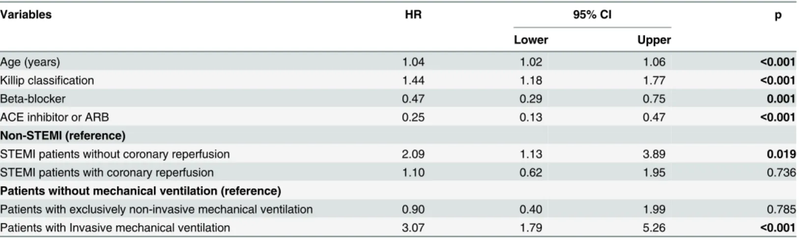
In the adjusted models (Table 3), compared with the subgroup without MV: (1) non-inva-sive MV was not associated with in-hospital death (adjusted HR = 0.90, 95% CI 0.40–1.99, P= 0.79); and (2) invasive MV was associated with a 3-fold increase in the risk of in-hospital death (adjusted HR = 3.07, 95% CI 1.79–5.26,P<0.001). Adjusted survival curves according to ventilation modality demonstrated an increased risk of in-hospital death in patients treated with invasive MV (Fig 1).
Discussion
In our study, we found that among patients with AMI, 18.2% needed treatment with MV. Approximately half of these patients were managed non-invasively. Patients treated exclusively with non-invasive MV had a relatively favorable prognosis, while patients who needed to be treated with invasive MV had a poor prognosis with a three-fold increase in the risk of death.
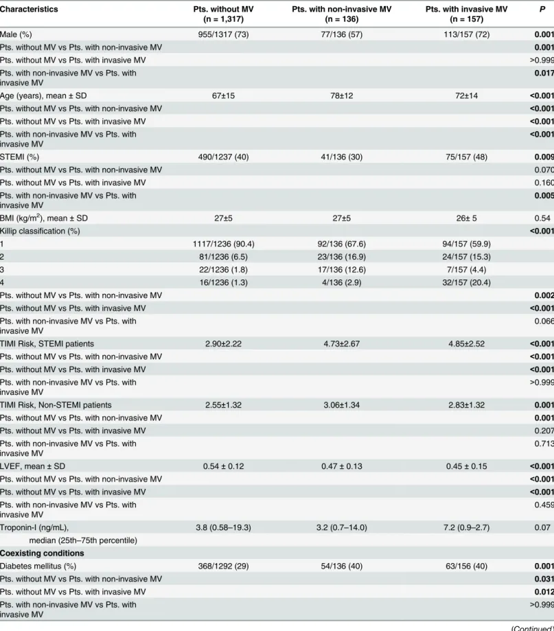
Table 1. Baseline characteristics of the patients according to mechanical ventilation treatment.
Characteristics Pts. without MV
(n = 1,317)
Pts. with non-invasive MV (n = 136)
Pts. with invasive MV (n = 157)
P
Male (%) 955/1317 (73) 77/136 (57) 113/157 (72) 0.001
Pts. without MV vs Pts. with non-invasive MV 0.001
Pts. without MV vs Pts. with invasive MV >0.999
Pts. with non-invasive MV vs Pts. with invasive MV
0.017
Age (years), mean±SD 67±15 78±12 72±14 <0.001
Pts. without MV vs Pts. with non-invasive MV <0.001
Pts. without MV vs Pts. with invasive MV <0.001
Pts. with non-invasive MV vs Pts. with invasive MV
<0.001
STEMI (%) 490/1237 (40) 41/136 (30) 75/157 (48) 0.009
Pts. without MV vs Pts. with non-invasive MV 0.070
Pts. without MV vs Pts. with invasive MV 0.160
Pts. with non-invasive MV vs Pts. with invasive MV
0.005
BMI (kg/m2), mean±SD 27±5 27±5 26±5 0.54
Killip classification (%) <0.001
1 1117/1236 (90.4) 92/136 (67.6) 94/157 (59.9)
2 81/1236 (6.5) 23/136 (16.9) 24/157 (15.3)
3 22/1236 (1.8) 17/136 (12.6) 7/157 (4.4)
4 16/1236 (1.3) 4/136 (2.9) 32/157 (20.4)
Pts. without MV vs Pts. with non-invasive MV 0.002
Pts. without MV vs Pts. with invasive MV <0.001
Pts. with non-invasive MV vs Pts. with invasive MV
0.066
TIMI Risk, STEMI patients 2.90±2.22 4.73±2.67 4.85±2.52 <0.001
Pts. without MV vs Pts. with non-invasive MV <0.001
Pts. without MV vs Pts. with invasive MV <0.001
Pts. with non-invasive MV vs Pts. with invasive MV
>0.999
TIMI Risk, Non-STEMI patients 2.55±1.32 3.06±1.34 2.83±1.32 0.001
Pts. without MV vs Pts. with non-invasive MV 0.001
Pts. without MV vs Pts. with invasive MV 0.207
Pts. with non-invasive MV vs Pts. with invasive MV
0.713
LVEF, mean±SD 0.54±0.12 0.47±0.13 0.45±0.15 <0.001
Pts. without MV vs Pts. with non-invasive MV <0.001
Pts. without MV vs Pts. with invasive MV <0.001
Pts. with non-invasive MV vs Pts. with invasive MV
0.459
Troponin-I (ng/mL), 3.8 (0.58–19.3) 3.2 (0.7–14.0) 7.2 (0.9–2.7) 0.07
median (25th–75th percentile)
Coexisting conditions
Diabetes mellitus (%) 368/1292 (29) 54/136 (40) 63/156 (40) 0.001
Pts. without MV vs Pts. with non-invasive MV 0.031
Pts. without MV vs Pts. with invasive MV 0.012
Pts. with non-invasive MV vs Pts. with invasive MV
>0.999
Table 1. (Continued)
Characteristics Pts. without MV
(n = 1,317)
Pts. with non-invasive MV (n = 136)
Pts. with invasive MV (n = 157)
P
Smoking (%) 286/1314 (22) 18/136 (13) 34/157 (22) 0.066
COPD (%) 33/1316 (2.5) 8/136 (5.9) 5/157 (3.2) 0.128
Previous AMI (%) 203/1280 (15.9) 28/132 (21.2) 24/152 (15.8) 0.28
Previous stroke (%) 48/1310 (3.7) 13/136 (9.6) 8/157 (5.1) 0.005
Pts. without MV vs Pts. with non-invasive MV 0.066
Pts. without MV vs Pts. with invasive MV >0.999
Pts. with non-invasive MV vs Pts. with invasive MV
0.439
Data are expressed as mean±standard deviation (SD), median (25th–75th percentile), or number (%).Pvalue was calculated for the comparison among the three groups. MV, mechanical ventilation; BMI, body mass index; STEMI, ST-segment elevation myocardial infarction; LVEF, left ventricular ejection fraction. TIMI, thrombolysis in myocardial infarction.
doi:10.1371/journal.pone.0151302.t001
Table 2. Medical treatment and coronary reperfusion according to mechanical ventilation treatment.
Characteristics Pts. without MV
(n = 1,317)
Pts. with non-invasive MV (n = 136)
Pts. with invasive MV (n = 157)
P
Pharmacotherapy during hospitalization
Aspirin (%) 1241/1309 (94.8) 124/136 (91.2) 137/157 (87.3) <0.001
Pts. without MV vs Pts. with non-invasive MV 0.444
Pts. without MV vs Pts. with invasive MV 0.017
Pts. with non-invasive MV vs Pts. with invasive MV 0.832
Thienopyridine (%) 1069/1289 (82.9) 95/134 (70.9) 88/146 (60.3) <0.001
Pts. without MV vs Pts. with non-invasive MV 0.009
Pts. without MV vs Pts. with invasive MV <0.001
Pts. with non-invasive MV vs Pts. with invasive MV 0.179
Beta-blocker (%) 1047/1308 (80,0) 97/136 (71,3) 88/157 (56,1) <0.001
Pts. without MV vs Pts. with non-invasive MV 0.092
Pts. without MV vs Pts. with invasive MV <0.001
Pts. with non-invasive MV vs Pts. with invasive MV 0.018
ACE inhibitor or ARB (%) 519/1310 (39.6) 72/136 (52.9) 50/157 (31.8) <0.001
Pts. without MV vs Pts. with non-invasive MV 0.009
Pts. without MV vs Pts. with invasive MV 0.148
Pts. with non-invasive MV vs Pts. with invasive MV 0.001
Reperfusion Therapy
Primary PCI or Fibrinolysis/STEMI patients (%) 394/485 (81.2) 28/41 (68.3) 51/75 (68.0) 0.008
Pts. without MV vs Pts. with non-invasive MV 0.251
Pts. without MV vs Pts. with invasive MV 0.059
Pts. with non-invasive MV vs Pts. with invasive MV >0.999
Primary PCI or Fibrinolysis/STEMI pts elegible for reperfusion*(%)
343/369 (93.0) 27/34 (79.4) 44/50 (88.0) 0.039
Pts. without MV vs Pts. with non-invasive MV 0.027
Pts. without MV vs Pts. with invasive MV 0.665
Pts. with non-invasive MV vs Pts. with invasive MV 0.871
MV, mechanical ventilation; STEMI, ST-segment elevation myocardial infarction; ACE, angiotensin converting enzyme; ARB, angiotensin II receptor blocker. PCI, percutaneous coronary intervention.
*STEMI patients on appropriate time window for reperfusion.
Table 3. Adjusted Cox proportional hazards regression model.
Variables HR 95% CI p
Lower Upper
Age (years) 1.04 1.02 1.06 <0.001
Killip classification 1.44 1.18 1.77 <0.001
Beta-blocker 0.47 0.29 0.75 0.001
ACE inhibitor or ARB 0.25 0.13 0.47 <0.001
Non-STEMI (reference)
STEMI patients without coronary reperfusion 2.09 1.13 3.89 0.019
STEMI patients with coronary reperfusion 1.10 0.62 1.95 0.736
Patients without mechanical ventilation (reference)
Patients with exclusively non-invasive mechanical ventilation 0.90 0.40 1.99 0.785
Patients with Invasive mechanical ventilation 3.07 1.79 5.26 <0.001
ACE, angiotensin converting enzyme; ARB, angiotensin II receptor blocker; HR, hazard ratio. 95% CI, 95% confidence interval; STEMI, acute myocardial infarction with ST-segment elevation. Adjusted for age, gender, diabetes, troponin levels, Killip classification on admission, left ventricular ejection fraction, body mass index, acute myocardial infarction with ST-segment elevation, previous stroke, use of aspirin, thienopyridines, beta-blockers, angiotensin converting enzyme inhibitors/angiotensin II receptor blockers and coronary reperfusion (for patients with acute myocardial infarction with ST-segment elevation).
doi:10.1371/journal.pone.0151302.t003
Fig 1. Adjusted mortality curves by ventilation modality.
study, the incidence of MV was 7.6%, and the reasons for intubation included cardiogenic shock in 64 patients (60.4%), ventricular fibrillation in 32 patients (30.1%), and acute pulmo-nary edema in 10 patients (9.5%).
We demonstrated that compared to patients treated without MV, patients treated with inva-sive or non-invainva-sive MV had a higher Killip class at hospital admission and had lower levels of LVEF. Interestingly, troponin peak levels were relatively modest and similar between groups. This finding suggests that the differences in LVEF levels and heart failure symptoms between groups were not linked with the extension of the acute myocardial necrosis. Additionally, we found that advanced age was also associated with the use of both types of MV. Besides heart failure, aging is particularly related to non-cardiac factors that may increase the risk of respira-tory failure, such as dysphagia, delirium, and muscle weakness [13,14]. Recently, Lazerri et al. [12] also observed that patients who received MV were older and more often had had a previ-ous episode of AMI.
Our rate of invasive MV was similar to that previously reported [12]. We demonstrated that AMI patients treated with invasive MV had a poor prognosis with a longer hospital length of stay and high short-term mortality rates reaching almost 40%. Invasive MV was associated with a three-fold increase in the risk of in-hospital death. These data are also similar to previ-ous findings [3,4,12]. Whether this unfavorable prognosis was more related to cardiac or non-cardiac complications (e.g., infections, septic shock, and pulmonary complications of MV) we could not determine. In fact, a cause-and-effect relationship between invasive MV and clinical outcomes could not be established, and our findings might have been influenced by complica-tions of invasive MV such as ventilator-associated pneumonia, delirium and acute respiratory distress syndrome [15,16,17,18]. Importantly, compared to patients treated without MV, the use of cardiovascular pharmacotherapy was less common in the invasive MV subgroup and reperfusion therapy was less common in both, invasive and non-invasive MV subgroups. Whether these differences were related to drug contraindications associated to acute comorbid-ities (e.g., bleeding, thrombocytopenia, hypotension, acute renal failure and hyperkalemia), delayed admission that may have contraindicated reperfusion on STEMI patients, or presence of type 2 AMI (secondary to ischemic imbalance), we could not determine. Nevertheless, con-founders related to cardiovascular pharmacotherapy and coronary reperfusion were included in our adjusted analysis.
On the other hand, clinical and randomized data on AMI patients treated with non-inva-sive MV are significantly more limited. The efficacy and safety of non-invanon-inva-sive MV in AMI have been tested in randomized small trials with patients who had cardiogenic pulmonary edema of multiple etiologies besides myocardial ischemia. In these trials, compared with stan-dard therapy, non-invasive MV reduced mortality and need for intubation [19–21]. Of note, the effect was more prominent in trials in which myocardial ischemia or infarction was the cause of pulmonary edema in higher proportions of patients [22]. More recently, one small trial has specifically compared non-invasive MV between AMI and non-AMI patients [9]. This study showed comparable benefits in both groups in terms of hemodynamic and respira-tory parameters, but was lacking in clinical outcomes data. In our study, we found that almost 10% of the entire population was treated with exclusively non-invasive MV and had relatively favorable outcomes with moderate in-hospital mortality rates. Moreover, exclusively non-invasive MV was not associated with in-hospital death in the adjusted analysis. Our findings suggest that in real-world practice, non-invasive MV is common and may be safe in patients with AMI.
not adjust our results by global risk scores, such as the APACHE II, by frailty scales, creatinine, albumin or some other prognostic biomarkers since this information was not available in this registry. Nonetheless, besides important clinical and treatment characteristics, we have adjusted our results to troponin levels. This fact further strength our results, since troponin has been considered the most important biomarker in AMI trials and correlates with the extent of myocardial necrosis. It is known that in AMI, other biomarkers such as the B-type natriuretic peptide and C-reactive protein might add prognostic information in selected patients. How-ever, they are not routinely collected in clinical practice, or recommended by guidelines and are not traditionally used in adjusted statistical models in AMI studies
Also, intention to treat analysis was not possible in this study, considering that we have no information on the chronology of the use of invasive and non-invasive MV in those patients that used both strategies. For instance, patients treated with the combination of both types of MV were considered as part of the invasive MV group. This criterion was applied to identify patients who were suitable to be exclusively treated by non-invasive strategies. Thus, we could not identify patients whose clinical status failed to improve after the initial non-invasive treat-ment and who subsequently needed invasive MV. We also did not have access to data on malignant arrhythmias, defibrillation and cardio-pulmonary resuscitation. Despite the impor-tance of these variables, it is unlikely that adjusting our results for them would change our find-ings, considering that cardio-pulmonary resuscitation are often associated with other than AMI etiologies of respiratory failure such as pulmonary edema, cardiogenic chock and systemic infections. Lastly, information on the etiology, timing, and medical treatment of the respiratory failure as well as MV duration were not available. Thus, we could not explore mechanisms of respiratory failure and treatment aspects of this group of patients. Consequently, we could not determine whether the high mortality in the invasive MV subgroup was more related to cardiac or non-cardiac complications.
Likewise, we don’t have information about the reasons for lack of coronary reperfusion among some patients in both groups treated with MV. It is possible that a significant part of these patients was not in the appropriate time window for reperfusion. In fact, considering only patients“eligible for reperfusion”(STEMI patients on appropriate time window for reperfusion), reperfusion rates were considerably increased. Nevertheless, both treatment groups had similar rates of reperfusion. Finally, we recognize that our findings do not demonstrate a causality relation between invasive-MV and death. Based on the observational nature of our study, we have only assessed an association between MV and outcomes, and therefore, we could not adjust for unmeasured confounders. Thus, our findings are hypothesis-generating that highlight the need of a large, well-powered, prospective trial comparing the effectiveness of invasive and non-invasive MV in AMI patients with respira-tory failure.
In conclusion, in AMI setting, 18% of the patients required MV. Almost half of these patients were treated with exclusively non-invasive MV. This subgroup had a relatively favor-able prognosis with moderate rates of short-term death. On the other hand, patients who needed to be treated invasively had poor outcomes and a three-fold increase in the risk of in-hospital death, compared to patients who did not use MV. Thus, future prospective random-ized trials are needed to compare the effectiveness of invasive and non-invasive MV for the ini-tial approach of respiratory failure in AMI patients.
Supporting Information
S1 Dataset. Dataset for the entire population.
Acknowledgments
This research received no specific grant from any funding agency in the public, commercial, or not-for-profit sectors. We would like to acknowledge Rogério Ruscitto Prado for his support with statistics.
Author Contributions
Conceived and designed the experiments: AEPP MK MF CP CVS RDL. Performed the experi-ments: AEPP MK MM AGC MF CP RDL. Analyzed the data: AEPP MK JNK CSVB MM MF CVS RDL. Wrote the paper: AEPP MK JNK CSVB MM AGC MF CP CVS RDL.
References
1. Tsai TH, Chua S, Hussein H, Leu S, Wu CJ, Hang CL, et al: Outcomes of patients with Killip class III acute myocardial infarction after primary percutaneous coronary intervention. Crit Care Med. 2011; 39:436–442. doi:10.1097/CCM.0b013e318206ccc3PMID:21242801
2. Park M, Sangean MC, Volpe Mde S, Feltrim MI, Nozawa E, Leite PF, et al. Randomized, prospective trial of oxygen, continuous positive airway pressure, and bilevel positive airway pressure by face mask in acute cardiogenic pulmonary edema. Crit Care Med. 2004; 32:2407–2415. PMID:15599144 3. Lesage A, Ramakers M, Daubin C, Verrier V, Beynier D, Charbonneau P, et al. Complicated acute
myocardial infarction requiring mechanical ventilation in the intensive care unit: prognostic factors of clinical outcome in a series of 157 patients. Crit Care Med. 2004; 32:100–105. PMID:14707566 4. Kouraki K, Schneider S, Uebis R, Tebbe U, Klein HH, Janssens U, et al. Characteristics and clinical
out-come of 458 patients with acute myocardial infarction requiring mechanical ventilation. Results of the BEAT registry of the ALKK-study group. Clin Res Cardiol. 2011; 100:235–239. doi: 10.1007/s00392-010-0235-6PMID:20878411
5. Bersten AD, Holt AW, Vedig AE, Skowronski GA, Baggoley CJ. Treatment of severe cardiogenic pul-monary edema with continuous positive airway pressure delivered by face mask. N Engl J Med. 1991; 325:1825–1830. PMID:1961221
6. Lin M, Yang YF, Chiang HT, Chang MS, Chiang BN, Cheitlin MD. Reappraisal of continuous positive airway pressure therapy in acute cardiogenic pulmonary edema. Short-term results and long-term fol-low-up. Chest. 1995; 107:1379–1386. PMID:7750335
7. Peter JV, Moran JL, Phillips-Hughes J, Graham P, Bersten AD. Effect of non-invasive positive pressure ventilation (NIPPV) on mortality in patients with acute cardiogenic pulmonary edema: a meta-analysis. Lancet. 2006; 367:1155–1163. PMID:16616558
8. Takeda S, Nejima J, Takano T, Nakanishi K, Takayama M, Sakamoto A, et al. Effect of nasal continu-ous positive airway pressure on pulmonary edema complicating acute myocardial infarction. Jpn Circ J. 1998; 62:553–558. PMID:9741730
9. Yamamoto T, Takeda S, Sato N, Akutsu K, Mase H, Nakazato K, et al. Noninvasive ventilation in pul-monary edema complicating acute myocardial infarction. Circ J. 2012; 76:2586–2591. PMID:
22850288
10. Alpert JS, Thygesen K, Antman E, Bassand JP. Myocardial infarction redefined—a consensus docu-ment of The Joint European Society of Cardiology/American College of Cardiology Committee for the redefinition of myocardial infarction. J Am Coll Cardiol. 2000; 36:959–969. PMID:10987628 11. Makdisse M, Katz M, Corrêa Ada G, Forlenza LM, Perin MA, de Brito FS Júnior, et al. Effect of
imple-menting an acute myocardial infarction guideline on quality indicators. Einstein (Sao Paulo). 2013; 11:357–363.
12. Lazzeri C, Valente S, Chiostri M, AttanàP, Mattesini A, Gensini GF. Mechanical ventilation in the early phase of ST elevation myocardial infarction treated with mechanical revascularization. Cardiol J. 2013; 20:612–617. doi:10.5603/CJ.2013.0161PMID:24338538
13. Sevransky JE, Haponik EF. Respiratory failure in elderly patients. Clin Geriatr Med. 2003; 19:205–224. PMID:12735123
14. Serra-Prat M, Hinojosa G, López D, Juan M, Fabré E, Voss DS, et al. Prevalence of oropharyngeal dys-phagia and impaired safety and efficacy of swallow in independently living older persons. J Am Geriatr Soc. 2011; 59:186–187. doi:10.1111/j.1532-5415.2010.03227.xPMID:21226704
16. Van Rompaey B, Elseviers MM, Schuurmans MJ, Shortridge-Baggett LM, Truijen S, Bossaert L. Risk factors for delirium in intensive care patients: a prospective cohort study. Crit Care. 2009; 13:R77. doi:
10.1186/cc7892PMID:19457226
17. Valente Barbas CS, Neto AS. Changing the focus in acute respiratory distress syndrome: treating is mandatory, but preventing is imperative. Crit Care Med. 2013; 41:2058–2059. doi:10.1097/CCM. 0b013e31828c25f2PMID:23863250
18. Serpa Neto A, Cardoso SO, Manetta JA, Pereira VG, Espósito DC, Pasqualucci Mde O, et al. Associa-tion between use of lung-protective ventilaAssocia-tion with lower tidal volumes and clinical outcomes among patients without acute respiratory distress syndrome: a meta-analysis. JAMA. 2012; 308:1651–1659. doi:10.1001/jama.2012.13730PMID:23093163
19. Vital FM, Saconato H, Ladeira MT, Sen A, Hawkes CA, Soares B, et al. Non-invasive positive pressure ventilation (CPAP or bilevel NPPV) for cardiogenic pulmonary edema. Cochrane Database Syst Rev. 2008; 16:CD005351.
20. Peter JV, Moran JL, Phillips-Hughes J, Graham P, Bersten AD. Effect of non-invasive positive pressure ventilation (NIPPV) on mortality in patients with acute cardiogenic pulmonary oedema: a meta-analysis. Lancet. 2006; 367:1155–1163. PMID:16616558
21. Winck JC, Azevedo LF, Costa-Pereira A, Antonelli M, Wyatt JC. Efficacy and safety of non-invasive ventilation in the treatment of acute cardiogenic pulmonary edema—a systematic review and meta-analysis. Crit Care. 2006; 10:R69. PMID:16646987