Endoscopic Resection of Sinonasal
Malignancy: A Systematic Review and
Meta-analysis
Otolaryngology– Head and Neck Surgery 2016, Vol. 155(3) 376–386 ÓAmerican Academy of Otolaryngology—Head and Neck Surgery Foundation 2016 Reprints and permission:
sagepub.com/journalsPermissions.nav DOI: 10.1177/0194599816646968 http://otojournal.org
Rounak B. Rawal, MD
1, Zainab Farzal
1,
Jerome J. Federspiel, MD, PhD
2,3, Satyan B. Sreenath, MD
1,
Brian D. Thorp, MD
1, and Adam M. Zanation, MD
1,4Sponsorships or competing interests that may be relevant to content are dis-closed at the end of this article.
Abstract
Objectives. The use of endoscopic approaches for sinonasal malignancy resection has increased, but survival data are lim-ited secondary to disease rarity and new surgical technique. Here we present a systematic review and meta-analysis of endoscopic endonasal resection of sinonasal malignancy.
Data Sources. MEDLINE, PubMed Central, NCBI Bookshelf, Cochrane Library, clinicaltrials.gov, National Guideline Clearinghouse.
Review Methods. PRISMA/MOOSE guidelines were followed. MeSH terms were ‘‘endoscopic’’ AND (‘‘esthesioneuroblastoma’’ OR ‘‘sinonasal adenocarcinoma’’ OR ‘‘squamous cell carcinoma’’ OR ‘‘sinonasal undifferentiated carcinoma’’). For studies in which individual-level data were available, results were obtained by direct pooling. For studies in which only summary Kaplan-Meier curves were available, numerical data were extracted, traced, and aggregated by fitting a Weibull model.
Results.Of 320 studies identified, 35 case series were included (n = 952 patients), with 15 studies analyzed via aggregate mod-eling and 20 studies analyzed via direct pooling. Two- and 5-year survival rates for patients in aggregate modeling were 87.5% and 72.3%, respectively (mean follow-up: 32.9 months). Two- and 5-year survival for patients in direct pool-ing were 85.8% and 83.5%, respectively (mean follow-up: 43.0 6 19.5 months). Significant overall survival difference was found between low- and high-grade cancers (P = .015) but not between low- and high-stage cancers (P= .79).
Conclusion. Overall 2- and 5-year survival rates are compara-ble and sometimes greater than those from open craniofacial resection. Survival rates significantly differ by cancer grade but not stage. Journals and investigators should be encour-aged to publish retrospective and prospective case series with staged survival updates based on established guidelines.
Keywords
sinonasal malignancy, endoscopic, meta-analysis, systematic review
Received June 28, 2015; revised March 8, 2016; accepted April 8, 2016.
T
he expanded endonasal approach has allowed for extir-pation of benign sinonasal tumors via minimally inva-sive techniques since the first series published by Jho and Carrau in 1996.1 While expanded endonasal approach indications continue to broaden for benign disease processes, open craniofacial resection continues to be the gold standard for extirpation of malignant sinonasal tumors. The preference of open technique is often driven by the desire for an en bloc resection of tumor with negative margins rather than a progres-sive resection with negative margins. Additional concerns include ensuring adequate hemostasis while minimizing vascu-lar, soft tissue, and dural injury, along with operator familiarity with endoscopic technology.2-4 However, nearly 2 decades have passed since the introduction of endoscopic technology, and with continued advancement in endoscopic hemostatic techniques, reconstructive options, and standardized safety pre-cautions, its use in malignant disease resection continues to rise.5-9Malignant sinonasal cancer comprises a variety of rare heterogeneous disease processes, with reported incidences
\1.5 per 100,000 in men and\1 per 100,000 in women.10 The rarity and high degree of heterogeneity in these cancers result in a paucity of evidence-based management. Published data are relegated to case series with various sample populations, staging, follow-up periods, surgical
1
Department of Otolaryngology–Head and Neck Surgery, University of North Carolina at Chapel Hill, Chapel Hill, North Carolina, USA
2
Department of Health Policy and Management, UNC Gillings School of Global Public Health, Chapel Hill, North Carolina, USA
3
University of North Carolina at Chapel Hill School of Medicine, Chapel Hill, North Carolina, USA
4
Department of Neurosurgery, University of North Carolina at Chapel Hill, Chapel Hill, North Carolina, USA
Corresponding Author:
Rounak B. Rawal, MD, Department of Otolaryngology–Head and Neck Surgery, University of North Carolina at Chapel Hill, 170 Manning Drive, CB #7070, Physician’s Office Building Room G-190, Chapel Hill, NC, 27599, USA.
techniques, and adjuvant therapies. To study the effect of endoscopic endonasal management of patients with these rare malignancies in a quantifiable generalizable method, we conducted a meta-analysis and systematic review of the current literature.
Methods
Search Methodology
A meta-analysis and systematic review of the literature was
conducted in accordance with PRISMA guidelines
(Preferred Reporting Items for Systematic Reviews and Meta-analyses).11,12 As many studies were found to be observational, MOOSE guidelines (Meta-analysis of Observational Studies in Epidemiology) were also used.13
MEDLINE, PubMed Central, NCBI Bookshelf, Cochrane Library, clinicaltrials.gov, and the National Guideline Clearinghouse databases were searched for the following MeSH terms: ‘‘endoscopic’’ AND (‘‘esthesioneuroblastoma’’ OR ‘‘sinonasal adenocarcinoma’’ OR ‘‘squamous cell carci-noma’’ OR ‘‘sinonasal undifferentiated carcicarci-noma’’). In addi-tion, references of retrieved studies were searched to identify all relevant articles. Studies with the following criteria were included for analysis: English-language studies with overall survival outcome data for patients having undergone endo-scopic or endoendo-scopic-assisted resection of sinonasal malig-nancy with a mean follow-up of at least 24 months. Studies with the following criteria were excluded from analysis: previ-ously reported patient data with significant overlap, pooled data with patients who had open craniofacial resection that could not be extracted, patients operated on with only pallia-tive intent, patients presenting with recurrent cancer, and case series with\3 patients, as authors of such series may have insufficient experience with surgical management. If studies also included results of patients who had open approaches for resection of sinonasal malignancy, only those patients who had endoscopic resection were included. A schematic flow diagram detailing the systematic search is included (Figure 1).
Statistical Analysis
Data were aggregated according to the nature of the available source data. For studies in which individual-level data were pro-vided, results were obtained by direct pooling of observations. For studies in which individual data were not available and only summary Kaplan-Meier curves were provided, 2 authors extracted numerical data from the provided graphical data (R.B.R. and S.B.S.) by tracing via DigitizeIt 2.0 software (DigitizeIt, Braunschweig, Germany), as previously described.14 These curves were then aggregated by fitting a Weibull model to the resultant data points. A random effects framework allowed for interstudy heterogeneity in outcomes.15 If a study provided both Kaplan-Meier curves and individual data, the latter were preferentially collected over numeric data extraction and model creation. For all studies, accompanying demographic data were collected for individual or pooled samples of patients. Given the greater detail of the pooled individual data subset, additional data points were extracted from these
studies, including cancer staging. Cancer staging was then separated into ‘‘low stage,’’ defined as American Joint Committee on Cancer (AJCC) stages T1/T2 or Kadish stages A/B, and ‘‘high stage,’’ defined as AJCC stages T3/T4 or Kadish stages C/D.16Overall mortality was compared on the basis of stage via a log-rank test. Histopathologic grading was also studied. ‘‘High grade’’ malignancies included the follow-ing: squamous cell carcinoma (including that originating from inverted papilloma), melanoma, sinonasal undifferentiated car-cinoma, meibomian gland carcar-cinoma, sarcoma, and spindle cell carcinoma. Overall mortality was compared according to histopathologic grading via a log-rank test. Differences in his-topathology between aggregate model and pooled analyses were tested with a 2-tailed Fisher’s exact test. Differences between patients in surgery and surgery and radiotherapy groups in regard to staging and grading were tested with a 2-tailed Fisher’s exact test. All data analyses were performed in Stata 13.1 (StataCorp, College Station, Texas).
Results
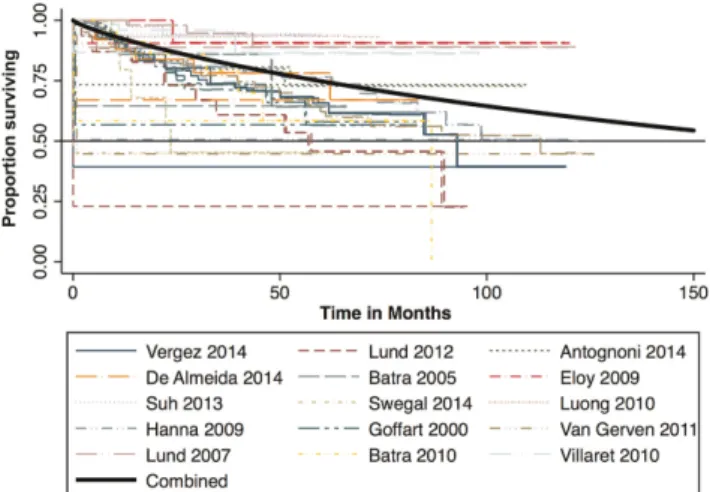
Of 320 initial search results, 61 full-text articles were assessed for eligibility, and 35 studies were ultimately included for systematic review and meta-analysis.17-52 A total of 952 patients were included. Fifteen studies (n = 759, 79.7%) provided only summary Kaplan-Meier curves, allowing only for aggregate model analysis, summarized in
Table 1 and illustrated in an aggregated Kaplan-Meier
curve in Figure 2. Twenty studies (n = 193 patients, 20.2%) provided individual-level data, allowing for direct pooling of observations, summarized in Table 2 and illu-strated in a pooled Kaplan-Meier curve in Figure 3 (for complete individual-level data, see Table S1 at www.oto-journal.org/supplemental). Individual-level data were then further classified into low- and high-stage disease and strati-fied by histopathology (Table 3).
Overall 2- and 5-year survival rates for patients in the aggregate model analysis were 87.5% and 72.3%, respec-tively. Mean follow-up for studies in the aggregate model analysis was 33.9 months. Of 759 patients, 684 patients had purely endoscopic surgical management of disease (90%), while 75 patients had endoscopic-assisted surgical manage-ment (9.9%). The majority of patients were male (64%), and the mean age was 61.4. The most prevalent histopathol-ogies were sinonasal adenocarcinoma (56%), sinonasal mel-anoma (13%), and squamous cell carcinoma (11%).
Overall 2- and 5-year survival rates for patients in the direct pooled analysis were 85.8% and 83.5%, respectively. Follow-up (mean 6 SD) for studies in the pooled analysis was 43.0 619.5 months. Of 193 patients, 157 patients had purely endoscopic surgical management of disease (78%), while 36 had endoscopic-assisted surgical management (19%). The majority of patients were male (64%), and the mean age was 56.6 6 8.1 years. The most prevalent histo-pathologies were esthesioneuroblastoma (32%), sinonasal adenocarcinoma (28%), and sinonasal melanoma (18%).
Given the greater detail of the pooled individual data subset, additional analysis was done on this group of patients.
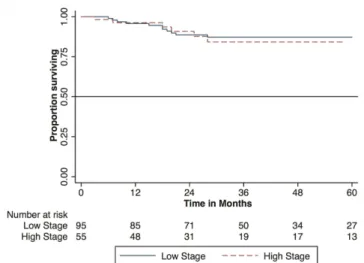
The majority of patients in the pooled analyses had low-stage cancer (63%), although staging data were not available for 22% of the malignancies (Table 3). The majority of esthesio-neuroblastomas (61%), sinonasal adenocarcinomas (73%), and squamous cell carcinomas6 inverted papilloma (74%) were low-stage malignancies. Sinonasal undifferentiated cancers rep-resented the only disease process with a majority of high-stage malignancies (67%). For the majority of melanomas and more uncommon malignancies included, staging data were unavail-able. No significant difference in overall survival between low- and high-stage cancers was found (P = .79; Figure 4). Histopathologic grading was also studied. Among the high-grade malignancies, the most common were squamous cell carcinoma (51.9%), melanoma (22.6%), and sarcoma (7.7%), while among the low-grade malignancies, the most common were sinonasal adenocarcinoma (61.0%) and esthesioneuro-blastoma (12.7%; Table 4). A significant survival difference between high- and low-grade cancer was found (P = .015;
Figure 5).
Last, survival analysis was performed on patients who received radiotherapy in addition to surgery. No statistically significant difference in overall survival was found between those patients who only underwent surgery and those patients who underwent surgery and radiotherapy (P = .85;
Figure 6). These 2 groups (surgery only versus surgery1
radiotherapy) were compared to assess whether an unequal distribution of patients was confounding survival rates based on tumor stage and grade. The 2 groups were signifi-cantly different according to stage: in the surgery-only cohort, 11.6% of tumors were high stage, and 88.4% of tumors were low stage, while in the surgery 1radiotherapy cohort, 46.7% of tumors were high stage, and 53.3% of tumors were low stage (P\.001). The 2 groups were not significantly different in respect to grading: in the surgery-only cohort, 33.3% of tumors were high grade, and 66.7% of tumors were low grade, while in the surgery1 radiother-apy cohort, 28.3% of tumors were high grade, and 71.7% of tumors were low grade (P= .602).
Histopathology differed greatly between analyses. The aggregate model analyses had a significantly greater preva-lence of SNAC (sinonasal adenocarcinoma; 56% vs 28%,P\
.0001). The reason for this skew was the large prevalence of SNAC described in a single multicenter study from France (n = 159, 49%).17 Because of this high prevalence of a single pathology, we sought to reanalyze our data after exclusion of SNAC. An attempt at data analysis excluding SNAC was made for studies in the aggregate model, but this was not feasible, as many studies included Literature Search:
Databases- MEDLINE, PubMed Central, NCBI Bookshelf, Cochrane Library, clinicaltrials.gov, and The Naonal Guideline Clearinghouse
Limits:English language only
Search results combined n = 320
Arcles screened on basis of tle and abstract
Full-text arcles assessed for eligibility
n = 61
Pooled: 30 Individual: 31
Excluded (n = 259)
Not pertaining to outcomes of endoscopic or endoscopic-assisted surgery for newly diagnosed malignant
anterior skull base cancers: 248 Case reports: 6 Previously reported data: 4
Full text unavailable: 1
Included n = 35
Pooled : 15 Individual : 20
Excluded (n = 26)
Unable to extract endoscopic -specific data: 3 No overall survival (OS) Kaplan-Meier curve or
OS individual data: 11 Previously reported data: 3 Mean follow-up <2 years for pernent
paents: 4
Only paents with recurrenct cancer: 1 Only surgery with palliave intent: 1 Case series < 3 pernent paents: 3
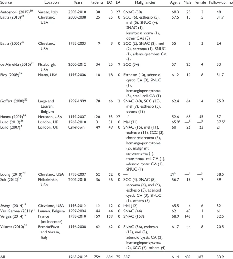
Table 1.Studies Included for Aggregate Model Analysis.a
Sex
Source Location Years Patients EO EA Malignancies
Mean
Age, y Male Female
Mean Follow-up, mo
Antognoni (2015)20 Varese, Italy 2003-2010 30 3 27 SNAC (30) 68.3 28 2 48
Batra (2010)32 Cleveland,
USA
2000-2008 25 25 0 SCC (6), esthesio (5), mel (5), SNUC (4), SNAC (1),
leiomyosarcoma (1), other CAs (3)
57.5 10 15 31.7
Batra (2005)45 Cleveland, USA
1995-2003 9 9 0 SCC (2), SNAC (2), mel
(2), sarcoma (1), SNUC (1), adenosquamous CA (1)
55 6 3 24
de Almeida (2015)21 Pittsburgh, USA
2000-2012 34 25 9 SCC (34) 57 20 14 33
Eloy (2009)36 Miami, USA 1997-2006 18 18 0 Esthesio (10), adenoid
cystic CA (3), SNUC (1),
hemangiopericytoma (3), small cell CA (1)
61.2 10 8 31.7
Goffart (2000)52 Liege and Leuven, Belgium
1992-1999 78 66 12 SNAC (40), SCC (13),
mel (7), esthesio (5), others (13)
62.4 64 14 25.9
Hanna (2009)34 Houston, USA 1992-2007 120 93 27 —b 52.6 65 55 37
Lund (2012)26 London, UK 1963-2010 31 31 0 Mel (31) 65.9b —b —b 37.5b
Lund (2007)41 London, UK Unknown 49 49 0 SNAC (15), mel (11),
esthesio (11), SCC (3), chondrosarcoma (3), hemangiopericytoma (2), malignant schwannoma (1), transitional cell CA (1), adenoid cystic CA (1), SNUC (1)
60 26 23 21
Luong (2010)29 Cleveland, USA 1998-2007 52 52 0 —b 59b —b —b 38.5
Suh (2013)24 Philadelphia, USA
2002-2010 36 36 0 SCC (4), SNAC (8),
sarcoma (6), mel (4), esthesio (5), adenoid cystic CA (3), SNUC (1), others (5)
56.7 19 17 39
Swegal (2014)19 Cleveland, USA 1998-2012 12 12 0 Mel (12) 65.5 6 6 32
Van Gerven (2011)31 Leuven, Belgium 1992-2004 44 44 0 SNAC (44) 62 43 1 61
Vergez (2014)17 France (multicenter)
1998-2010 159 159 0 SNAC (159) 68.9 148 11 32.5
Villaret (2010)30 Brescia/Pavia
and Varese, Italy
1996-2008 62 62 0 SNAC (36), esthesio
(13), mel (3),
adenoid cystic CA (2), hemangiopericytoma (2), SCC (2), others (4)
61.7 44 18 20.5
All 1963-2012c 759 684 75 587 61.4 489 187 33.9
Abbreviations: CA, carcinoma; EA, endoscopically assisted; EO, endoscopic only; esthesio, esthesioneuroblastoma; IP, inverted papilloma; mel, melanoma; SCC, squamous cell carcinoma; SNAC, sinonasal adenocarcinoma
a
Values presented as n or mean. Number of patients reviewed includes only those who met inclusion criteria. The listed studies reviewed more patients than the number included in our analysis.
b
Demographic data for endoscopic population unable to be extracted from overall patient population.
c
Studies may predate advent of endoscopic techniques (1991) because patients with open resection were included.
SNAC among other histopathologies, making non-SNAC data inextricable. This was easily extracted in the pooled group, however. In the pooled group, no significant differ-ence in overall survival between low- and high-stage can-cers was found even once SNAC was excluded (P = .67;
Figure 7). In the pooled group, there was still a
statisti-cally significant difference in survival between high- and low-grade malignancies even once SNAC was excluded (P= .010;Figure 8).
Interextractor reliability in data extraction was excellent. Estimated 2-year survival was 87.0% with data only from extractor 1 versus 87.5% for data from extractor 2, while estimated 5-year survival was 74.7% with extractor 1’s data versus 72.3% with extractor 2’s data.
Discussion
The Kaplan-Meier overall survival curves and percentages at 2 and 5 years in patients in the pooled analysis and the aggregate model analysis provide strong evidence for con-tinued use and further adoption of endoscopic endonasal resection of sinonasal malignancy. While open craniofacial resection remains the gold standard surgical technique for extirpation of sinonasal malignancy,53 endoscopic resection may allow for decreased cosmetic deformity, associated hospital stay, and complication rates. The endoscopic sur-geon must be able and willing to sacrifice these advantages and convert to an open approach when necessary to adhere to oncologic principles, especially that of margin-negative en bloc resection. Our data support the use of endoscopic techniques only with adherence to this caveat. Our analysis does not compare endoscopic resection with open resection, as no randomized controlled trials exist comparing the 2 surgical modalities. We therefore sought only to quantify the existing data to determine overall survival rates and explore possible predictors for increased survival.
Comparing endoscopic management with open craniofa-cial management is difficult. In the preendoscopic era, a pre-vious systematic review by Dulguerov et al in 2001 outlined overall survival rates of sinonasal malignancy throughout the
decades, showing survival rates of 28%613% in the 1960s, 36% 6 13% in the 1970s, 43% 6 15% in the 1980s, and 51% 6 14% in the 1990s.54 Patients treated prior to the 1990s could not have been treated endoscopically; as such, our data show excellent continuation of this upward trend of overall survival rates. Comparisons with such historical data may be misleading, however, as multiple confounding factors undoubtedly played a role, such as increased training stan-dards, differences in staging, standardized measures (preo-perative, o(preo-perative, and postoperative), adjunctive therapies, and better diagnostic modalities. A more recent systematic review by Higgins et al in 2011 shows overall 5-year survival rates of open craniofacial resection at 55.2% (n = 101).16Our 5-year survival rates of 72.3% and 83.5% are therefore encouraging.
Our aggregate and pooled model overall survival out-comes were similar for the 2-year benchmark (87.5% vs 85.8%) but somewhat different for the 5-year benchmark (72.3% vs 83.5%). As noted earlier, the aggregate analysis had a significantly increased proportion of SNAC as com-pared with the pooled analysis largely due to one French multicenter study (56% vs 28%, P\.0001). In this study, the overall 5-year survival rate was 62% and likely skewed the overall survival rate of the aggregate model.17 SNAC has historically been further classified into intestinal-type adenocarcinomas and nonintestinal-type adenocarcinomas, with varying survival outcomes for each histologic type, but the authors did not explicitly stratify their results by these groupings, possibly also confounding survival out-comes.53,55 To determine whether SNAC prevalence con-founded the results for our pooled analysis, we repeated survival analysis by stage (Figures 4, 7) and grade
(Figures 5, 8) excluding patients with SNAC, but our
results were comparable for these secondary subanalyses. Based on the smaller, pooled analysis of patients, stage had a nonsignificant effect on overall survival outcomes when based on endoscopic surgical techniques. Upon closer inspection, however, this may be misleading for 2 reasons: sinonasal undifferentiated carcinoma was the only histo-pathology type with a majority of high-stage cancers (67%), and staging for sinonasal melanoma, a particularly fatal dis-ease, was unavailable for 18 of the 20 patients and therefore could not be included in this analysis.56 Indeed, mucosal melanoma by definition is high stage, as staging begins at T3 according to the latest AJCC guidelines.57
According to our data, histopathologic grade does have a significant effect on overall survival outcomes. As noted earlier, skull base malignancies encompass a variety of his-topathology, and classifying these malignancies into low-and high-grade subtypes may allow for better counseling for patients in regard to survival outcomes.10,53
Other variables were also initially considered for study in the pooled data analysis, including disease-specific survival, recurrence-free survival, previous therapy, metastasis/nodal status, and extent/location of disease process. However, published data were too variable in reporting of these out-comes; therefore, we were confined in our ability to draw
Figure 2.Kaplan-Meier overall survival curve of patients in
T able 2. Studies Included for P ooled Model Analysis. a Sex Sour ce Location Y ears Patients EO EA Malignancies Age, y Male Female Follow-up , mo Castelnuov o (2010) 28 V ar ese/Pisa/Br escia, Italy 1997-2008 4 4 0 Adenoid cystic C A (2), papillar y A CC (1), SNUC (1) 49.75 6 12.09 4 0 34.25 6 34.23 Chen (2006) 42 Changhua, T aiwan 2000-2004 7 7 0 A C C (4 ), SC C (1 ), SN U C (1 ), sa rc o m a (1 ) 57 6 16.57 5 2 30.7 6 16.84 Constantinidis (2004) 46 Erlangen/ Nur emberg, German y 1975-2000 11 6 5 Esthesio (12) 51 6 13.53 — b — b 102.55 6 78.39 De vaiah (2003) 49 Boston/Kansas City , USA 1991-2002 7 0 7 Esthesio (7) 47.71 6 7.87 4 3 62.29 6 34.49 Gallia (2013) 23 Baltimor e, USA 2005-2012 9 9 0 Esthesio (9) 51.22 6 10.21 6 3 30.44 6 16.45 Huber (2011) 50 Zurich, Switzerland 1992-2007 12 11 1 A CC (12) 61.5 6 19.26 9 3 16.08 6 8.33 Jar deleza (2009) 35 Adelaide, Australia 1999-2008 12 12 0 A CC (12) 65.83 6 10.18 9 3 42 6 32.12 Kim (2008) 39 Seoul, South K ore a 1989-2006 8 0 8 Esthesio (8) 46.13 6 14.40 5 3 18.25 6 11.78 Mohindra (2014) 18 Chandigarh, India 2005-2009 8 6 2 Esthesio (8) 43 6 9.62 6 2 35.5 6 11.11 Nicolai (2007) 51 Br escia/V ar ese, Italy 1999-2003 16 16 0 A CC (12), SCC (4) 62.19 6 17.71 9 7 47.25 6 12.97 Or vidas (2005) 47 Rochester , USA 1980-2001 3 3 0 A CC (3) 71.33 6 11.02 1 2 36.33 6 32.52 P odboj (2007) 40 Ljubljana, Slov enia 1991-2006 16 16 0 A CC (3), SNUC (6), Leiom yosar coma (1), papillar y adenocar cinoma (2), mel (2), esthesio (1), chondr osar coma (1) 56.69 6 21.76 8 8 66.25 6 40.71 P oetkar (2005) 43 Milwauk ee, USA 1993-2003 16 14 2 Esthesio (5), SCC (5), A CC (2), adenoid cystic C A (1), mel (1), chondr osar coma (1), hemangiopericytoma (1) 57 6 1.45 — b — b 51.5 6 38.11 Re venaugh (2011) 27 Cle veland, USA 2002-2009 7 6 1 SNUC (7) 42.86 6 17.24 — b — b 32.29 6 19.65 Roh (2004) 48 Cle veland / Philadelphia, USA 1996-2003 19 19 0 SCC (1), SCC 1 IP (5), A CC (3), mel (4), esthesio (1), leiom yosar coma (1), Others (4) 56.95 6 17.24 10 9 26.42 6 21.85 Roth (2010) 33 Zurich, Switzerland 1992-2007 13 13 0 Mel (13) 65.23 6 12.81 — b — b 46.46 6 42.11 Shipchandler (2005) 37 Cle veland, USA 1996-2004 9 6 3 SCC (5), SCC 1 IP (4) 62.78 6 10.99 7 2 26.56 6 25.64 T ojima (2012) 25 Otsu, Japan 2000-2009 6 6 0 SCC (1), SCC 1 IP (1), esthesio (1), others (3) 65 6 14.67 4 2 43.33 6 39.94 Y uen (2005) 44 Hong K ong, Hong K ong 1996-2003 6 0 6 Esthesio (6) 51 6 17.79 4 2 32.83 6 22.42 Zafer eo (2008) 38 Cle veland, USA 1980-2004 4 3 1 Esthesio (4) 60.25 6 11.44 2 2 54 6 62.55 All 1975-2014 c 193 157 36 193 56.64 6 8.12 93 53 42.97 6 19.52 Abbr e viat ions: A CC , ade nocar cino ma; C A, car cinoma; EA, endo scopi cally assisted; EO , endosco pic only ; esthe sio , esthe sioneur oblasto ma; IP , in verted papilloma; mel, melano ma; SCC , squam ous cell car cinoma. a V alu es pr esen ted as n or mean 6 SD . Nu mber of patie nts re vie w ed includes only th ose w ho met inclusion criteria. Th e listed stud ies re vie w ed mor e pat ients than the number included in our anal ysis. bDem ographic data for endo scopic populat ion unabl e to be ext racted fr om ov erall patient pop ulation. cStudies ma y pr edate adv ent of endo scopic techni ques (199 1) bec ause pat ients with ope n resec tion w er e included in st udy . 381 at SOCIEDADE BRASILEIRA DE CIRUR on September 2, 2016
conclusions solely based on overall survival and cancer stage.
It is important to note that 112 patients included in the study (10.9%) had ‘‘endoscopic assisted’’ surgery. The defini-tion of endoscopic-assisted surgery was also variable, some-times including orbital incision, frontal/subfrontal craniotomy, or anterior craniotomy.34,49,52 It is theoretically possible that these endoscopic-assisted techniques allowed for greater access and visualization of the sinonasal malignancy, artificially increasing overall survival rates for some studies. It is difficult to generalize conclusions for this subset of the population.
The effect of radiation therapy with or without chemotherapy after surgery cannot be underestimated. Although indications for adjuvant therapy differ from institution to institution, it is gener-ally reserved for patients with high-grade tumors, advanced tumor stage, bone invasion, perineural spread, intracranial
extension, dural or brain involvement, and/or positive mar-gins.34In addition, controversy exists for elective neck dis-section or elective radiotherapy.58
At first glance, our data seem to indicate that adjuvant radiation therapy did not result in a statistically significant survival benefit, but there were significantly higher numbers of patients with high-stage tumors in the surgery1radiotherapy
Table 4. Histopathology of Tumors in Pooled Model Analysis
Stratified by Grade.a
Studies, n (%)
Grade Aggregate (n = 693) Pooled (n = 186)
Low
Esthesioblastoma 74 61
SNAC 355 54
Adenoid cystic carcinoma 18 3
Hemangiopericytoma 7 2
Chondrosarcoma 5 3
Total 459 (66.2) 123 (66.1)
High
Squamous cell carcinoma 127 27
Melanoma 67 20
SNUC 12 15
Sarcoma 23 1
Neuroendocrine 5 0
Total 234 (33.8) 63 (33.9)
Abbreviations: SNAC, sinonasal adenocarcinoma; SNUC, sinonasal undiffer-entiated carcinoma.
aMore uncommon malignancies that were present but excluded from analysis
of tumor grade included the following: leiomyosarcoma, clear cell carcinoma, basal cell carcinoma, angiomyxoid neoplasm, angiosarcoma, osteosarcoma, spindle cell carcinoma, nonspecified carcinoma, carcinoma ex pleomorphic adenoma, poorly differentiated carcinoma, teratocarcinoma, plasmacytoma, teratocarcinoma, fibrosarcoma, extrapleural solitary fibrous tumor, small cell carcinoma, malignant schwannoma, and transitional cell carcinoma.
Table 3. Histopathology of Tumors in Pooled Model Analysis
Stratified by Stage.
Low Stage High Stage N/A Total
Esthesioneuroblastoma 36 23 2 61
SNAC 37 14 3 54
SCC 12 3 2 17
SCC1IP 2 2 6 10
Melanoma 2 0 18 20
SNUC 5 10 0 15
Adenoid cystic carcinoma 1 2 0 3
Othera 0 1 12 13
Total 95 55 43 193
Abbreviations: IP, inverted papilloma; N/A, not applicable; SCC, squamous cell carcinoma; SNAC, sinonasal adenocarcinoma; SNUC, sinonasal undiffer-entiated carcinoma.
a
Other malignancy types include chondrosarcoma (3), chordoma (1), hemangiopericytoma (2), leiomyosarcoma (2), malignant peripheral nerve sheath tumor (1), Meibomian gland carcinoma (1), plasmocytoma (1), sar-coma (1), and spindle cell carcinoma (1).
Figure 3.Kaplan-Meier overall survival curve of patients in pooled
individual data analysis (n = 193 patients).
Figure 4.Kaplan-Meier overall survival curve of patients in pooled
cohort as compared with the surgery-only cohort. Our results therefore continue to support the use of multimodality therapy for low- and high-stage tumors. There were no differences in patient distribution in regard to tumor grade. Further prospective trials are therefore needed to evaluate whether patients with low-grade tumors would benefit from multimodality therapy. For now, the most important factor for multimodality therapy remains the presence of a multidisciplinary skull base team to decide on therapeutic options for patients with these rare malig-nancies in a case-by-case scenario.
Conclusions based on systematic reviews and meta-analyses are limited by several factors. Publication bias may have allowed for investigators with the largest case series to be published. rather than those with smaller case series and less experience, as has been noted.59If so, our data set may have an artificially inflated survival rate due to high-volume experience with resection of sinonasal malignancy. When compounded with the inherent referral bias of tertiary and
quaternary skull base centers, our published survival rates may not be generalizable to smaller skull base practices. In addition, during data collection, all attempts were made to not include studies that had a significant amount of previ-ously reported data, but it is possible that results of several patients may have been repeated, especially as authors may have changed institutions and/or reported results twice, as some included studies were multi-institutional.
Sinonasal malignancy is heterogeneous, and our data sup-port epidemiologic studies describing variations in preva-lence and presentation throughout the world.55 Pooled analyses offer an advantage in allowing increased sample sizes for data analysis and a disadvantage in that conclu-sions may not always be applicable to populations with a high degree of heterogeneity in cancer subtypes.13 This pooled analysis attempts to mitigate the retrospective limita-tions of nonrandomized treatment selection by offering a global perspective.54
Figure 8.Kaplan-Meier overall survival curve of patients in pooled
individual data analysis stratified by cancer grade excluding sinona-sal adenocarcinoma (n = 193 patients).
Figure 7.Kaplan-Meier overall survival curve of patients in pooled
individual data analysis stratified by cancer stage excluding sinonasal adenocarcinoma (n = 193 patients).
Figure 6.Kaplan-Meier overall survival curve of patients in pooled
individual data analysis stratified by type of intervention (n = 193 patients).
Figure 5.Kaplan-Meier overall survival curve of patients in pooled
individual data analysis stratified by cancer grade (n = 193 patients).
Although retrospective comparisons between open and endoscopic surgical methods have been attempted, an ideal comparison would include a multicenter prospective rando-mized controlled trial for open versus endoscopic techniques with stratification by histopathology and staging. This is unlikely to happen due to worldwide rarity of disease, sur-geon preference and comfort with surgical technique, and variation in presentation and anatomic location of disease. Instead, we urge investigators to publish further prospective and retrospective case series with staged survival updates concerning patient cohorts. As journals have done by requir-ing investigators to adhere to the CONSORT guideline for reporting randomized controlled trials, they should require investigators to report observational studies in a standardized fashion using PRISMA or MOOSE guidelines.12,13,60 In this way, information may not be missed, such as adjuvant ther-apy, location, staging, margin status, and recurrence. Journals may require investigators to summarize their results for the body of the journal article, but they should encourage investi-gators to explicitly submit individual data in appendices and supplements. Only in this way will we be able to continue to draw evidence-based conclusions regarding endoscopic surgi-cal management of sinonasal malignancy.
Conclusion
Overall 2- and 5-year survival rates of endoscopic endonasal resection of sinonasal malignancy are comparable and sometimes greater than the published literature for open cra-niofacial resection of sinonasal malignancy. Survival rates of endoscopic endonasal resection appear to significantly correlate with cancer grading but not with cancer staging. Journals and investigators should be encouraged to publish retrospective and prospective case series with staged sur-vival updates based on established guidelines to provide outcomes that may be used in future systematic reviews and meta-analysis.
Author Contributions
Rounak B. Rawal, conception of work,
acquisition/analysis/inter-pretation of data, drafting/critical revision, final approval, agree-ment to be accountable for all aspects of the work;Zainab Farzal, conception of work, acquisition/analysis/interpretation of data, drafting/critical revision, final approval, agreement to be accounta-ble for all aspects of the work;Jerome J. Federspiel, conception of work, analysis/interpretation of data, drafting/critical revision, final approval, agreement to be accountable for all aspects of the work;Satyan B. Sreenath, conception of work, acquisition/analy-sis of data, critical revision, final approval, agreement to be accountable for all aspects of the work;Brian D. Thorp, concep-tion of work, analysis/interpretaconcep-tion of data, critical revision, final approval, agreement to be accountable for all aspects of the work;
Adam M. Zanation, conception of work, analysis/interpretation of
data, critical revision, final approval, agreement to be accountable for all aspects of the work.
Disclosures
Competing interests:Brian Thorp, Medtronic Energy—consultant;
Adam Zanation Acclarent, Stryker, Medtronic—consultant.
Sponsorships:None.
Funding source: Adam M. Zanation, supported by a grant from
the National Institute on Deafness and Other Communicative Disorders (2T32DC005360-11A1); Jerome J. Federspiel, supported by a grant from the National Heart Lung and Blood Institute (F30-HL110483); Adam M. Zanation, supported by the Cochrane ENT and American Academy of Otolaryngology—Head and Neck Surgery Cochrane Scholars Program. None of the 3 funding sources had any role in study design and conduct; collection, anal-ysis, or interpretation of the data; or writing or approval of the manuscript.
Supplemental Material
Additional supporting information may be found at http://otojournal .org/supplemental.
References
1. Jho HD, Carrau RL. Endoscopy assisted transsphenoidal sur-gery for pituitary adenoma: technical note. Acta Neurochir (Wien). 1996;138:1416-1425.
2. Snyderman CH, Carrau RL, Kassam AB, et al. Endoscopic skull base surgery: principles of endonasal oncological sur-gery.J Surg Oncol. 2008;97:658-664.
3. Banhiran W, Casiano RR. Endoscopic sinus surgery for benign and malignant nasal and sinus neoplasm.Curr Opin Otolaryngol Head Neck Surg. 2005;13:50-54.
4. Samant S, Kruger E. Cancer of the paranasal sinuses. Curr Oncol Rep. 2007;9:147-151.
5. Patel MR, Stadler ME, Snyderman CH, et al. How to choose? Endoscopic skull base reconstructive options and limitations.
Skull Base. 2010;20:397-404.
6. Patel MR, Taylor RJ, Hackman TG, et al. Beyond the nasosep-tal flap: outcomes and pearls with secondary flaps in endo-scopic endonasal skull base reconstruction. Laryngoscope. 2014;124:846-852.
7. Eloy JA, Svider PF, Setzen M. Clinical pearls in endoscopic sinus surgery: key steps in preventing and dealing with com-plications.Am J Otolaryngol. 2014;35:324-328.
8. Schaberg MR, Anand VK, Schwartz TH.10 pearls for safe endoscopic skull base surgery. Otolaryngol Clin North Am. 2010;43:945-954.
9. Hanasono MM, Silva A, Skoracki RJ, et al. Skull base recon-struction: an updated approach. Plast Reconstr Surg. 2011; 128:675-686.
10. Barnes LEJ, Reichart P, Sidransky D, eds.WHO Classification of Tumours: Pathology and Genetics of Head and Neck Tumours. 3rd ed.Lyon, France: IARC Press; 2005.
11. Moher D, Cook DJ, Eastwood S, et al. Improving the quality of reports of meta-analyses of randomised controlled trials: the QUOROM statement. Quality of reporting of meta-analyses.
Lancet. 1999;354:1896-1900.
12. Liberati A, Altman DG, Tetzlaff J, et al. The PRISMA state-ment for reporting systematic reviews and meta-analyses of studies that evaluate health care interventions: explanation and elaboration.PLoS Med. 2009;6:e1000100.
of observational studies in epidemiology: a proposal for report-ing.JAMA. 2000;283:2008-2012.
14. Guyot P, Ades AE, Ouwens MJ, Welton NJ.Enhanced second-ary analysis of survival data: reconstructing the data from pub-lished Kaplan-Meier survival curves.BMC Med Res Methodol. 2012;12:9.
15. Arends LR, Hunink MG, Stijnen T. Meta-analysis of summary survival curve data.Stat Med. 2008;27:4381-4396.
16. Higgins TS, Thorp B, Rawlings BA, Han JK. Outcome results of endoscopic vs craniofacial resection of sinonasal malignan-cies: a systematic review and pooled-data analysis.Int Forum Allergy Rhinol. 2011;1:255-261.
17. Vergez S, du Mayne MD, Coste A, et al. Multicenter study to assess endoscopic resection of 159 sinonasal adenocarcinomas.
Ann Surg Oncol. 2014;21:1384-1390.
18. Mohindra S, Dhingra S, Kumar N, Gupta B. Esthesioneuroblastoma: good local control of disease by endoscopic and endoscope assisted approach. Is it possible?Indian J Otolaryngol Head Neck Surg. 2014; 66:241-247.
19. Swegal W, Koyfman S, Scharpf J, et al. Endoscopic and open surgical approaches to locally advanced sinonasal melanoma: comparing the therapeutic benefits. JAMA Otolaryngol Head Neck Surg. 2014;140:840-845.
20. Antognoni P, Turri-Zanoni M, Gottardo S, et al. Endoscopic resection followed by adjuvant radiotherapy for sinonasal intestinal-type adenocarcinoma: retrospective analysis of 30 consecutive patients.Head Neck. 2015;37:677-684.
21. de Almeida JR, Su SY, Koutourousiou M, et al. Endonasal endoscopic surgery for squamous cell carcinoma of the sinona-sal cavities and skull base: Oncologic outcomes based on treat-ment strategy and tumor etiology.Head Neck. 2015;37:1163-1169.
22. Saedi B, Aghili M, Motiee M, et al. Surgical outcomes of malignant sinonasal tumours: open versus endoscopic surgical approaches.J Laryngol Otol. 2014;128(9):784-790.
23. Gallia GL, Reh DD, Lane AP, et al. Endoscopic resection of esthesioneuroblastoma.J Clin Neurosci. 2012;19:1478-1482. 24. Suh JD, Ramakrishnan VR, Chi JJ, Palmer JN, Chiu AG.
Outcomes and complications of endoscopic approaches for malignancies of the paranasal sinuses and anterior skull base.
Ann Otol Rhinol Laryngol. 2013;122:54-59.
25. Tojima I, Ogawa T, Kouzaki H, et al. Endoscopic resection of malignant sinonasal tumours with or without chemotherapy and radiotherapy.J Laryngol Otol. 2012;126:1027-1032. 26. Lund VJ, Chisholm EJ, Howard DJ, Wei WI. Sinonasal
malig-nant melanoma: an analysis of 115 cases assessing outcomes of surgery, postoperative radiotherapy and endoscopic resec-tion.Rhinology. 2012;50:203-210.
27. Revenaugh PC, Seth R, Pavlovich JB, Knott PD, Batra PS. Minimally invasive endoscopic resection of sinonasal undiffer-entiated carcinoma.Am J Otolaryngol. 2011;32:464-469. 28. Castelnuovo P, Dallan I, Bignami M, et al. Nasopharyngeal
endoscopic resection in the management of selected malignan-cies: ten-year experience.Rhinology. 2010;48:84-89.
29. Luong A, Citardi MJ, Batra PS. Management of sinonasal malignant neoplasms: defining the role of endoscopy. Am J Rhinol Allergy. 2010;24:150-155.
30. Villaret AB, Yakirevitch A, Bizzoni A, et al. Endoscopic transnasal craniectomy in the management of selected sinona-sal malignancies.Am J Rhinol Allergy. 2010;24:60-65. 31. Van Gerven L, Jorissen M, Nuyts S, Hermans R, Vander
Poorten V. Long-term follow-up of 44 patients with adenocar-cinoma of the nasal cavity and sinuses primarily treated with endoscopic resection followed by radiotherapy. Head Neck. 2011;33:898-904.
32. Batra PS, Luong A, Kanowitz SJ, et al. Outcomes of mini-mally invasive endoscopic resection of anterior skull base neo-plasms.Laryngoscope. 2010;120:9-16.
33. Roth TN, Gengler C, Huber GF, Holzmann D. Outcome of sinonasal melanoma: clinical experience and review of the lit-erature.Head Neck. 2010;32:1385-1392.
34. Hanna E, DeMonte F, Ibrahim S, et al. Endoscopic resection of sinonasal cancers with and without craniotomy: oncologic results.
Arch Otolaryngol Head Neck Surg. 2009;135:1219-1224. 35. Jardeleza C, Seiberling K, Floreani S, Wormald PJ. Surgical
outcomes of endoscopic management of adenocarcinoma of the sinonasal cavity.Rhinology. 2009;47:354-361.
36. Eloy JA, Vivero RJ, Hoang K, et al. Comparison of transnasal endoscopic and open craniofacial resection for malignant tumors of the anterior skull base. Laryngoscope. 2009;119: 834-840.
37. Shipchandler TZ, Batra PS, Citardi MJ, Bolger WE, Lanza DC.Outcomes for endoscopic resection of sinonasal squamous cell carcinoma.Laryngoscope. 2005;115:1983-1987.
38. Zafereo ME, Fakhri S, Prayson R, et al. Esthesioneuroblastoma: 25-year experience at a single institution. Otolaryngol Head Neck Surg. 2008;138:452-458.
39. Kim BJ, Kim DW, Kim SW, et al. Endoscopic versus tradi-tional craniofacial resection for patients with sinonasal tumors involving the anterior skull base. Clin Exp Otorhinolaryngol. 2008;1:148-153.
40. Podboj J, Smid L. Endoscopic surgery with curative intent for malignant tumors of the nose and paranasal sinuses. Eur J Surg Oncol. 2007;33:1081-1086.
41. Lund V, Howard DJ, Wei WI.Endoscopic resection of malig-nant tumors of the nose and sinuses.Am J Rhinol. 2007;21:89-94.
42. Chen MK. Minimally invasive endoscopic resection of sinona-sal malignancies and skull base surgery. Acta Otolaryngol. 2006;126:981-986.
43. Poetker DM, Toohill RJ, Loehrl TA, Smith TL. Endoscopic management of sinonasal tumors: a preliminary report.Am J Rhinol. 2005;19:307-315.
44. Yuen AP, Fan YW, Fung CF, Hung KN. Endoscopic-assisted cranionasal resection of olfactory neuroblastoma. Head Neck. 2005;27:488-493.
45. Batra PS, Citardi MJ, Worley S, Lee J, Lanza DC. Resection of anterior skull base tumors: comparison of combined traditional and endoscopic techniques.Am J Rhinol. 2005;19:521-528. 46. Constantinidis J, Steinhart H, Koch M, et al. Olfactory
neuro-blastoma: the University of Erlangen-Nuremberg experience 1975-2000.Otolaryngol Head Neck Surg. 2004;130:567-574. 47. Orvidas LJ, Lewis JE, Weaver AL, Bagniewski SM, Olsen
KD. Adenocarcinoma of the nose and paranasal sinuses: a
retrospective study of diagnosis, histologic characteristics, and outcomes in 24 patients.Head Neck. 2005;27:370-375. 48. Roh HJ, Batra PS, Citardi MJ, et al. Endoscopic resection of
sinonasal malignancies: a preliminary report. Am J Rhinol. 2004;18:239-246.
49. Devaiah AK, Larsen C, Tawfik O, O’Boynick P, Hoover LA. Esthesioneuroblastoma: endoscopic nasal and anterior craniot-omy resection.Laryngoscope. 2003;113:2086-2090.
50. Huber GF, Gengler C, Walter C, et al. Adenocarcinoma of the nasal cavity and paranasal sinuses: single-institution review of diagnosis, histology, and outcome. J Otolaryngol Head Neck Surg. 2011;40:34-39.
51. Nicolai P, Castelnuovo P, Lombardi D, et al. Role of endo-scopic surgery in the management of selected malignant epithelial neoplasms of the naso-ethmoidal complex. Head Neck. 2007;29:1075-1082.
52. Goffart Y, Jorissen M, Daele J, et al. Minimally invasive endo-scopic management of malignant sinonasal tumours. Acta Otorhinolaryngol Belg. 2000;54:221-232.
53. Lund VJ, Stammberger H, Nicolai P, et al. European position paper on endoscopic management of tumours of the nose, paranasal sinuses and skull base.Rhinol Suppl. 2010;22:1-143.
54. Dulguerov P, Jacobsen MS, Allal AS, Lehmann W, Calcaterra T. Nasal and paranasal sinus carcinoma: are we making prog-ress? A series of 220 patients and a systematic review.
Cancer. 2001;92:3012-3029.
55. Rawal RB, Gore MR, Harvey RJ, Zanation AM. Evidence-based practice: endoscopic skull base resection for malig-nancy.Otolaryngol Clin North Am. 2012;45:1127-1142. 56. Thompson LD, Wieneke JA, Miettinen M. Sinonasal tract and
nasopharyngeal melanomas: a clinicopathologic study of 115 cases with a proposed staging system. Am J Surg Pathol. 2003;27:594-611.
57. Edge SBD, Compton CC, Fritz AG, Greene FL, Trotti A.AJCC Cancer Staging Manual. 7th ed.New York, NY: Springer; 2010. 58. Zanation AM, Ferlito A, Rinaldo A, et al. When, how and why to treat the neck in patients with esthesioneuroblastoma: a review.Eur Arch Otorhinolaryngol. 2010;267:1667-1671. 59. Easterbrook PJ, Berlin JA, Gopalan R, Matthews DR.
Publication bias in clinical research.Lancet. 1991;337:867-872. 60. Moher D, Hopewell S, Schulz KF, et al. CONSORT 2010