Transplantation of human villous trophoblasts preserves
cardiac function in mice with acute myocardial infarction
Zegen Wang
a, Ningzheng Dong
a, b, Yayan Niu
a, Zhiwei Zhang
a, Ce Zhang
a, Meng Liu
a,
Tiantian Zhou
a, Qingyu Wu
a, c,*, Ke Cheng
a, d, e,*
a
Cyrus Tang Hematology Center and Collaborative Innovation Center of Hematology, Soochow University, Suzhou, China
b
Thrombosis and Hemostasis Key Laboratory, Ministry of Education Engineering Center for Hematological Disease, Jiangsu
Institute of Hematology, the First Affiliated Hospital of Soochow University, Suzhou, China
c
Molecular Cardiology, Cleveland Clinic, Cleveland, OH, USA
d
Department of Molecular Biomedical Sciences and Comparative Medicine Institute, North Carolina State University, Raleigh,
NC, USA
e
Joint Department of Biomedical Engineering, University of North Carolina at Chapel Hill and North Carolina State University,
Chapel Hill, NC, USA
Received: October 27, 2016; Accepted: February 5, 2017
Abstract
Over the past decade, cell therapies have provided promising strategies for the treatment of ischaemic cardiomyopathy. Particularly, the
benefi-cial effects of stem cells, including bone marrow stem cells (BMSCs), endothelial progenitor cells (EPCs), mesenchymal stem cells (MSCs),
embryonic stem cells (ESCs), and induced pluripotent stem cells (iPSCs), have been demonstrated by substantial preclinical and clinical
stud-ies. Nevertheless stem cell therapy is not always safe and effective. Hence, there is an urgent need for alternative sources of cells to promote
cardiac regeneration. Human villous trophoblasts (HVTs) play key roles in embryonic implantation and placentation. In this study, we show that
HVTs can promote tube formation of human umbilical vein endothelial cells (HUVECs) on Matrigel and enhance the resistance of neonatal rat
cardiomyocytes (NRCMs) to oxidative stress
in vitro.
Delivery of HVTs to ischaemic area of heart preserved cardiac function and reduced
fibro-sis in a mouse model of acute myocardial infarction (AMI). Histological analyfibro-sis revealed that transplantation of HVTs promoted angiogenefibro-sis
in AMI mouse hearts. In addition, our data indicate that HVTs exert their therapeutic benefit through paracrine mechanisms. Meanwhile,
injec-tion of HVTs to mouse hearts did not elicit severe immune response. Taken together, our study demonstrates HVT may be used as a source for
cell therapy or a tool to study cell-derived soluble factors for AMI treatment.
Keywords:
Human villous trophoblasts
myocardial infarction
cell therapy
paracrine effects
Introduction
AMI, also known as heart attack, leads to loss of cardiomyocytes,
scar formation, ventricular remodelling and subsequent incurable
heart failure [1, 2]. Despite great advances in pharmacological and
invasive treatment regimens, AMI causes high mortality and
morbid-ity worldwide [3].
Cell-based
therapies
offer
a
promising
option
for
the
treatment of AMI [4]. And the beneficial effects of various cell
types in AMI have been demonstrated by basic researches and
clinical studies, which include skeletal myoblasts, BMSCs, EPCs,
MSCs, ESCs, iPSCs, cardiac stem cells (CSCs) and cardiospheres
[4
–
13].
Stem cell therapy, however, is not always safe and effective
[14]. For example, transplantation of skeletal myoblasts in injured
hearts can lead to severe arrhythmia which limits the further
inves-tigation of this cell type [15]. Intramyocardial injection of
pluripo-tent stem cells such as ESCs and iPSCs can lead to tumorigenicity
and immunogenicity [16, 17]. On the other hand, BMSC therapy is
generally safe but recent meta-analysis has revealed that the
*Correspondence to: Ke CHENG, Ph.D. E-mail: [email protected]
Qingyu WU, M.D., Ph.D. E-mail: [email protected]
benefits of such therapy for AMI are only marginal [18]. Intrinsic
heart stem cells were identified one and half decades ago and such
research has propelled the recent clinical trials using ckit-positive
CSCs or cardiosphere cells [11, 19]. However, ckit-positive cells
are rare in the heart and the expansions of CSCs and cardiosphere
cells are time-consuming.
In consideration of all this, it is imperative to seek for new cell
types to promote cardiac regeneration. Placental trophoblasts play
pivotal roles at the maternal and foetal interface that is essential for
nutrient and oxygen transfer [20]. Trophoblasts can secrete a wide
range of angiogenic factors and cytokines, such as vascular
endothe-lial growth factor (VEGF) and insulin-like growth factor (IGF) [21, 22].
These factors have been reported to play essential roles in heart
regeneration [23]. Moreover, trophoblasts can produce Fas ligand to
induces immune cell apoptosis [24]. Furthermore, the expression of
FSTL1 was also detected in primary term human villous [25]. It is
intriguing that the recombinant bacterial-synthetized human Fstl1
pro-tein promotes myocardial repair and regeneration by stimulating
pre-existing cardiomyocytes to re-enter into cell cycle entry and division
[26
–
28]. In addition, human trophoblast injection did not cause
tumour formation in nude mice [29].
Hence, we suggested that HVTs could secrete pro-angiogenic and
pro-cardiogenic growth factors to promote heart regeneration after
AMI. The data presented here provide new insights into the
mecha-nisms of HVT regenerative biology and also form the foundation of
future development of HVT-based therapy for MI treatment.
Materials and methods
HVT culture and paracrine assay
HVTs (ScienCell Research Laboratories, San Diego, CA, USA) were maintained with trophoblast medium (ScienCell Research Laboratories) plus 5% FBS, 1% trophoblast growth supplement and 1% penicillin/ streptomycin solution under 5% CO2at 37°C, as described previously [30]. When the cells reached 90% confluence, the culture medium was changed to serum-free IMDM for 3 days for conditioning. Then, the conditioned medium (CM) from HVTs was harvested. Meanwhile, 59105 NRCMs isolated from 48-hrs-old Sprague Dawley rat pups [12] were plated onto fibronectin-coated 12-well plates (Corning, New York, USA) with 1 ml IMDM (Gibco, Waltham, MA, USA) containing
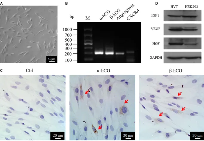
Fig. 1Characterization of HVTs. (A) Morphology of HVTs in trophoblast medium (TM) after 24 hrs. (B) Transcription of RNAs by RT-PCR in HVTs. (C,D) Protein expression of HVTs by immunofluorescence or Western blotting. HEK293 cell lysate is a positive control in D.
and apoptotic cells were detected by terminal deoxynucleotidyl trans-ferase dUTP nickend labelling (TUNEL) assay using the In Situ Cell Death Detection Kit (Roche Diagnostics, Indianapolis, IN, USA).
The pro-angiogenic effects of HVT-CM were studied in an endothelial cell tube formation assay. HUVECs; from ATCC were seeded onto growth factor-reduced MatrigelTM
(BD Biosciences, New York, USA) in 96-well plates at a density of 29104cells per well. 100ll HVT-CM or DMEM (as control) was added into the wells. Within 6 hrs, the wells were imaged with a Leica (Wetzlar, Germany) white light microscope. The average tube length was then measured with NIH Image J Software (NIH, Bethesda, MD, USA).
The presence of various proteins in the CM was assayed with a human angiogenesis antibody array (RayBiotech, Norcross, GA, USA).
RT-PCR
Total RNA was extracted from HVT cell pellets using a High Pure RNA Isolation Kit (OMEGA bio-tek, Norcross, GA, USA), according to the manufacturer’s instructions. The first strand of cDNA synthesis from RNA template was performed with cDNA reverse transcription kit (Thermo, Logan, UT, USA), according to manufacturer’s instructions. The sequences of the primers used in this study were listed as follows: for a-hCG, forward primer AACCCATTCTTCTCCCAGCC, reverse primer GCCGTGTGGTTCTCCACTTT; forb-hCG, forward primer ATGTGCGCTTC-GAGTCCATC, reverse primer GGGCCTTTGAGGAAGAGGAG; for angio-genin, forward primer CCTGACCTCACCCTGCAAAG, reverse primer GCT CGGTACTGGCATGGAG; for CXCR4, forward primer TCATCACGCTTCCC TTCTGG, reverse primer CCACCTTTTCAGCCAACAGC.
Animal procedures
All animal experiments were performed in accordance with the experi-mental protocol approved by Soochow University Laboratory Animal Center. The AMI model was performed as described before [31]. Briefly, male C57BL6 mice were anaesthetized with 3% isoflurane inhalation. A small skin cut and a purse suture were made over the left thoracic wall. After that, a small incision was made at the fourth intercostal space. The heart was then gently popped out through the window. The left main descending coronary artery (LCA) was ligated at a site 3 mm from its origin. After the ligation, one million of HVTs were intramyocardially injected into the border zone of the mice immediately. Then the heart was placed back into the intrathoracic space with fast air evacuation and chest wall closure, by the previously placed purse-string suture. All animals received buprenorphine for analgesia after the surgery.
Mice were anaesthetized with a 1.5% isoflurane–oxygen mixture at baseline (4 hrs post-MI) and 4 weeks afterwards. And two-dimensional short axis images were recorded using Vevo2100 System (Visual Son-ics, Toronto, ON, Canada).
Tissue harvesting
Mice were killed at various time-points (24 hrs for TUNEL staining, 7 days for CD8 staining and 28 days for histology) after treatment, and then the hearts were harvested and snap-frozen in optimal cutting tem-perature (OCT) compound (Leica) at 30°C for cryosectioning and his-tology or immersed to liquid nitrogen for Western blotting.
Western blotting
Heart tissues stored in liquid nitrogen were lysed using RIPA buffer (Beyotime, Nantong, Jiangsu, China). Protein concentrations were mea-sured by the BCA method (Thermo). To separate proteins, equal amounts of protein samples were loaded to SDS-PAGE gels, separated and then transferred onto PVDF membranes (Millipore, Billerica, MA, USA). Then the membranes were incubated with primary antibodies against VEGF, hepatocyte growth factor (HGF), insulin-like growth fac-tor-1 (IGF1) and GAPDH, respectively. This was followed by blotting with HRP-conjugated secondary antibodies. Immunoreactive bands were visualized by enhanced chemiluminescence on exposed X-ray films.
Masson’s trichrome staining
Masson’s trichrome stain was performed as described [32]. In brief, sections were immersed in Bouin’s solution overnight. Slides were then rinsed for 10 min. under running water to remove excessive picric acid. Then the slides were transferred to Weigert’s haematoxylin solution for 5 min. Slides were then rinsed and stained with scarlet-acid fuchsin for 5 min. and rinsed again. Slides were then stained with phospho-tungstic/phosphomolybdic for 10 min. and transferred directly into ani-line blue solution for 5 min., and 2% acetic acid for 2 min. each. Slides were then rinsed, dried, and mounted using DPX mounting media.
Histology
Hearts were sectioned at a thickness of 3lm for immunofluorescence stain-ing. The sections were first blocked with 5% BSA in PBS and then stained with
NC, USA) were used in conjunction with these primary antibodies. Images were taken by a Zeiss (Olympus, Tokyo, Japan) confocal microscopy system. Apoptotic cells in heart tissues were detected by In Situ Cell Apoptosis Detec-tion Kit, FITC (Sangon, Shanghai, China).
Statistical analysis
Results are presented as meanstandard deviation (S.D.). Compar-isons between PBS and HVT groups were performed with two-tailed unpaired Student’st-test. Differences were considered statistically sig-nificant whenP<0.05.
Results
Characterization of human HVTs
To confirm the identity of human HVTs, the RNA and protein
expres-sions of widely accepted HVT markers were examined. As indicated in
Figure 1A, HVT cells exhibited a typical multiangular morphology as
descriptions by Graham
et al.
[33] under our culture conditions.
Expressions of
a
/
b
-hCG, CXCR4 and angiogenin were detected by
RT-PCR (Fig. 1B). Meanwhile, immunocytochemical staining confirmed
the
a
/
b
-hCG expression in HVTs (Fig. 1C). Moreover, VEGF, HGF and
IGF-1 proteins were detected in HVT cell lysates (Fig. 1D) by Western
blotting. These compound data sets confirmed the identity of HVTs.
Paracrine assays on HVTs
A cytokine array (Fig. 2A) revealed that HVTs secreted pro-angiogenic
factors such as angiogenin, angiopoietin-1 (ANGPT1), angiopoietin-2
(ANGPT2) and metalloproteinase inhibitor-1/2 (TIMP-1/2). Given the
presence of the pro-angiogenic growth factors in HVT cell lysates
(Fig. 1D) and the result of paracrine assays of HVT-derived CM, we
showed that CM from HVTs could promote tube formation of endothelial
cells
in vitro
. When incubated with HVT-CM, the tube formation of
HUVECs on Matrigel
TMwas enhanced within 6 hrs (Fig. 2B), as
com-pared to the tubes formed in control DMEM. Furthermore, HVT-CM
enhanced the resistance of NRCMs to oxidative stress. After exposure to
100
l
M H
2O
2for 24 hrs, the number of TUNEL-positive cells was lower
in the HVT-CM group than that in the control IMDM group (Fig. 2C).
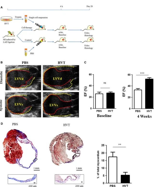
Intramyocardial injection of HVTs improves
cardiac function and decreases infarct size
The animal study design is outlined in Figure 3A. To determine the
cardiac function, echocardiography was performed at 4 hrs
indicated that HVT treatment protected cardiac functions while the
cardiac functions in PBS-treated mice continued to deteriorate
(Fig. 3C). We reasoned that part of HVT therapeutic effects on
car-diac function may come from decreased scar size. Thus, fibrosis
was analysed by Masson’s trichrome staining. Compared to
PBS-injected hearts, HVT treatment significantly inhibited scar formation
(Fig. 3D). These data conferred the therapeutic benefits of HVTs in
our mouse models of AMI.
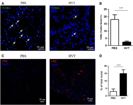
HVTs attenuate tissue apoptosis and promote
angiogenesis
To test the cardioprotective effects of HVTs, we performed TUNEL
staining to evaluate myocardial apoptosis 24 hrs after the treatment.
HVT injection reduced TUNEL-positive nuclei (Figs. 4A and B). These
data sets manifest a cardioprotective effect of HVT treatment post-MI.
As CM from HVTs promoted the tube formation of HUVECs
in vitro
(Fig. 2A), we showed that transplantation of HVTs can
stimu-late neovascularization in post-MI hearts. vWF-positive capillary
den-sity was strikingly higher in the HVT-treated group than that in the
PBS control-treated group (Figs. 4C and D).
HVTs do not graft permanently or trigger
immune reactions in the mouse heart
Although a few HNA
+cells could be detected in the cell-injected hearts
(Fig. 5A), the small numbers of engrafted cells seemed not sufficient
to support the observed therapeutic benefits. This suggested that
HVTs exerted cardiac protection through paracrine effects rather than
direct engraftment and differentiation [34].
To evaluate the immunogenicity of such xenogenic transplantation
of HVTs [35], immunofluorescent staining of immune cells was
per-formed 7 days after cell injection. The numbers of CD8-positive T
cells were indistinguishable between the HVT and PBS treatment
groups (Fig. 5B and C), suggesting that injection of human HVTs did
not trigger T-cell infiltration in the mouse heart.
Discussion
Fig. 3Cardiac function and fibrosis. (A) Schematic diagram showing the animal procedures. (B) Representative echocardiography images at 4 weeks after treatment. The pericardium and endocardium are outlined with yellow and red dotted lines, respectively. LVVd, left ventricular volume in dias-tole; LVVs, left ventricular volume in systole. (C) Left ventricular ejection fraction (LVEF) measured by echocardiography at baseline (left) and 4 weeks afterwards (right) in PBS or HVT groups (n=7–9 mice per group). Baseline LVEFs were indistinguishable between the two groups. (D) Representative Masson’s trichrome-stained images and quantification of fibrotic area of the infarcted myocardium 4 weeks after treatments (n=4 mice per group). Scar tissue and viable myocardium are identified by the blue and red colours, respectively. Snapshots of the infarct border zone (black box area) are presented beneath each group.**indicates P<0.01 when HVT group compared to PBS.,***indicates P<0.001 when HVT group compared to PBS. Data are presented as meanS.D.
group).***indicates P<0.001 when HVT group compared to PBS. Data are presented as meanS.D.
patients suffering from progressive heart failure as a result of
myo-cardium injury.
The last decade witnessed a burst of cell therapy trials for heart
diseases. Various sources of cells were studied to treat MI. These
cells include telocytes, skeletal myoblasts, BMSCs, EPCs, MSCs,
ESCs and iPSCs [1, 37, 38]. Intrinsic heart stem cells in MI treatment
research have propelled the recent clinical trials using ckit-positive
CSCs or cardiosphere-derived cells [11, 19].
Nevertheless, there is an urgent need for alternative cells sources
to promote cardiac regeneration. HVTs can be readily isolated from
human placental villous. Comparing to stem cells, HVTs can be readily
grown into a large quantity, avoiding ethical and logistical issues as
harvesting of HVTs does not harm the mother or the infant. Overall,
the HVTs encompass several properties, as described above, which
encourage their utilization in the cell transplantation for cardiac repair.
Our present results demonstrated that HVTs are capable of
pro-moting angiogenesis, reducing apoptosis and fibrosis, and improving
cardiac function in mice with AMI.
We found that CM collected from HVTs can promote the tube
for-mation of HUVECs on Matrigel
TMand enhance the resistance of rat
cardiomyocytes to oxidative stress
in vitro
. After injection, only a few
of HVTs engrafted in the mouse heart. This might indicate that
injected cells contributed to therapeutic benefits mainly through
para-crine effects, for example secretion of regenerative factors. It has
been reported that trophoblasts produce various factors including
VEGF, SDF-1, HGF and IGF-1 [39]. These factors have been shown to
play essential roles in heart regeneration. VEGF is a pro-angiogenic
agent [40]. SDF-1 augments EPCs recruitment and haematopoietic
stem cells (HSCs) mobilization and homing to ischaemic sites,
subse-quent promoting neovascularization in infarct area [41, 42]. HGF has
mitogenic, morphogenic, and anti-apoptotic effects and plays pivotal
roles in regeneration and protection of organs such as liver, kidney
and lung and reduces ischaemia/reperfusion injury in AMI hearts
directly by suppressing cardiomyocyte cell death [43], and IGF-1 is a
potent pro-survival factor, which has been shown to reduce myocyte
apoptosis and ventricular dilation after infarction [44]. In addition, it
has been reported that genes related to apoptosis inhibition were
up-regulated in first trimester placental villi [45], which may contribute to
cardiomyocyte survival after ischaemia.
Xenogenic/allogeneic cell transplantation can lead to potential
graft-versus-host disease [35]. Here we showed that there were no
significant differences in the numbers of CD8-positive T cells between
the HVT and PBS treatment groups in immunocompetent (normal)
mice. This is consistent with the previous observations regarding the
immunomodulatory effects of HVTs. During pregnancy, to prevent
deleterious immune response towards the conceptus, trophoblasts
induce tolerance of the foetal allograft against maternal immune
sys-tem through Fas
–
FasL interactions [46, 47].
Conclusion
Taken together, this study demonstrates the regenerative potential
of HVTs in AMI. Such therapeutic benefits are mediated by the
pro-angiogenic, anti-apoptotic and anti-fibrotic effects of HVTs.
Therefore, HVTs may be used an alternative cell source for the
treatment of MI.
Acknowledgements
This work was supported by grants from National Natural Science Foundation of China (Grant No. 81370216; 81671485; 81570457; 81301844 and 31500636), and Natural Science Foundation of Jiangsu Province (Grant No. BK20150319) and the Priority Academic Program Development of Jiangsu Higher Education Institutes.
Conflicts of interest
The authors confirm that there are no conflicts of interest.
References
1. Hou J, Wang L, Jiang J,et al.Cardiac stem cells and their roles in myocardial infarction.
Stem Cell Rev. 2013; 9: 326–38.
2. Lalit PA, Hei DJ, Raval AN,et al.Induced pluripotent stem cells for post-myocardial infarction repair: remarkable opportunities and challenges.Circ Res. 2014; 114: 1328–45. 3. Yang H, Sun W, Quan N,et al.
Cardiopro-tective actions of Notch1 against myocardial infarction via LKB1-dependent AMPK signal-ing pathway. Biochem Pharmacol. 2016; 108: 47–57.
4. Singla DK.Stem cells and exosomes in car-diac repair.Curr Opin Pharmacol. 2016; 27: 19–23.
5. Antanaviciute I, Ereminiene E, Vysockas V, et al. Exogenous connexin43-expres-sing autologous skeletal myoblasts ame-liorate mechanical function and electrical activity of the rabbit heart after experi-mental infarction. Int J Exp Pathol. 2015; 96: 42–53.
6. Orlic D, Kajstura J, Chimenti S,et al.Bone marrow cells regenerate infarcted myocar-dium.Nature. 2001; 410: 701–5.
7. Sanganalmath SK, Bolli R.Cell therapy for heart failure: a comprehensive overview of experimental and clinical studies, current challenges, and future directions.Circ Res. 2013; 113: 810–34.
8. Barad L, Schick R, Zeevi-Levin N, et al. Human embryonic stem cells vs human induced pluripotent stem cells for cardiac repair.Can J Cardiol. 2014; 30: 1279–87. 9. Li Q, Guo Y, Ou Q,et al.Intracoronary
admin-istration of cardiac stem cells in mice: a new, improved technique for cell therapy in murine models.Basic Res Cardiol. 2011; 106: 849–64. 10. Matsuura K, Honda A, Nagai T,et al. Trans-plantation of cardiac progenitor cells amelio-rates cardiac dysfunction after myocardial infarction in mice.J Clin Invest. 2009; 119: 2204–17.
11. Makkar RR, Smith RR, Cheng K, et al. Intracoronary cardiosphere-derived cells for
895–904.
12. Hensley MT, de Andrade J, Keene B,et al. Cardiac regenerative potential of cardio-sphere-derived cells from adult dog hearts.J Cell Mol Med. 2015; 19: 1805–13. 13. Gavira JJ, Nasarre E, Abizanda G, et al.
Repeated implantation of skeletal myoblast in a swine model of chronic myocardial infarction.Eur Heart J. 2010; 31: 1013–21. 14. Lodi D, Iannitti T, Palmieri B.Stem cells in
clinical practice: applications and warnings.
J Exp ClinCancer Res. 2011; 30: 1–20. 15. Murry CE, Wiseman RW, Schwartz SM,
et al.Skeletal myoblast transplantation for repair of myocardial necrosis.J Clin Invest. 1996; 98: 2512–23.
16. Jeong J-O, Han JW, Kim J-M,et al. Malig-nant tumor formation after transplantation of short-term cultured bone marrow mes-enchymal stem cells in experimental myocardial infarction and diabetic neuropa-thy.Circ Res. 2011; 108: 1340–7. 17. Riggs JW, Barrilleaux BL, Varlakhanova N,
et al.Induced pluripotency and oncogenic transformation are related processes.Stem Cells Dev. 2013; 22: 37–50.
18. Breitbach M, Bostani T, Roell W, et al. Potential risks of bone marrow cell trans-plantation into infarcted hearts.Blood. 2007; 110: 1362–9.
19. Bolli R, Chugh AR, D’Amario D,et al. Car-diac stem cells in patients with ischaemic cardiomyopathy (SCIPIO): initial results of a randomised phase 1 trial.Lancet. 2011; 378: 1847–57.
20. Capellini I.The evolutionary significance of placental interdigitation in mammalian reproduction: contributions from compara-tive studies.Placenta. 2012; 33: 763–8. 21. Cooper JC, Sharkey AM, McLaren J,et al.
Localization of vascular endothelial growth factor and its receptor, flt, in human placenta and decidua by immunohistochemistry. J Reprod Fertil. 1995; 105: 205–13. 22. Han VK, Bassett N, Walton J, et al.The
expression of insulin-like growth factor (IGF) and IGF-binding protein (IGFBP) genes in the human placenta and membranes: evi-dence for IGF-IGFBP interactions at the feto-maternal interface.J Clin Endocrinol Metab. 1996; 81: 2680–93.
23. Dvir T, Kedem A, Ruvinov E,et al. Prevas-cularization of cardiac patch on the
omen-Guller S, et al.First trimester trophoblast cells secrete Fas ligand which induces immune cell apoptosis. Mol Hum Reprod. 2004; 10: 55–63.
25. Mouillet JF, Mishima T, Paffaro AM,et al. The expression and post-transcriptional regulation of FSTL1 transcripts in placental trophoblasts.Placenta. 2015; 36: 1231–8. 26. Wei K, Serpooshan V, Hurtado C,et al.
Epi-cardial FSTL1 reconstitution regenerates the adult mammalian heart.Nature. 2015; 525: 479–85.
27. Vunjak-Novakovic G.Cardiac biology: a pro-tein for healing infarcted hearts. Nature. 2015; 525: 461–2.
28. van Rooij E.Cardiac Repair after Myocardial Infarction.N Engl J Med. 2016; 374: 85–7. 29. Wang YL, Qiu W, Feng HC,et al.
Immortal-ization of normal human cytotrophoblast cells by reconstitution of telomeric reverse transcriptase activity. Mol Hum Reprod. 2006; 12: 451–60.
30. Cui Y, Wang W, Dong N,et al.Role of corin in trophoblast invasion and uterine spiral artery remodelling in pregnancy. Nature. 2012; 484: 246–50.
31. Gao E, Lei YH, Shang X,et al.A novel and efficient model of coronary artery ligation and myocardial infarction in the mouse.Circ Res. 2010; 107: 1445–53.
32. Ibrahim AG, Cheng K, Marban E.Exosomes as critical agents of cardiac regeneration triggered by cell therapy.Stem Cell Reports. 2014; 2: 606–19.
33. Graham CH, Lysiak JJ, McCrae KR,et al. Localization of transforming growth factor-beta at the human fetal-maternal interface: role in trophoblast growth and differentia-tion.Biol Reprod. 1992; 46: 561–72. 34. Chimenti I, Smith RR, Li TS,et al.Relative
roles of direct regenerationversusparacrine effects of human cardiosphere-derived cells transplanted into infarcted mice. Circ Res. 2010; 106: 971–80.
35. Griffin MD, Ritter T, Mahon BP. Immunolog-ical aspects of allogeneic mesenchymal stem cell therapies.Hum Gene Ther. 2010; 21: 1641–55.
36. Kofidis T, de Bruin JL, Hoyt G,et al. Inject-able bioartificial myocardial tissue for large-scale intramural cell transfer and functional recovery of injured heart muscle.J Thorac Cardiovasc Surg. 2004; 128: 571–8.
38. Varga I, Danisovic L, Kyselovic J,et al.The functional morphology and role of cardiac telocytes in myocardium regeneration.Can J Physiol Pharmacol. 2016; 1–5.
39. Sokolov DI, Furaeva KN, Stepanova OI, et al.Changes in Functional Activity of JEG-3 Trophoblast Cell Line in the Presence of Factors Secreted by Placenta. Arch Med Res. 2015; 46: 245–56.
40. Banai S, Jaklitsch MT, Shou M, et al. Angiogenic-induced enhancement of collat-eral blood flow to ischemic myocardium by vascular endothelial growth factor in dogs.
Circulation. 1994; 89: 2183–9.
41. Yamaguchi J, Kusano KF, Masuo O, et al. Stromal cell-derived factor-1 effects on ex vivo expanded endothelial progeni-tor cell recruitment for ischemic neovas-cularization. Circulation. 2003; 107: 1322–
8.
42. Walter DH, Haendeler J, Reinhold J,et al. Impaired CXCR4 signaling contributes to the reduced neovascularization capacity of endothelial progenitor cells from patients with coronary artery disease. Circ Res. 2005; 97: 1142–51.
43. Nakamura T, Mizuno S, Matsumoto K, et al. Myocardial protection from ische-mia/reperfusion injury by endogenous and exogenous HGF. J Clin Invest. 2000; 106: 1511–9.
44. Davis ME, Hsieh PC, Takahashi T, et al. Local myocardial insulin-like growth factor 1 (IGF-1) delivery with biotinylated peptide nanofibers improves cell therapy for myocardial infarction. Proc Natl Acad Sci USA. 2006; 103: 8155–60.
45. Khan MA, Manna S, Malhotra N, et al. Expressional regulation of genes linked to immunity & programmed development in human early placental villi. Indian J Med Res. 2014; 139: 125–40.
46. Uckan D, Steele A, Cherry, et al. Tro-phoblasts express Fas ligand: a proposed mechanism for immune privilege in placenta and maternal invasion. Mol Hum Reprod. 1997; 3: 655–62.