!
COMPARATIVE EFFECTIVENESS AND COST OF PREOPERATIVE BREAST MRI IN ELDERLY BREAST CANCER PATIENTS
Alice K. Fortune-Greeley, BA
A dissertation submitted to the faculty of the University of North Carolina at Chapel Hill in partial fulfillment of the requirements for the degree of Doctor of Philosophy in the Department of Health Policy and Management in the Gillings School of Global Public Health.
Chapel Hill 2013
Approved by:
William R. Carpenter, PhD
Andrea K. Biddle, PhD Stephanie B. Wheeler, PhD
Hyman Muss, MD
© 2013
Alice K. Fortune-Greeley
ALL RIGHTS RESERVED
iii! ABSTRACT
ALICE K. FORTUNE-GREELEY: Comparative effectiveness and cost of preoperative breast MRI in elderly breast cancer patients
(Under the direction of William R. Carpenter, PhD)
Preoperative breast magnetic resonance imaging (MRI) has been used increasingly
in the preoperative evaluation of women with newly diagnosed breast cancer. Despite its
rapid adoption, limited evidence exists to support the routine use of breast MRI, creating
controversy in breast cancer management. Existing evidence suggests that breast MRI may
change treatment patterns, leading to surgical treatment delay and more extensive surgeries
but may not improve patient outcomes. This study is one of the first to examine the
association between preoperative breast MRI and surgical planning (i.e., time to complete
surgery and type of initial surgery), short-term outcomes (i.e., re-excision and second breast
cancer event rates), and cost in the elderly women using a large, population-based dataset.
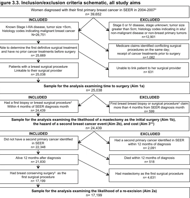
In this observational, retrospective analysis, we identified women diagnosed with
early-stage (I-IIB), operable breast cancer from 2004-2007 in the Surveillance,
Epidemiology, and End Results (SEER)-Medicare dataset. Medicare claims were used to
define the initial treatment phase, to identify breast cancer treatments, and to categorize
Medicare payments. Second breast cancer events (i.e., recurrence or a second primary
breast cancer) were identified through an algorithm validated in breast cancer patients. To control for measured confounders, we used propensity score methods.
Twelve percent of our sample had a preoperative breast MRI. Compared to women who did not undergo breast MRI, we found that receipt of breast MRI was associated with a
median 15-day delay in complete surgery and an increased likelihood of a mastectomy as
was associated with an increased hazard of a second breast cancer event. Women who
received a breast MRI had higher total all-cause and breast cancer-attributable costs during
the initial treatment phase than those women who did not undergo a breast MRI.
Since findings from this dissertation indicate that breast MRI was associated with a
slight surgical delay and an increased likelihood of a mastectomy in the absence of
evidence for improved short-term outcomes, healthcare providers and elderly breast cancer
patients should consider these factors when making informed decisions about whether the
v!
DEDICATION
This dissertation is dedicated to my parents. Mom, thank you for the encouragement to
pursue whatever my heart desires and, Dad, thank you for inspiring the passion and work
ethic to see these dreams come to fruition. I am profoundly grateful for your support as I
strive to define my career and am deeply appreciative to both of you for demonstrating the
importance of having a vocation that is meaningful, just, and has a positive impact on
ACKNOWLEDGMENT
I would like to express my profound appreciation and heartfelt gratitude to the people
who have made my doctoral education and research possible. First and foremost, I would
like to thank my mentor, advisor, and committee chair, Bill Carpenter. In addition to his
positive encouragement and sound advice throughout the dissertation process, I credit Bill
with helping me lay the essential groundwork that made this dissertation a success.
Furthermore, I thank him for helping to shape my career path as a health services
researcher. When I started working with Bill after my first semester at UNC, I was struggling
to carve out my niche and find my place in health services research. Early on, Bill helped
foster my passion for cancer outcomes research by recruiting me to comparative
effectiveness research projects and connecting me with the cancer research community at
UNC. He encouraged me to apply to the Cancer Care Quality Training Program (CCQTP)
and provided invaluable insight when I was developing my dissertation research topic. He
also helped me establish relationships with great resource people for methodological,
epidemiological, or clinical questions. All of these elements were essential in making my
dissertation a success.
I am also indebted to the other members of my doctoral committee for their valuable
time, constrictive criticism, thoughtful advice, and sincere support. Specifically, I am thankful to Andrea Biddle for her encouragement, ability to keep me on track, and expert editorial
skills. I am grateful to Stephanie Wheeler for her insightful comments and honest practical suggestions that strengthened my study design and improved my work. I am thankful to
Anne-Marie Mayer for teaching me to really explore and understand my variables and data,
vii!
I am appreciative for the clinical insight and enthusiasm for my topic that Hy Muss
contributed to my dissertation, which was a source of motivation and inspiration.
I would also like to thank other individuals in the UNC community that helped be
during my doctoral education. First, it is difficult for me to express my gratitude and respect
for Morris Weinberger. From the day I met him at Duke almost six years ago, his door has
always been open and his genuine interest in my well-being and future unwavering. I am
thankful for his support and encouragement of my decision to apply to medical school, and
my long-term personal and career goals. I also am appreciative of Katie Reeder-Hayes for
being my “go-to” breast cancer clinician, thoughtfully answering any questions I had on
topics ranging from breast cancer histologies to SEER-Medicare breast cancer variables. I
am also grateful to Keith Amos for taking the time to review surgical codes, explain technical
details of surgeries, and provide insights on the surgical decision-making process. I would
also like to thank my friends and colleagues in our dissertation working group and
programmers at ICISS, specifically Huan Liu and Seth Tyree, who helped create my
datasets.
I am appreciative of the training, mentorship, and financial support provided by the
CCQTP Pre-doctoral Fellowship Program (5R25CA116339) sponsored by the UNC
Department of Health Policy and Management. While funding provided by the National Cancer Institute enabled me to focus on completing my dissertation, the valuable training
component of the CCQTP strengthened my understanding of breast cancer management. Shadowing clinicians, sitting in tumor board meetings, and volunteering at the breast
imaging clinic gave me a much deeper appreciation for the complexities and nuances of
breast cancer diagnosis and treatment that could not have been captured with claims and
registry research.
Finally, I would like to thank my friends and family whose presence, love, and
my siblings, Hannah, Jake, and Nell. At least one of you inspires me, grounds me, motivates
me, or makes me laugh on a daily basis. I love and am so grateful for each of you. And
Doug, thank you for your ceaseless encouragement and belief my in my success. Also,
thanks for willingly putting up with me throughout the dissertation process. I am deeply
ix!
TABLE OF CONTENTS
LIST OF TABLES ... XII
LIST OF FIGURES ... XV
LIST OF ABBREVIATIONS ... XVII
CHAPTER 1: INTRODUCTION ... 1
References ... 10
CHAPTER 2: LITERATURE REVIEW ... 19
Breast cancer burden in the United States ... 19
Overview of breast cancer diagnosis, staging, and treatment ... 20
Conventional evaluation of breast cancer ... 24
Current breast MRI utilization ... 29
Limitations of current breast MRI research ... 31
References ... 71
CHAPTER 3: METHODS OVERVIEW ... 52
Overview and study design ... 52
Conceptual framework: Cancer Care Continuum ... 52
Research questions and hypotheses ... 54
Data ... 55
Study population and inclusion/exclusion criteria ... 56
Variables and measures ... 57
Analyses ... 60
CHAPTER 4: THE ASSOCIATION BETWEEN PREOPERATIVE BREAST MRI, SURGICAL DELAY, AND TYPE OF INITIAL SURGERY IN ELDERLY BREAST
CANCER PATIENTS ... 87
Overview ... 87
Introduction ... 88
Methods ... 68
Results ... 98
Discussion ... 101
References ... 119
CHAPTER 5: THE ASSOCIATION BETWEEN PREOPERATIVE BREAST MRI AND THE LIKELIHOOD OF A RE-EXCISION AND SECOND BREAST CANCER EVENT IN ELDERLY BREAST CANCER PATIENTS ... 127
Overview ... 127
Introduction ... 128
Methods ... 130
Results ... 140
Discussion ... 143
Limitations ... 146
Conclusion ... 148
References ... 160
CHAPTER 6: ESTIMATING THE COSTS OF INITIAL CANCER TREATMENT RELATED TO PREOPERATIVE BREAST MRI IN ELDERLY BREAST CANCER PATIENTS ... 168
Overview ... 168
Introduction ... 169
Methods ... 172
Results ... 179
Discussion ... 181
xi!
Conclusion ... 186
References ... 194
CHAPTER 7: DISCUSSION ... 201
Summary of findings ... 201
Contributions to existing literature, and policy and practice implications ... 204
Limitations ... 207
Future research ... 209
Conclusion ... 211
APPENDIX A: SUPPLEMENTAL TABLES AND FIGURES FOR CHAPTER 4 ... 213
APPENDIX B: SUPPLEMENTAL TABLES AND FIGURES FOR CHAPTER 5 ... 228
APPENDIX C: SUPPLEMENTAL TABLES AND FIGURES FOR CHAPTER 6 ... 247
APPENDIX D: TECHNICAL APPENDIX ... 254
LIST OF TABLES
Table 2.1. American Joint Committee on Cancer breast cancer staging
definitions ... 33
Table 2.2. Association between preoperative breast MRI and re-excision rates, a review of studies using a controlled design ... 36
Table 2.3. Association between preoperative breast MRI and recurrence rates, a review of studies using a controlled design ... 36
Table 3.1 SEER-Medicare inclusion/exclusion criteria ... 66
Table 3.2 Definition of study timeframe ... 68
Table 3.3 Identification of breast magnetic resonance imaging ... 69
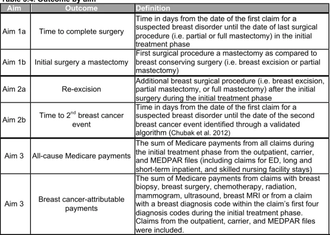
Table 3.4 Outcome by aim ... 69
Table 3.5 Patient characteristics ... 70
Table 3.6 Surgical facility and community characteristics ... 70
Table 4.1 Baseline characteristics of patients for the time to surgery analysis ... 110
Table 4.2 Baseline characteristics of patients in the analysis examining the first surgical procedure (main analysis) ... 112
Table 4.3 Multivariate logistic regression to generate preoperative breast MRI propensity scores... 114
Table 4.4 Impact of preoperative breast MRI on the time until complete surgery ... 118
Table 4.5 Impact of preoperative breast MRI on the likelihood of a mastectomy compared to BCS as the first surgical procedure (main analysis) ... 118
Table 4.6 Impact of preoperative breast MRI on the likelihood of a mastectomy compared to partial mastectomy as the first surgical procedure (sub-analysis) ... 118
Table 5.1 Baseline characteristics of patients for the re-excision analysis (main analysis) ... 152
Table 5.2 Baseline characteristics of patients for the second breast cancer event analysis ... 154
xiii!
Table 5.4 Impact of preoperative breast MRI on the likelihood of a re-excision
after partial mastectomy (sub-analysis) ... 157
Table 5.5 Association between breast MRI and a second breast cancer event ... 158
Table 6.1 Initial phase of care and cost definitions ... 187
Table 6.2 Baseline characteristics... 189
Table 6.3 Healthcare utilization and Medicare payments during the initial treatment phase, by patients with and without preoperative breast MRI ... 191
Table 6.4 Unadjusted Medicare payments, by breast MRI receipt ... 192
Table 6.5 Multivariate models examining the effect of preoperative breast MRI on Medicare payments and average marginal effect estimates ... 193
Table A.1 SEER-Medicare inclusion/exclusion criteria ... 213
Table A.2 Codes for identifying breast cancer events and surgical procedures ... 214
Table A.3 First breast-related diagnosis code distributions and the time to event by first breast-related diagnosis code ... 215
Table A.4 Multivariate proportional hazard model examining the time to complete surgery ... 218
Table A.5 Multivariate logistic regression examining the likelihood of a mastectomy as the first surgical procedure (main analysis) ... 220
Table A.6 Baseline characteristics of patients in the analysis examining the first surgical procedure (sub-analysis) ... 222
Table A.7 Multivariate logistic regression examining the likelihood of a mastectomy as the first surgical procedure (sub-analysis) ... 224
Table B.1 SEER-Medicare inclusion/exclusion criteria ... 228
Table B.2 Codes for identifying breast cancer events and treatments ... 229
Table B.3 Multivariate logistic regression to generate preoperative breast MRI propensity scores ... 231
Table B.4 Multivariate logistic regression examining the likelihood of a re-excision after BCS (main analysis) ... 233
Table B.6 Baseline characteristics of patients in the re-excision sub-analysis ... 241
Table B.7 Likelihood of a re-excision after a partial mastectomy (sub-analysis) ... 243
Table C.1 SEER-Medicare inclusion/exclusion criteria ... 247
Table C.2 Codes for identifying breast cancer events ... 248
Table C.3 Adjusted multiplicative effects on Medicare payments during the initial treatment phase ... 250
Table C.4 Sensitivity analysis: Adjusted multiplicative effects on Medicare payments during the first 12 months of diagnosis and treatment ... 252
Table D.1 Impact of preoperative breast MRI on the time until complete surgery, results of propensity score trimming ... 254
Table D.2. Impact of preoperative breast MRI on the likelihood of a mastectomy compared to BCS, results of propensity score trimming ... 254
Table D.3. Impact of preoperative breast MRI on the likelihood of a re-excision, results of propensity score trimming ... 255
xv!
LIST OF FIGURES
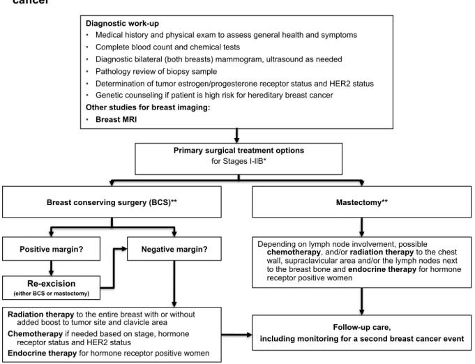
Figure 2.1 Initial tests and treatment pathways for early stage,
invasive breast cancer ... 34
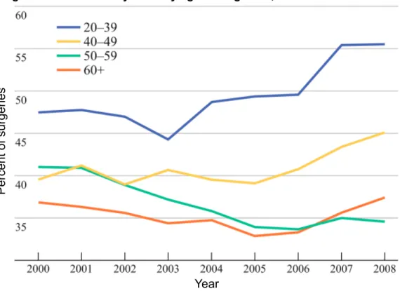
Figure 2.2 Mastectomy rates by age at diagnosis, 2000-2008 ... 35
Figure 2.3 Percent MRI use among elderly women diagnosed with ductal carcinoma in situ or locally invasive breast cancer from SEER-Medicare, 2002–2007 ... 37
Figure 3.1 The cancer care continuum ... 64
Figure 3.2 Levels and factors that impact the cancer care continuum ... 65
Figure 3.3 Inclusion/exclusion criteria schematic, all study aims ... 67
Figure 4.1 Inclusion/exclusion schematic for the time to surgery and initial type of surgery analyses ... 108
Figure 4.2 First surgical procedure comparison group definitions for the main and sub-analyses ... 109
Figure 4.3 Breast MRI propensity score distributions ... 116
Figure 4.4 Time until surgery in populations with and without preoperative breast MRI, unadjusted vs. adjusted using inverse probability weighting ... 117
Figure 5.1 Inclusion/exclusion schematic ... 149
Figure 5.2 Patient sample eligible for a re-excision in the main and sub-analyses ... 150
Figure 5.3 Algorithm for identifying a second breast cancer event ... 151
Figure 5.4 Breast MRI propensity score distributions ... 156
Figure 5.5 Rates of second breast cancer events in populations with and without preoperative breast MRI, unadjusted vs. adjusted with the use of inverse probability weighting ... 159
Figure 6.1 Inclusion/exclusion schematic ... 188
Figure A.1 Absolute standardized differences before and after adjusting with inverse probability weights for the time to surgery analysis ... 216
Figure A.3 Breast MRI propensity score distribution for the analysis
examining the first surgical procedure ... 226 Figure A.4 Absolute standardized differences before and after adjusting
with inverse probability weights for the first surgical procedure ... 227 Figure B.1 Absolute standardized differences before and after adjusting
with inverse probability weights for the re-excision analysis (main analysis) ... 237 Figure B.2 Absolute standardized differences before and after adjusting
with inverse probability weights for the second breast cancer event analysis ... 238 Figure B.3 Additional algorithms for identifying a second breast cancer event ... 239 Figure B.4 Breast MRI propensity score distribution (sub-analysis) ... 245 Figure B.5 Absolute standardized differences before and after adjusting
with inverse probability weights for the sub-analysis examining re-excision ... 246
xvii!
LIST OF ABBREVIATIONS ACOSOG American College of Surgeons Oncology Group
ACS American Cancer Society
ACRIN American College of Radiology Imaging Network
AHA American Heart Association
AJCC American Joint Committee on Cancer
ASCO American Society of Clinical Oncology
ARF Area Resource File
BCT Breast Conserving Therapy
BCS Breast Conserving Surgery
CER Comparative Effectiveness Research
COMICE Comparative Effectiveness of Magnetic Resonance Imaging in Breast Cancer
CMS Centers for Medicare & Medicaid Services
CI Confidence interval
CALGB Cancer and Leukemia Group B
CPT Current Procedural Terminology DCIS Ductal carcinoma in situ
ECOG Eastern Cooperative Oncology Group EDB Enrollment Data Base
ER Estrogen Receptor
EUSOMA European Society of Breast Cancer Specialists
GLM Generalized linear models
HCPCS Healthcare Common Procedure Coding System
HER2 Human Epidermal Growth Factor Receptor 2
HR Hazard ratio
ICD-9-CM International Statistical Classification of Diseases and Related Health Problems,
9th revision, clinical modification
ILC Invasive lobular carcinoma
IOM Institute of Medicine
MEDPAR Medicare Provider Analysis and Review
MONET Mammography of non-palpable breast tumors trial
MRI Magnetic Resonance Imaging
MSA Metropolitan Statistical Area
NCCN National Comprehensive Cancer Network
NCI National Cancer Institute
NSABP National Surgical Adjuvant Breast and Bowel Project
OR Odds ratio
PEDSF Patient Entitlement and Diagnosis Summary File
PR Progesterone Receptor
RCT Randomized Control Trial
SEER Surveillance, Epidemiology and End Results
SWOG Southwest Oncology Group TNM Tumor, Node, Metastasis System
!
CHAPTER 1: INTRODUCTION
Advanced imaging modalities in cancer care are being adopted rapidly, but scientific
evidence about their impact in real world settings lags behind.1-4 As new and advanced imaging modalities that assist with diagnosing, staging, and treating cancer are introduced
into clinical practice, it is important to examine their impact on treatment planning and to
develop evidence about their influence on short- and long-term outcomes. This evidence
can be used, in turn, to determine their appropriateness and to inform dissemination into
clinical practice. Breast magnetic resonance imaging (MRI) is one type of advanced
imaging that is being used to a greater extent in practice with limited evidence
demonstrating its benefit.
During the past ten years, breast MRI has been used increasingly as part of
preoperative planning for patients with early stage invasive breast cancer.1,2,5-16 The percentage of elderly breast cancer patients with preoperative breast MRI increased from
1.2% in 2002 to 18.8% in 2007.13 Breast MRI has been used in addition to conventional assessment, which includes clinical examination of the breasts, mammography and
ultrasound, and pathological assessment of suspicious lesions, to measure the extent of
disease and provide enhanced cancer detection. Preoperative breast MRI is highly sensitive
and capable of detecting suspicious lesions not visible with mammography or ultrasound,
population. Additional research is needed to examine the influence of breast MRI on
outcomes in the real world setting to inform its appropriate use.
Existing evidence examining the association between breast MRI and surgical
planning, short-term outcomes, and healthcare costs is limited in several aspects. First, much of the evidence is based on multiple studies from single institutions and two
randomized controlled trials (RCT). To our knowledge, few studies examine the effect of
MRI on surgical planning, outcomes, and cost in a population comparable to the US elderly
population. Patients and medical groups participating in the RCTs and single institution
studies were highly selective and the study populations were not comparable to the US
elderly population. For example, the median and mean ages for the RCTs were 5720 and 55.5 years,21 two decades younger than the average age of breast cancer patients enrolled in Medicare28 and the average patient in our study (76.1 years). Second, to our knowledge, only one study29 has examined the impact of breast MRI on surgical planning or short-term outcomes in older women, a population that is least likely to benefit from breast MRIs
because older patients are more likely to have less dense breasts and, therefore, fewer
occult tumors with conventional assessment.30-35 Third, the two randomized controlled trials were conducted in the Netherlands21 and the United Kingdom20,36 where physician practice patterns, payment and referral structures, health insurance coverage, fiscal considerations,
and patient preferences for mastectomy over breast conserving surgery (BCS) differ
significantly from the US.37-39 Fourth, no study, to our knowledge, has examined the
association between breast MRI and the cost of initial treatment in the US. The only study to
examine the cost of breast MRI was a randomized controlled trial conducted in the United
Kingdom.36
This dissertation addressed this gap in the literature by examining preoperative
3!
short-term outcomes, and Medicare costs in older, newly-diagnosed breast cancer patients
using a large, population-based dataset. It is worth noting that, although breast MRI is used
for other, non-preoperative indications such as screening high-risk women12,40-47 or
monitoring the response to neoadjuvant therapy,48-54 these indications for breast MRI are not the focus of this study.
Our central hypothesis was that preoperative breast MRI would be associated with a
surgical treatment delay and an increased likelihood of a mastectomy as the initial surgery
without evidence for improved short-term outcomes. Additionally, we hypothesized that
breast MRI would be associated with increased costs to treat the incident breast cancer.
Specific aims include:
1. Examine the association between preoperative breast MRIs and surgical
planning:
a. Examine the association between preoperative breast MRI and the time
elapsed from the date of the first claim for a suspected breast disorder
to the date of complete surgical treatment; and
b. Examine the association between of preoperative breast MRI and initial
type of breast surgery (i.e., mastectomy vs. breast conserving surgery).
2. Examine the association between preoperative breast MRIs and short-term outcomes:
a. Examine the association between preoperative breast MRI and whether a patient had a re-excision; and
b. Examine the association between preoperative breast MRI and whether
a patient had a second breast cancer event.
3. Examine the costs associated with breast MRI during the initial treatment phase
Aim 1a examined the time elapsed from the date of the first claim for a suspected
breast disorder (e.g., lump or mass in breast or abnormal mammogram) until the date of
complete surgical treatment for women with and without breast MRI. Examining the time to
complete surgical treatment is important because a surgical delay of 3 - 8 months may have
detrimental effects on breast cancer outcomes.55,56 Moreover, any delay in treatment may be anxiety-provoking for patients, lead to uncertainties related to the interpretation and
management of additional findings, and delay the start of adjuvant therapy.57 Previous single institution studies have found that breast MRI was associated with a 22- to 41-day
delay.5,15,23
Aim 1b examined the association between breast MRI and initial type of breast
surgery (i.e., BCS vs. mastectomy). Recent studies have reported that mastectomy rates for
early-stage breast cancer patients have increased, and more patients with early-stage breast cancer are undergoing aggressive surgical treatment.11,58-62 Although evidence has shown that breast conserving therapy (BCT), or BCS followed by radiation, and mastectomy
yield equivalent survival outcomes63-65 and the decision regarding the type of initial surgery has been considered a “preference-sensitive decision,”66,67 it is important to examine what other factors, such as breast MRI, may influence the likelihood of a mastectomy over BCT.
In multiple recent single institutional studies,10,23,26,68-77 meta-analyses,17,18,22 and two
European RCTs,20,21 breast MRI was found to be associated with an increased likelihood of
mastectomy compared to breast conserving surgery due to additional lesions detected by MRI that were occult with conventional assessment. However, there is a dearth of literature
examining the association between breast MRI and surgical planning on a sample
comparable to the US elderly population.
In the observational, retrospective analyses for Aim 1, we identified women
diagnosed with early stage (I-IIB), operable breast cancer from 2004-2007 whose data
5!
was linked to Medicare claims. Cancer treatment and breast MRI receipt were identified
from the Medicare claims. Time from the first claim for a suspected breast disorder to
complete surgical treatment was defined as the number of days from the first claim for a
suspected breast disorder diagnosis to the last surgical procedure in the initial treatment
phase. The first surgical procedure was defined as the first surgery that was either a BCS
(partial mastectomy or a breast excision) or a full mastectomy. To control for measured
confounders, we used propensity score methods.78,79 Based on previous findings,13,17-22 we hypothesized that women who received preoperative breast MRI would be associated with:
(a) a longer time to complete surgery; and (b) more extensive initial surgeries (i.e.,
mastectomy) than those women who did not receive a breast MRI.
In Aims 1a and 1b, we used multivariate logistic regression to determine each
patient’s propensity for receipt of preoperative breast MRI on the basis of observed patient
and surgical facility characteristics.80 We selected variables based on their hypothesized relationship with breast MRI receipt and the outcome (either time to surgery or type of first
surgery). Patient-level covariates included year of diagnosis (2004-2007), age group at
diagnosis (in five-year intervals), marital status, Medicare state buy-in coverage status, race,
and co-morbidities using the National Cancer Institute (NCI) combined index.81 We included person-level tumor characteristics such as stage, grade, histology, and tumor size as well as hormone receptor status. Surgical provider variables were included, such as whether or not
the surgical facility was affiliated with NCI Cooperative Groups with breast cancer research portfolios, a designated NCI Cancer Center, a teaching hospital, or had high breast cancer
surgical volume. We also included indicators for SEER region and education level as the
percentage of high school graduates in the patient’s zip code of residence.
Aim 2a examined the association between breast MRI and the likelihood of
re-excision after a patient’s initial surgery. Re-re-excision rates after initial breast conserving
60% of breast conserving surgeries.82-86 Re-excisions have multiple negative consequences,
including a worsened cosmetic outcome, delay in adjuvant therapy, and higher cost
associated with additional treatment.84,85,87-89 Reoperation rates are also an important clinical issue in the elderly population. Operations and re-excisions are more problematic for elderly
women who are more likely to have co-morbidities and for whom it is riskier to undergo
anesthesia. Furthermore, recovery from surgery is much more difficult for these women.
Because of its sensitivity and enhanced imaging, researchers and clinicians have begun to
investigate whether or not breast MRI can increase the likelihood of identifying clear margins
after the initial surgery and reduce the need for re-excisions. Multiple single institution
studies, meta-analyses, and two RCTs found no difference in re-excision rates between
patients with and without preoperative breast MRI.10,17,18,20-23,26,27,68
Aim 2b examined the likelihood of whether the women had a second breast cancer
event, either a recurrence or a second cancer in the contralateral breast. Studies have
shown that recurrence rates after the completion of adjuvant therapy are 6–13%,90-92 and the likelihood of recurrence has been reported to peak within the first five years after primary
treatment.93-95 A second breast cancer event is an important short-term outcome due to its effect on overall survival96 as well as the physiological and physical distress of additional cancer treatments. Additionally, anxiety from fear of recurrence or a second breast cancer is one of the most prevalent long-term psychological consequences of breast cancer.97-99
Because of breast MRI sensitivity and enhanced imaging, researchers and clinicians have examined whether or not breast MRI can reduce the likelihood of a second breast cancer
event. One RCT from United Kingdom20 found no significant difference in one-year, local recurrence-free interval rates, which is consistent with other retrospective, single institution
studies reporting that preoperative breast MRI was not associated with reduced local
recurrence rates.10,24-27 Despite existing evidence examining the effectiveness of
7!
breast MRI and the likelihood of a re-excision or second breast cancer event in a sample
comparable to the US elderly population.
Aims 2a and 2b used a similar cohort of elderly women described in Aim 1. However,
for Aim 2a, we limited our sample to women eligible for re-excision by including only women
with BCS as their initial surgery. Cancer treatment and breast MRI receipt were again
identified from the Medicare claims. A re-excision was defined as a claim for a breast
surgical procedure (a breast excision, partial mastectomy, or conversion to mastectomy)
after the initial surgery but during the initial treatment phase, which ended on the last day of
treatment before a treatment gap of 90 days. Second breast cancer events were identified
through an algorithm validated in breast cancer patients using information regarding
secondary cancers and surgical procedures from claims data and SEER registries.100 To control for measured confounders, we used propensity score methods78,79 to estimate the association of breast MRI on the likelihood of a re-excision and the hazard of a second
breast cancer event. Based on previous findings,10,17,20-22,24-27 we hypothesized that the group of women who received a preoperative breast MRI would have a similar likelihood of
a re-excision (Aim 2a) and a second breast cancer event (Aim 2b) as the group of women
who did not have a preoperative breast MRI.
As with Aim 1, we used multivariate logistic regression in the analyses for Aim 2 to determine each patient’s propensity for receipt of preoperative breast MRI on the basis of
observed patient and surgical facility characteristics.80 Again, we selected from the previously mentioned variables based on their hypothesized relationship with breast MRI
receipt and outcome (re-excision or second breast cancer event).
The third study aim focused on the costs associated with breast MRI for elderly
breast cancer patients in the US from the perspective of Medicare. Funds spent on breast
cancer, the most expensive cancer diagnosis, are estimated at $16.5 billion, which comprise
estimated that national expenditures for breast cancer will increase by 24.2% and reach
$20.5 billion by 2020.101 Spending on breast cancer is not only growing due to increased prevalence, but also due to escalating treatment costs per patient.102-104 Studies also have estimated that the inflation-adjusted cost of initial care for each breast cancer patient
increased by 25% from $16,775 in 1991 to $20,964 in 2002.105 Stakeholders view the increasing cost of cancer care as a major societal issue that may impact the health of the
US population and exacerbate disparities in care and outcomes.106 Rising costs have prompted the American Society of Clinical Oncology (ASCO) to create a task force on the
cost of cancer care to examine the drivers of increasing cancer costs, their impact, and
strategies to modulate them in order to sustain progress against cancer and universal
access to high-quality care.106,107
The rising cost of breast cancer treatment has been attributed to changes in
treatment patterns and the increased use of targeted therapies and supportive medicine. 102-104 Additionally, it has been estimated that the growing use of advanced imaging contributes
to the accelerating cost of breast cancer care, with the costs of advanced imaging
increasing at a greater rate than total costs among Medicare beneficiaries with cancer.2,3 Preoperative breast MRI is an example of an advanced imaging modality that is being
rapidly adopted, and research has shown that breast MRI may change treatment patterns in ways that may affect the cost of the initial treatment phase through additional diagnostic
work-up21,108,109 and lead to extensive surgical treatment without reducing the likelihood and cost of re-excision surgeries.17,18,20-22 Because of breast MRI’s influence on treatment patterns and limited evidence for improved outcomes, it is important to examine the effect
this imaging has on the overall cost to care for women with breast cancer. No study, to our
knowledge, has examined the association between breast MRI and the cost of initial
treatment in the US. The only study to examine the cost of breast MRI was a RCT
9!
For the third study aim, we used the same patient sample from Aim 1, or women
diagnosed with early stage (I-IIB), operable breast cancer from 2004-2007 in the SEER
registries. Using claims from 2004 through 2009, we identified Medicare payments during
the initial treatment phase, which we defined to include the diagnostic, preoperative,
surgical, and adjuvant therapy stages of care. For women with and without a preoperative
breast MRI, we examined unadjusted all-cause and breast cancer-attributed Medicare
payments during the initial treatment phase. Further, we used multivariate generalized linear
models, controlling for the same independent variables as in Aims 1 and 2, to generate the
adjusted multiplicative and marginal effects of preoperative breast MRI on all-cause or
breast cancer-attributed Medicare payments. We hypothesized that the overall cost of care
for older women with breast cancer would be higher for those women who received a
preoperative breast MRI than for women who did not.20
The subsequent chapters are organized as follows: (a) Chapter 2 provides an
overview of the breast cancer burden and a review of the current standards for diagnosis,
staging, and treatment, including conventional and breast MRI evaluation of breast cancer;
(b) Chapter 3 describes the methods used in this dissertation, detailing the conceptual
framework, research questions and hypotheses, data sources, study population, variables
and measurement, and statistical analyses employed; (c) Chapters 4, 5, and 6 comprise Aims 1, 2, and 3, respectively, and each of these chapters is written as a self-contained
journal manuscript and, hence, includes an overview, introduction, methods, results, and discussion (because they are to be submitted for publication, there are some redundancies
across papers); and (d) Chapter 7 summarizes this dissertation’s strengths and limitations,
References
1. Sommer CA, Stitzenberg KB, Tolleson-Rinehart S, et al: Breast MRI utilization in older patients with newly diagnosed breast cancer. J Surg Res 170:77-83, 2011 2. Dinan MA, Curtis LH, Hammill BG, et al: Changes in the Use and Costs of Diagnostic
Imaging Among Medicare Beneficiaries With Cancer, 1999-2006. JAMA 303:1625, 2010
3. Hu YY, Kwok AC, Jiang W, et al: High-cost imaging in elderly patients with stage IV cancer. J Natl Cancer Inst 104:1164-72, 2012
4. Crivello ML, Ruth K, Sigurdson ER, et al: Advanced Imaging Modalities in Early Stage Breast Cancer: Preoperative Use in the United States Medicare Population. Ann Surg Oncol 20:102-10, 2013
5. Krishnan M, Thorsteinsson D, Horowitz N, et al: The influence of preoperative MRI in the timing and type of therapy in women newly diagnosed with breast cancer. AJR Am J Roentgenol 190:A31-4, 2008
6. Hollingsworth AB, Stough RG, O'Dell CA, et al: Breast magnetic resonance imaging for preoperative locoregional staging. Am J Surg 196:389-397, 2008
7. Silverstein MJ, Lagios MD, Recht A, et al: Image-detected breast cancer: state of the art diagnosis and treatment. Journal of the American College of Surgeons 209:504-520, 2009
8. Bassett LW, Dhaliwal SG, Eradat J, et al: National trends and practices in breast MRI. AJR Am J Roentgenol 191:332-9, 2008
9. Dang CM, Zaghiyan K, Karlan SR, et al: Increased use of MRI for breast cancer surveillance and staging is not associated with increased rate of mastectomy. The American Surgeon 75:937-940, 2009
10. Hwang N, Schiller DE, Crystal P, et al: Magnetic resonance imaging in the planning of initial lumpectomy for invasive breast carcinoma: its effect on ipsilateral breast tumor recurrence after breast-conservation therapy. Ann Surg Oncol 16:3000-3009, 2009
11. Katipamula R, Degnim AC, Hoskin T, et al: Trends in Mastectomy Rates at the Mayo Clinic Rochester: Effect of Surgical Year and Preoperative Magnetic Resonance Imaging. J Clin Oncol 27:4082-4088, 2009
12. Morrow M, Waters J, Morris E: MRI for breast cancer screening, diagnosis, and treatment. Lancet 378:1804-11, 2011
13. Tuttle TM, Jarosek S, Durham S, et al: Use of Preoperative MRI Among Older
11!
Minnesota DEcIDE Center, under Contract No. HHSA29020100013I ), AHRQ Publication No. 12-EHC086-EF, 2012
14. Killelea BK, Lannin DR, Horvath LJ, et al: Factors Associated with Breast MRI Use: A Population-Based Analysis. Ann Surg Oncol epub, 2012
15. Hulvat M, Sandalow N, Rademaker A, et al: Time from diagnosis to definitive operative treatment of operable breast cancer in the era of multimodal imaging. Surgery 148:746-751, 2010
16. Breslin TM, Banerjee M, Gust C, et al: Trends in advanced imaging use for women undergoing breast cancer surgery. Cancer 119:1251-6, 2013
17. Plana MN, Carreira C, Muriel A, et al: Magnetic resonance imaging in the
preoperative assessment of patients with primary breast cancer: systematic review of diagnostic accuracy and meta-analysis. Eur Radiol 22:26-38, 2012
18. Houssami N, Ciatto S, Macaskill P, et al: Accuracy and surgical impact of magnetic resonance imaging in breast cancer staging: systematic review and meta-analysis in detection of multifocal and multicentric cancer. J Clin Oncol 26:3248-58, 2008 19. Brennan ME, Houssami N, Lord S, et al: Magnetic Resonance Imaging Screening of
the Contralateral Breast in Women With Newly Diagnosed Breast Cancer:
Systematic Review and Meta-Analysis of Incremental Cancer Detection and Impact on Surgical Management. J Clin Oncol 27:5640-5649, 2009
20. Turnbull L, Brown S, Harvey I, et al: Comparative effectiveness of MRI in breast cancer (COMICE) trial: a randomised controlled trial. Lancet 375:563-571, 2010 21. Peters NH, van Esser S, van den Bosch MA, et al: Preoperative MRI and surgical
management in patients with nonpalpable breast cancer: the MONET - randomised controlled trial. Eur J Cancer 47:879-86, 2011
22. Houssami N, Turner R, Morrow M: Preoperative Magnetic Resonance Imaging in Breast Cancer: Meta-Analysis of Surgical Outcomes. Ann Surg 257:249-255, 2013 23. Bleicher RJ, Ciocca RM, Egleston BL, et al: Association of routine pretreatment
magnetic resonance imaging with time to surgery, mastectomy rate, and margin status. Journal of the American College of Surgeons 209:180-187, 2009
24. Solin LJ, Orel SG, Hwang W-T, et al: Relationship of Breast Magnetic Resonance Imaging to Outcome After Breast-Conservation Treatment With Radiation for Women With Early-Stage Invasive Breast Carcinoma or Ductal Carcinoma in Situ. J Clin Oncol 26:386-391, 2008
25. Fischer U, Zachariae O, Baum F, et al: The influence of preoperative MRI of the breasts on recurrence rate in patients with breast cancer. Eur Radiol 14:1725-1731, 2004
27. Shin HC, Han W, Moon HG, et al: Limited value and utility of breast MRI in patients undergoing breast-conserving cancer surgery. Ann Surg Oncol 19:2572-9, 2012 28. Kanapuru B, Ershler WB, Hesdorffer C, et al: Long-term survival of older breast
cancer patients: population-based estimates over three decades. Breast Cancer Res Treat 134:853-7, 2012
29. Wang S-Y, Kuntz KM, Tuttle TM, et al: The association of preoperative breast magnetic resonance imaging and multiple breast surgeries among older women with early stage breast cancer. Breast Cancer Res Treat, epub 2013
30. Biglia N, Bounous VE, Martincich L, et al: Role of MRI (magnetic resonance imaging) versus conventional imaging for breast cancer presurgical staging in young women or with dense breast. Eur J Cancer 37:199-204, 2011
31. Bluemke DA, Gatsonis CA, Chen MH, et al: Magnetic resonance imaging of the breast prior to biopsy. JAMA 292:2735-42, 2004
32. Sardanelli F, Giuseppetti GM, Panizza P, et al: Sensitivity of MRI versus
mammography for detecting foci of multifocal, multicentric breast cancer in fatty and dense breasts using the whole-breast pathologic examination as a gold standard. American Journal of Roentgenology 183:1149-57, 2004
33. Van Goethem M, Schelfout K, Kersschot E, et al: Enhancing area surrounding breast carcinoma on MR mammography: comparison with pathological examination. Eur Radiol 14:1363-1370, 2004
34. Boyd NF, Guo H, Martin LJ, et al: Mammographic density and the risk and detection of breast cancer. N Engl J Med 356:227-236, 2007
35. Kerlikowske K: Efficacy of screening mammography among women aged 40 to 49 years and 50 to 69 years: comparison of relative and absolute benefit. JNCI Monographs 1997:79-86, 1997
36. Turnbull L, Brown S, Olivier C, et al: Multicentre randomised controlled trial examining the cost-effectiveness of contrast-enhanced high field magnetic
resonance imaging in women with primary breast cancer scheduled for wide local excision (COMICE). Health technology assessment (Winchester, England) 14:1-182, 2010
37. Garcia-Etienne CA, Tomatis M, Heil J, et al: Mastectomy trends for early-stage breast cancer: A report from the EUSOMA multi-institutional European database. Eur J Cancer 48:1947-56, 2012
38. Habermann E, Abbott A, Parsons H: Are Mastectomy Rates Really Increasing in the United States? J Clin Oncol 28:3437-3441, 2010
13!
40. Riedl CC, Ponhold L, Flöry D, et al: Magnetic resonance imaging of the breast improves detection of invasive cancer, preinvasive cancer, and premalignant lesions during surveillance of women at high risk for breast cancer. Clinical cancer research 13:6144-6152, 2007
41. Kuhl CK, Schrading S, Leutner CC, et al: Mammography, breast ultrasound, and magnetic resonance imaging for surveillance of women at high familial risk for breast cancer. J Clin Oncol 23:8469-8476, 2005
42. Saslow D, Boetes C, Burke W, et al: American Cancer Society guidelines for breast screening with MRI as an adjunct to mammography. CA: a cancer journal for clinicians 57:75-89, 2007
43. Lord S, Lei W, Craft P, et al: A systematic review of the effectiveness of magnetic resonance imaging (MRI) as an addition to mammography and ultrasound in screening young women at high risk of breast cancer. Eur J Cancer 43:1905-1917, 2007
44. Warner E, Messersmith H, Causer P, et al: Systematic review: using magnetic resonance imaging to screen women at high risk for breast cancer. Annals of Internal Medicine 148:671-9, 2008
45. Kriege M, Brekelmans CT, Boetes C, et al: Efficacy of MRI and mammography for breast-cancer screening in women with a familial or genetic predisposition. N Engl J Med 351:427-437, 2004
46. Leach MO, Boggis C, Dixon A, et al: Screening with magnetic resonance imaging and mammography of a UK population at high familial risk of breast cancer: a prospective multicentre cohort study (MARIBS). Lancet 365:1769-78, 2005 47. National Comprehensive Cancer Network: NCCN Clinical Practice Guidelines in
Oncology: Breast Cancer Screening and Diagnosis.!Retrieved February 19, 2013, from nccn.org, 2013
48. Pickles MD, Lowry M, Manton DJ, et al: Role of dynamic contrast enhanced MRI in monitoring early response of locally advanced breast cancer to neoadjuvant
chemotherapy. Breast Cancer Res Treat 91:1-10, 2005
49. Sardanelli F, Boetes C, Borisch B, et al: Magnetic resonance imaging of the breast: recommendations from the EUSOMA working group. Eur J Cancer 46:1296-1316, 2010
50. Croshaw R, Shapiro-Wright H, Svensson E, et al: Accuracy of Clinical Examination, Digital Mammogram, Ultrasound, and MRI in Determining Postneoadjuvant
Pathologic Tumor Response in Operable Breast Cancer Patients. Ann Surg Oncol 18:3160-3163, 2011
52. Chen JH, Feig B, Agrawal G, et al: MRI evaluation of pathologically complete response and residual tumors in breast cancer after neoadjuvant chemotherapy. Cancer 112:17-26, 2008
53. Marinovich ML, Sardanelli F, Ciatto S, et al: Early prediction of pathologic response to neoadjuvant therapy in breast cancer: systematic review of the accuracy of MRI. Breast 21:669-77, 2012
54. Lobbes MB: Treatment response evaluation by MRI in breast cancer patients receiving neoadjuvant chemotherapy: there is more than just pathologic complete response prediction. Breast Cancer Res Treat 136: 313-4, 2012
55. Richards MA, Westcombe AM, Love SB, et al: Influence of delay on survival in patients with breast cancer: a systematic review. Lancet 353:1119-1126, 1999 56. Barber MD, Jack W, Dixon JM: Diagnostic delay in breast cancer. British journal of
surgery 91:49-53, 2004
57. Colbert K: The longer the delay, the greater the anxiety. Delay in treatment for breast cancer. Prof Nurse 9:517-20, 1994
58. Mcguire KP, Santillan AA, Kaur P, et al: Are Mastectomies on the Rise? A 13-Year Trend Analysis of the Selection of Mastectomy Versus Breast Conservation Therapy in 5865 Patients. Ann Surg Oncol 16:2682-2690, 2009
59. Sorbero MES, Dick AW, Beckjord EB, et al: Diagnostic breast magnetic resonance imaging and contralateral prophylactic mastectomy. Ann Surg Oncol 16:1597-1605, 2009
60. Dragun AE, Huang B, Tucker TC, et al: Increasing Mastectomy Rates Among all Age Groups for Early Stage Breast Cancer: A 10‐Year Study of Surgical Choice. Breast J. 18:318-25, 2012
61. Mahmood U, Hanlon AL, Koshy M, et al: Increasing National Mastectomy Rates for the Treatment of Early Stage Breast Cancer. Ann Surg Oncol epub:1-8, 2012
62. Gomez SL, Lichtensztajn D, Kurian AW, et al: Increasing mastectomy rates for early-stage breast cancer? Population-based trends from California. J Clin Oncol 28:e155-7; author reply e158, 2010
63. Morris AD, Morris RD, Wilson JF, et al: Breast-conserving therapy vs mastectomy in early-stage breast cancer: a meta-analysis of 10-year survival. Cancer J Sci Am 3:6-12, 1997
64. Fisher B, Jeong JH, Anderson S, et al: Twenty-five-year follow-up of a randomized trial comparing radical mastectomy, total mastectomy, and total mastectomy followed by irradiation. N Engl J Med 347:567-75, 2002
15!
66. Lee CN, Chang Y, Adimorah N, et al: Decision Making about Surgery for Early-Stage Breast Cancer. Journal of the American College of Surgeons 214:1-10, 2012
67. Wennberg JE, Fisher ES, Skinner JS: Geography and the debate over Medicare reform. Health Aff (Millwood) Suppl Web Exclusives: W96-114, 2002
68. Pengel KE, Loo CE, Teertstra HJ, et al: The impact of preoperative MRI on breast-conserving surgery of invasive cancer: a comparative cohort study. Breast Cancer Res Treat 116:161-169, 2009
69. Fan XC, Nemoto T, Blatto K, et al: Impact of presurgical breast magnetic resonance imaging (MRI) on surgical planning - a retrospective analysis from a private radiology group. Breast J 19:134-41, 2013
70. Barchie MF, Clive KS, Tyler JA, et al: Standardized pretreatment breast MRI— accuracy and influence on mastectomy decisions. J Surg Oncol 104:741-745, 2011 71. Godinez J, Gombos EC, Chikarmane SA, et al: Breast MRI in the evaluation of
eligibility for accelerated partial breast irradiation. AJR Am J Roentgenol 191:272-7, 2008
72. Grobmyer S, Mortellaro V, Marshall J: Is There a Role for Routine Use of MRI in Selection of Patients for Breast-Conserving Cancer Therapy? Journal of the American College of Surgeons 206:1045-1050, 2008
73. Braun M, Polcher M, Schrading S, et al: Influence of preoperative MRI on the surgical management of patients with operable breast cancer. Breast Cancer Res Treat 111:179-87, 2008
74. Ha GW, Yi MS, Lee BK, et al: Clinical Outcome of Magnetic Resonance Imaging-Detected Additional Lesions in Breast Cancer Patients. Journal of Breast Cancer 14:213-218, 2011
75. Pediconi F, Miglio E, Telesca M, et al: Effect of Preoperative Breast Magnetic Resonance Imaging on Surgical Decision Making and Cancer Recurrence Rates. Investigative Radiology 47: 128-135, 2012
76. Lim HI, Choi JH, Yang JH, et al: Does pre-operative breast magnetic resonance imaging in addition to mammography and breast ultrasonography change the
operative management of breast carcinoma? Breast Cancer Res Treat 119:163-167, 2010
77. Angarita FA, Acuna SA, Fonseca A, et al: Impact of preoperative breast MRIs on timing of surgery and type of intervention in newly diagnosed breast cancer patients. Ann Surg Oncol 17 Suppl 3:273-9, 2010
79. Robins JM, Hernan MA, Brumback B: Marginal structural models and causal inference in epidemiology. Epidemiology 11:550-60, 2000
80. Rosenbaum P: The central role of the propensity score in observational studies for causal effects. Biometrika, 1983
81. Klabunde CN, Legler JM, Warren JL, et al: A refined comorbidity measurement algorithm for claims-based studies of breast, prostate, colorectal, and lung cancer patients. Annals of epidemiology 17:584-590, 2007
82. Waljee JF, Hu ES, Newman LA, et al: Predictors of re-excision among women undergoing breast-conserving surgery for cancer. Ann Surg Oncol 15:1297-303, 2008
83. McCahill LE, Single RM, Aiello Bowles EJ, et al: Variability in reexcision following breast conservation surgery. JAMA 307:467-75, 2012
84. Menes TS, Tartter PI, Bleiweiss I, et al: The consequence of multiple re-excisions to obtain clear lumpectomy margins in breast cancer patients. Ann Surg Oncol 12:881-885, 2005
85. Morrow M, Jagsi R, Alderman AK, et al: Surgeon recommendations and receipt of mastectomy for treatment of breast cancer. JAMA 302:1551-6, 2009
86. Wright MJ, Park J, Fey JV, et al: Perpendicular inked versus tangential shaved margins in breast-conserving surgery: does the method matter? Journal of the American College of Surgeons 204:541-549, 2007
87. Al-Ghazal SK, Blamey RW, Stewart J, et al: The cosmetic outcome in early breast cancer treated with breast conservation. Eur J Surg Oncol 25:566-70, 1999 88. Wazer DE, DiPetrillo T, Schmidt-Ullrich R, et al: Factors influencing cosmetic
outcome and complication risk after conservative surgery and radiotherapy for early-stage breast carcinoma. J Clin Oncol 10:356-63, 1992
89. Pleijhuis RG, Graafland M, De Vries J, et al: Obtaining adequate surgical margins in breast-conserving therapy for patients with early-stage breast cancer: current modalities and future directions. Ann Surg Oncol 16:2717-2730, 2009
90. Goodwin A, Parker S, Ghersi D, et al: Post-operative radiotherapy for ductal
carcinoma in situ of the breast--a systematic review of the randomised trials. Breast 18:143-9, 2009
91. Brewster AM, Hortobagyi GN, Broglio KR, et al: Residual risk of breast cancer recurrence 5 years after adjuvant therapy. J Natl Cancer Inst 100:1179-83, 2008 92. Boyages MBBS J, Delaney MBBS G, Taylor MBBS R: Predictors of local recurrence
17!
93. Stokes ME, Thompson D, Montoya EL, et al: Ten-Year Survival and Cost Following Breast Cancer Recurrence: Estimates from SEER-Medicare Data. Value in Health 11:213-220, 2008
94. Saphner T, Tormey DC, Gray R: Annual hazard rates of recurrence for breast cancer after primary therapy. J Clin Oncol 14:2738-46, 1996
95. Cheng L, Swartz MD, Zhao H, et al: Hazard of recurrence among women after primary breast cancer treatment--a 10-year follow-up using data from SEER-Medicare. Cancer Epidemiol Biomarkers Prev 21:800-9, 2012
96. Early Breast Cancer Trialists’ Collaborative Group: Effects of chemotherapy and hormonal therapy for early breast cancer on recurrence and 15-year survival: an overview of the randomised trials. Lancet 365:1687-1717, 2005
97. van den Beuken‐van Everdingen MHJ, Peters ML, de Rijke JM, et al: Concerns of former breast cancer patients about disease recurrence: a validation and prevalence study. Psycho-Oncology 17:1137-1145, 2008
98. Mellon S, Gold R, Janisse J, et al: Risk perception and cancer worries in families at increased risk of familial breast/ovarian cancer. Psycho-Oncology 17:756-766, 2008 99. Deimling GT, Bowman KF, Sterns S, et al: Cancer‐related health worries and
psychological distress among older adult, long‐term cancer survivors. Psycho-Oncology 15:306-320, 2006
100. Chubak J, Yu O, Pocobelli G, et al: Administrative Data Algorithms to Identify
Second Breast Cancer Events Following Early-Stage Invasive Breast Cancer. J Natl Cancer Inst 104:931-940, 2012
101. Mariotto AB, Yabroff KR, Shao Y, et al: Projections of the cost of cancer care in the United States: 2010-2020. J Natl Cancer Inst 103:117-28, 2011
102. Elkin EB, Bach PB: Cancer's next frontier: addressing high and increasing costs. JAMA 303:1086-7, 2010
103. Bach PB: Limits on Medicare's ability to control rising spending on cancer drugs. N Engl J Med 360:626-633, 2009
104. Yabroff KR, Warren JL: High-cost imaging in elderly patients with stage IV cancer: challenges for research, policy, and practice. J Natl Cancer Inst 104:1113-4, 2012 105. Warren JL, Yabroff KR, Meekins A, et al: Evaluation of trends in the cost of initial
cancer treatment. J Natl Cancer Inst 100:888-97, 2008
106. Meropol NJ, Schrag D, Smith TJ, et al: American Society of Clinical Oncology guidance statement: The cost of cancer care. J Clin Oncol 27:3868, 2009
108. Pilewskie M, Kennedy C, Shappell C, et al: Effect of MRI on the Management of Ductal Carcinoma In Situ of the Breast. Ann Surg Oncol, epub 2012
109. Elshof LE, Rutgers EJT, Deurloo EE, et al: A practical approach to manage
!
CHAPTER 2: LITERATURE REVIEW
Breast cancer burden in the United States
Cancer is a major public health problem in the United States (US), both in terms of
the number of life-years lost and the cost of treatment. Presently, one in four deaths in the
US are attributable to cancer, which is the leading cause of death in adults aged 40 to 79.110 In 2013, it has been estimated that more than 1.6 million Americans will be newly diagnosed
with cancer and more than half a million people will die from the disease.110 Additionally, cancer prevalence is predicted to increase over the next decade with the number of
estimated cancer survivors in the US increasing from 13.8 million in 2010 to 18.1 million in
2020.101
Over the past twenty years, the medical costs of cancer have nearly doubled.111 The overall costs of cancer in 2010 have been estimated at $263.8 billion, with direct medical
costs, indirect morbidity costs, and interect mortality costs comprising $102.8, $20.9, and
$140.1 billion, respectively.112 Elements contributing to the rising cost of cancer care include increasing cancer incidence rates as the population ages, increasing prices for novel
techniques and chemotherapy agents, and increasing intensity of cancer care. 102,103,105,111,113-115 Stakeholders view the increasing cost of cancer care as a major societal issue that may
impact the health of the population and exacerbate disparities in care and outcomes.106 Rising costs have prompted the American Society of Clinical Oncology to create a task force
on the cost of cancer care to examine drivers of increasing cancer costs, their impact, and
strategies to modulate them in order to sustain progress against cancer and universal
Breast cancer is the most common cancer among women, with an estimated 3.5
million breast cancer survivors in the US in 2010,101 and is the second most fatal cancer site.110 In 2013, an estimated 234,580 women have been diagnosed with breast cancer and an estimated 39,620 are expected to die from the disease.110 Elderly women are most at risk for developing breast cancer. The incidence among women age 65 or older is approximately
five times greater than the incidence among women younger than 65, and risk increases at
all ages until age 80.116 Breast cancer has a high treatment burden and, as a result of the disease and its treatments, health-related quality of life in breast cancer patients is
significantly diminished through compromised physical, psychological, and social
functioning.117 In addition, breast cancer presents a significant cost burden in national expenditures, which were estimated at $16.5 billion in 2010. This figure constitutes 13.3% of
total healthcare spending on cancer, more than any other cancer site.112,118 Based on US population changes alone, it has been estimated that national expenditures for breast
cancer will increase by 24.2% and reach $20.5 billion by 2020.101 Spending on breast cancer is not only growing due to increased prevalence, but also to escalating treatment
costs per patient.102-104 It has been estimated that the inflation-adjusted cost of initial care for each breast cancer patient increased 25% from 1991 ($16,775) to 2002 ($20,964).105 Thus, the significant morbidity, mortality, and cost of treating breast cancer patients result in a considerable breast cancer burden in the US.
Overview of breast cancer diagnosis, staging, and treatment
After breast cancer is detected, patients have the best chance of a successful
outcome if the extent of the cancer is accurately identified, diagnosed, staged, and treated
accordingly. The cancer stage at diagnosis determines the type and extent of disease in the
21!
providers to determine prognosis and treatment options.121 The AJCC system (Table 2.1), also referred to as the Tumor, Node, Metastasis (TNM) system, is used to ascertain whether
a cancer is invasive or non-invasive, the size of the tumor (T), how many lymph nodes are
involved (N), and if the cancer has spread to other parts of the body (M). Patients have a
better prognosis if cancer is diagnosed at an early stage. Five-year survival rates for women
diagnosed between 2000-2007 with localized (i.e., confined to primary site), regional (i.e.,
spread to regional lymph nodes), and distant (i.e., metastasized) tumors were 98.6%,
83.8%, 23.3%, respectively.122 In addition to staging, other tumor factors can influence treatment decisions and patient outcomes. Hormone receptor tests determine whether or
not the tumor is estrogen receptor (ER) or progesterone receptor (PR) positive. ER/PR
positive tumors are dependent on estrogen and progesterone to grow and, thus, hormone
therapy is often recommended to block the cancer cell from using these hormones. Testing
is available to determine the amount of Human Epidermal Growth Factor Receptor 2, also
known as HER2/neu, which is a protein that stimulates the growth of breast cancer cells in
cancerous tissue. If cancerous tissue has excessive amounts of the protein, the patient
might be eligible for a targeted therapy that blocks growth of HER2/neu.47 Additionally, the nature of the tumor, such as whether or not it is multifocal (i.e., smaller cancer spots
occurring in the same quadrant of the breast as the main tumor) or multicentric (i.e., smaller cancer spots in other quadrants than the one containing the main tumor), can influence
treatment decisions.
! Multiple treatment guidelines and practice standards exist for the management of
patients with breast cancer, each with different treatment risks, costs, and outcomes. For
most women with operable breast cancer, treatment involves local therapy, such as surgery
and radiation, to remove or destroy the breast cancer, and/or systemic therapy, such as
undergo BCS or a mastectomy and may possibly have the lymph nodes under an arm
removed. After the initial surgery, a pathologist examines the excised tumor to see if
cancerous tissue remains at the margins. If the margins are positive, the patient may need a
re-excision to obtain negative tumor margins.
After surgery, many women receive adjuvant therapy to lower the chance of breast
cancer recurrence. The National Comprehensive Cancer Network (NCCN) recommends
that women who undergo BCS also receive adjuvant radiation therapy, or the combination of
the two, which is known as breast conserving therapy (BCT).47 Women may also receive hormone therapy, chemotherapy, targeted therapy, or a combination of these therapies to
eliminate any remaining cancer and reduce the risk of recurrence.
Treatment decision-making by a woman diagnosed with early-stage breast cancer is complex. Treatment decisions are based on a wide range of biological, physical, emotional, economic, and social factors.123-129 These factors include stage of disease, genetic
indicators, age, family history, and risk of local recurrence. They also include personal
experiences, concern about radiation exposure, the inconvenience of adding daily radiation to a complicated treatment regimen, social factors, educational status, insurance coverage, patient preferences regarding physical appearance, and patient fears of recurrence.
Geographic location and type of clinical care provider also play a role in treatment decisions.122
Since the early 1990s, evidence has shown that BCT and mastectomy are equally effective,130 and trials reporting long-term survival have found equivalent outcomes for both
surgeries.63,131-135 Additional research reported improved quality of life with BCT, which is often associated with improved psychosocial health in terms of body image, sexuality, and
23!
because it provides survival equivalent to total mastectomy while preserving the breast,” and
professional consensus began to favor the less-invasive surgery.64,132,140
After NCI’s 1990 publication, mastectomy rates for early-stage breast cancer
patients markedly decreased in the United States.141-144 However, recent studies have reported that this trend is changing and more patients with early-stage breast cancer are
undergoing more aggressive surgical treatment. Single institution studies indicate that
mastectomy rates for early-stage breast cancer patients increased from 35% in 2004 to 60%
in 2007 at Moffitt Cancer Center,58 from 35% of in 2004 to 60% at the Magee-Women’s' Hospital at the University of Pittsburgh,59 and from 31% in 2003 to 43% in 2006 at the Mayo
Clinic.11 Additional studies using SEER registry data (Figure 2.2) showed that mastectomies
for elderly women decreased from 2000-2005, but then increased in 2006-2008.38,61
Research using SEER registries and single institution data145-147 has also documented a trend toward more aggressive surgical treatment, which was exhibited by contralateral
prophylactic mastectomy rate in the US that more than doubled from 1998 to 2003.
These results have generated significant discussion in the lay press and medical
literature about how to explain these trends.39,148-151 Some authors have suggested that recent changes in preoperative management may explain the changing trend, such as
genetic testing for BRCA1 and BRCA2 mutations, increased education regarding surgical treatment options, concern over the long-term side effects of radiation, and the introduction
of new or improved imaging modalities (e.g., MRI or digital mammography).11,152-157
Increased mastectomy rates could also be due to improvements in mastectomy techniques
and reconstruction options,125,151,158,159 however, this explanation may be more likely in younger populations. The findings also suggest a possible global shift in breast surgical
treatment preferences toward mastectomy, and may reflect a growing patient choice for a
Conventional evaluation of breast cancer
Breast cancer can be discovered through screening mammograms, routine clinical
breast exams, breast self-exams, breast symptoms, or routine screening of high-risk
individuals with breast MRI.47 If a screening mammogram shows an abnormal area of the breast or the patient experiences unusual breast changes, such as a lump, pain, tissue
thickening, nipple discharge, or a change in breast size or shape, it is recommended that the
patient undergo a diagnostic mammogram. NCCN recommends that all women diagnosed
with breast cancer be evaluated using standard breast imaging with mammography and,
when indicated, diagnostic breast ultrasound.47 Suspicious lesions should then be biopsied and the presence of malignant cells confirmed by a pathologist.
Once the patient has a confirmed diagnosis, the cancer is then staged. According to
NCCN guidelines, clinical staging includes a physical examination of the skin of the breast,
mammary glands, and lymph nodes, as well as imaging and pathologic examination of the
breast or other tissues.47 Based on the physical examination and imaging results, the cancer is then staged based on the size of primary tumor, chest wall invasion, and presence or
absence of regional or distant metastasis according to the AJCC staging system described
earlier.
Breast MRI evaluation of breast cancer
Although the diagnostic mammogram has been the imaging gold standard to
determine the extent of disease, MRI has been used increasingly in the preoperative
evaluation of women with newly diagnosed breast cancer to provide enhanced cancer
detection during the past ten years.1,2,5-16 There is strong evidence demonstrating that breast MRI has improved sensitivity over mammography to detect additional cancerous