INVESTIGATING THE GLIAL CONTRIBUTION TO PERSISTENT NEUROPATHIC PAIN
Sarah Taves Whitley
A dissertation submitted to the faculty of the University of North Carolina at Chapel Hill in partial fulfillment of the requirements for the degree of Doctor of Philosophy in the
Curriculum in Neurobiology
Chapel Hill 2012
Approved by: Ken McCarthy PhD
William Maixner DDS PhD Edward Perl MD
Mark Zylka PhD
©2012
ABSTRACT
SARAH TAVES WHITLEY: Investigating the Glial Contribution to Persistent Neuropathic Pain
(Under the guidance of Ken McCarthy)
Persistent neuropathic pain is the coordinated activation and sensitization of glial and neuronal elements both peripherally and centrally. Here, we have investigated the role of glial fibrillary acidic protein (GFAP)-positive glial cells including astrocytes in the central nervous system and non-myelinating Schwann cells in the peripheral nervous system and their individual contributions to persistent neuropathic pain.
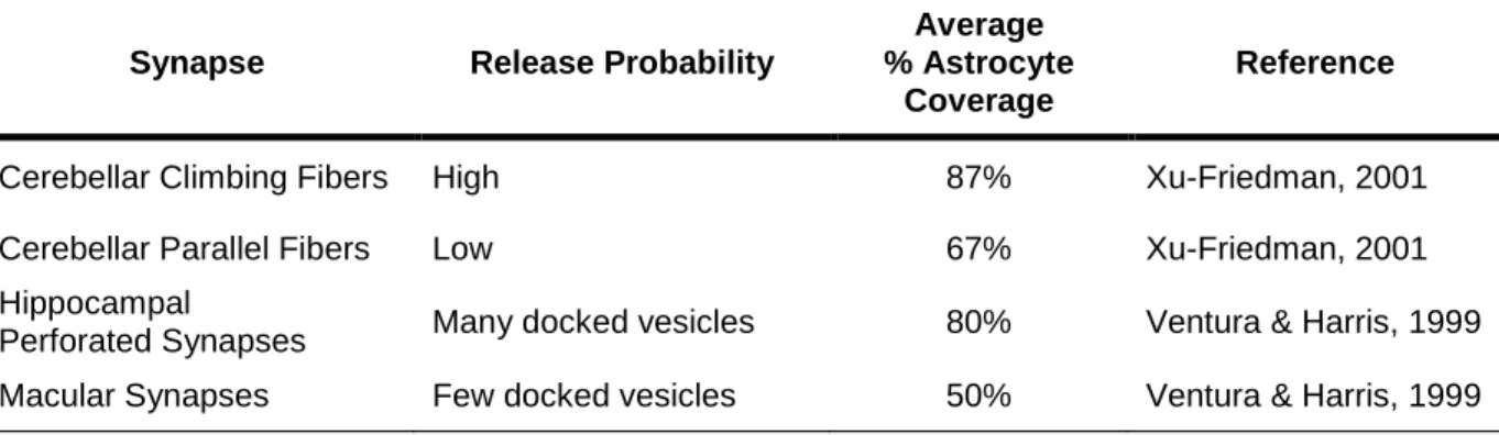
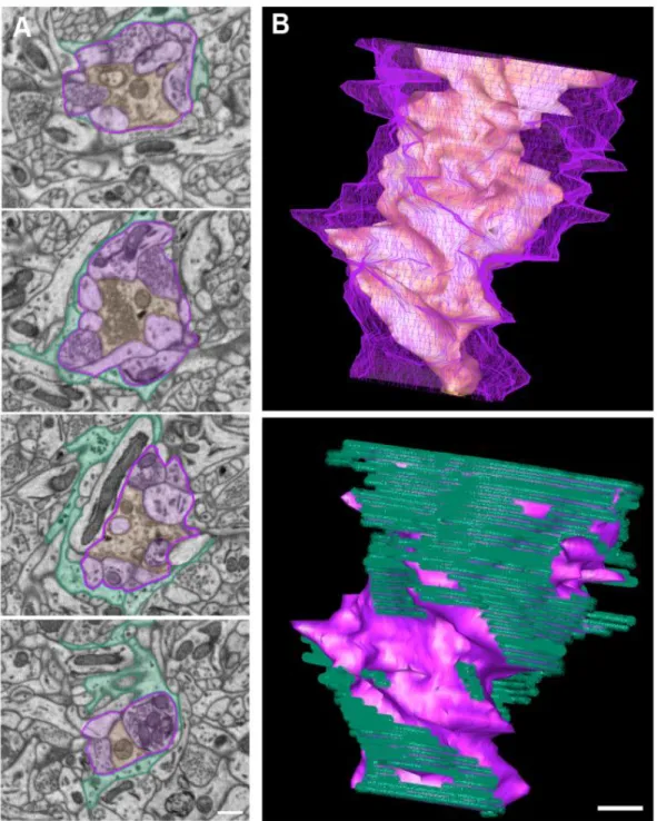
We used three-dimensional reconstruction of ultrastructural data to establish the morphological relationship between astrocyte processes and incoming C and A-delta fiber synapses with second-order pain neurons. We found that not only do astrocytes contact 100% of the C and A-delta fiber glomerular synapses, but they also provide a high degree of
ensheathment of each glomerulus. This encapsulation of the glomerular synapses puts astrocytes in a position to potentially modulate neuronal activity and synapse structure.
Next, we used transgenic and knockout mouse models to interfere vesicular
gliotransmitter release: a glial-specific IP3R2 conditional knockout mouse and a transgenic mouse expressing a dominant-negative SNARE protein. However, neither blocking IP3 dependent Ca2+ release or SNARE-dependent vesicle release have any effect on basal
inflammatory pathways known to be active in persistent pain conditions: a transgenic mouse with suppressed NFB activity and a COX2 conditional knockout mouse, both of which were selectively expressed in GFAP-positive cells. In both lines of mice, we observed a robust yet temporary alleviation of pain behavior from one to five weeks post-nerve injury. This finding indicates that the NFB-COX2 signaling pathway in GFAP-positive glia is critical to the maintenance of a specific phase of persistent neuropathic pain.
This work is dedicated to my husband, Jeremy Whitley, who has pushed me to keep my scientific passion alive while becoming a wife and a mother.
ACKNOWLEDGEMENTS
This document is the summation of an immense amount of time, energy and effort, not just by me, but by many individuals. First and foremost, I would first like to acknowledge my mentor, Dr. Ken McCarthy, for all the academic, financial and personal support throughout my graduate years. You have given me the opportunity to pursue my own interests and the guidance to learn to choose my interests wisely. The knowledge and experience I have gained from your mentorship will guide me for the rest of my career. I also deeply appreciate all of the scientific beer bets you graciously lost to me during my years of graduate school.
This work would not have been possible without the input of the entire McCarthy lab, both past and present. A special thanks goes to Kristi Boyt for her endless genotyping, Suzanne Minton who, with me, charged ahead into the field of pain research and Allison McMullen for her tireless editing of this document. I would also like to thank my student assistants Joe Gitt and Megan Hayworth, I think I learned as much from you about teaching as you learned from me about science.
I am also thankful for the support of my dissertation committee, Ed Perl, Bill Maixner, Mark Zylka and Glenn Matsushima, as well as Andrea Nackley and Juli
leading the curriculum during my years here and to Lori Blalock and Denise Kenney for administrative assistance.
I would like to acknowledge the unwavering support of numerous friends with whom I have celebrated our victories, commiserated over our setbacks, shared our complaints, voiced our encouragements and provided endless distractions. You have made the obstacles along the way molehills instead of mountains, and I cannot thank you enough.
TABLE OF CONTENTS
TABLE OF CONTENTS ... viii
LIST OF TABLES ... xii
LIST OF FIGURES ... xiii
LIST OF ABBREVIATIONS ... xiv
CHAPTER 1. INTRODUCTION TO NEUROPATHIC PAIN ...1
1.1. TREATMENT ...3
1.2. SOMATOSENSORY NEUROTRANSMISSION ...8
1.2.1. Peripheral Nervous System ...8
1.2.2. Central Nervous System ...11
1.3. SENSITIZATION ...14
1.3.1. Peripheral Sensitization ...14
1.3.2. Central Sensitization ...17
CHAPTER 2. ASTROCYTE MODULATION OF EXCITATORY NEURONAL ACTIVITY IN THE CENTRAL NERVOUS SYSTEM...23
2.1. TRIPARTITE SYNAPSE STRUCTURE...24
2.2. SYNAPSE FORMATION AND STRENGTH ...27
2.2.1. Thrombospondins ...28
2.2.2. Ephrins ...29
2.2.3. TNFα ...32
2.3. NEUROTRANSMITTER UPTAKE ...34
2.4. CALCIUM MEDIATED RELEASE OF GLIOTRANSMITTERS FROM ASTROCYTES ...35
2.5. THE IMPACT OF THE PHYSIOLOGICAL ROLES ASTROCYTES ON NEUROPATHIC PAIN ...39
CHAPTER 3. TRIPARTITE SYNAPSE STRUCTURE SURROUNDING GLOMERULAR SYNAPSES OF INCOMING PRIMARY AFFERENTS ...42
3.1. OVERVIEW ...42
3.2. INTRODUCTION ...42
3.3.1. Spinal cord tissue procurement for fixed tissue preparations ...44
3.3.2. Electron Microscopy Processing ...45
3.3.3. Stereology ...45
3.3.4. Synapse Reconstruction ...46
3.4. RESULTS ...46
3.4.1. Astrocyte content of lamina II in the dorsal horn. ...46
3.4.2. Tripartite Synapse Structure ...47
3.5. DISCUSSION ...51
CHAPTER 4. TRANSGENIC MANIPULATION OF GLIOTRANSMISSION DURING PERSISTENT NEUROPATHIC PAIN HAS NO EFFECT ON MECHANICAL SENSITIVITY ...56
4.1. OVERVIEW ...56
4.2. INTRODUCTION ...57
4.3. MATERIALS & METHODS: ...59
4.3.1. Animals ...59
4.3.2. Spinal cord tissue preparation for calcium imaging ...60
4.3.3. Calcium Imaging ...60
4.3.4. Spared Nerve Injury Surgery ...61
4.3.5. Behavioral testing ...61
4.3.6. Immunohistochemistry ...62
4.4. RESULTS ...62
4.4.1. Dorsal horn astrocytes from IP3R2 KO lack spontaneous and Gq-coupled GPCR Ca2+ increases, whereas cKO reduced the number of responding cells ...62
4.4.2. Reduction in IP3R2-dependent Ca2+ increases does not affect the development of mechanical allodynia following spared nerve injury ...66
4.4.3. The development of astrocyte hypertrophy is unaltered in IP3R2 cKO mice following spared nerve injury ...68
4.4.4. Transgenic dominant negative SNARE protein is expressed in astrocytes of the superficial laminae of the dorsal horn ...70
4.4.5. Dominant negative SNARE does not affect the development of mechanical allodynia following spared nerve injury ...70
4.5. DISCUSSION ...70
5.1. OVERVIEW ...74
5.2. CELLULAR ACTIVATION FOLLOWING NERVE INJURY ...75
5.2.1. Peripheral Cellular Activation Following Nerve Injury ...75
5.2.2. Central Cellular Activation Following Nerve Injury ...80
5.3. TEMPORALLY PHASIC RESPONSES OF SPECIFIC MOLECULES TO PERIPHERAL NERVE INJURY ...83
5.3.1. Matrix metalloproteases 9 and 2 ...83
5.3.2. Cytokine TNFalpha ...85
5.3.3. Chemokine MCP-1 ...88
5.3.4. Transcription factor NFB ...89
5.4. SUMMARY ...91
CHAPTER 6. THE INVOLVEMENT OF PERIPHERAL GLIA IN THE ALLEVIATION OF MECHANICAL ALLODYNIA BY INFLAMMATORY PATHWAY SUPPRESSION ...93
6.1. OVERVIEW ...93
6.2. INTRODUCTION ...94
6.3. METHODS ...96
6.3.1. Mice ...96
6.3.2. Generation of GFAP-specific COX2-deficient mice. ...96
6.3.3. Generation of GFAP-specific NFB constitutive activator or suppressor mice ...96
6.3.4. Spared nerve injury model of neuropathic pain ...97
6.3.5. Behavior ...97
6.3.6. Immunohistochemistry ...98
6.3.7. Electrophysiology ...98
6.3.8. Statistical analysis ...100
6.4. RESULTS ...100
6.4.1. Glial-specific transgenic COX2 knockout temporarily reduces mechanical sensitivity following peripheral nerve injury ...100
6.4.2. Glial-specific constitutive activation of NFB has no effect on mechanical sensitivity following peripheral nerve injury ...103
6.4.3. Glial-specific transgenic suppression of NFB temporarily reduces mechanical sensitivity following peripheral nerve injury ...104
6.4.5. Astrocyte-specific transgenic inhibition of NFB has no effect on
mechanical sensitivity following peripheral nerve injury ...112
6.4.6. Glial NF-B inhibition does not change the threshold or spontaneous firing of unmyelinated C-fibers ...114
6.5. DISCUSSION ...116
CHAPTER 7. SUMMARY AND DISCUSSION ...121
7.1. OVERVIEW ...121
7.2. TRIPARTITE SYNAPSE STRUCTURE SURROUNDING GLOMERULAR SYNAPSES OF INCOMING PRIMARY AFFERENTS IN THE DORSAL HORN ...122
7.2.1. Summary ...122
7.2.2. Future Directions ...124
7.3. TRANSGENIC MANIPULATION OF GLIOTRANSMISSION DURING PERSISTENT NEUROPATHIC PAIN ...124
7.3.1. Summary ...124
7.3.2. Future Directions ...127
7.4. NEUROINFLAMMATORY INTERACTIONS BETWEEN NEURONS AND GLIA DURING PERSISTENT NEUROPATHIC PAIN ...127
7.4.1. Summary ...127
7.4.2. Future Directions ...131
7.5. CONCLUDING REMARKS ...133
LIST OF TABLES
Table 1. The relationship between release probability and astrocyte synapse coverage. ... 27 Table 2. Stereological measurements of the astrocytic, neuropil and cell body/vasculature
LIST OF FIGURES
Figure 1. An example of a C1-type terminal forming a glomerular synapse.. ...48 Figure 2. An example of a C2-type terminal forming a glomerular synapse ...49 Figure 3. IP3R2 conditional knockout mice show no difference in the
development of mechanical allodynia post-spared nerve injury...63 Figure 4. Knockout of IP3R2 obliterates astrocyte calcium responses to a
Gq-coupled GPCR agonist cocktail; however, the conditional knockout only
reduced the number of responding cells ...65 Figure 5. Astrogliosis develops normally following SNI in IP3R2 knockouts
and littermate controls...67 Figure 6. In the dorsal horn of the spinal cord, transgenic dnSNARE was
expressed specifically astrocytes but did not affect mechanical sensitivity
in naïve or nerve-lesioned animals ...69 Figure 7. Withdrawal threshold and mechanical allodynia following SNI in
male and female COX2 cKO mice WT littermate controls and WT
littermate animals given tamoxifen ...101 Figure 8. A western blot showing higher protein levels of IKK in the spinal
cord and mid-brain lysates of IKKca mice compared to WT controls ...102 Figure 9. Withdrawal threshold and mechanical allodynia following SNI in
male and female IKKca mice and WT littermate controls ...103 Figure 10. GFAP-tTA tetO-eGFP reporter expression in naïve and SNI
animals ...105 Figure 11. Withdrawal threshold and mechanical allodynia following SNI in
male and female IKKdn mice and WT littermate controls ...106 Figure 12. A pre- and post-nerve injury timeline showing transgenic, eGFP
expression and GFAP immunohistochemical staining from mice on Oxy ...108 Figure 13. SNI activates NFB in non-myelinating Schwann cells which is
inhibited by the expression of IKKdn and reversed with Oxy
administration ...110 Figure 14. Withdrawal threshold and mechanical allodynia following SNI in
male and female IKKdn mice and WT littermate controls with and
LIST OF ABBREVIATIONS ACSF artificial cerebral-spinal fluid
AMPA α-amino-3-hydroxy-5-methylisoxazole-4-propionic acid ATP adenosine triphosphate
BBB blood-brain barrier
BDNF brain derived neurotrophic factor CaMK calmodulin-dependent kinases cAMP cyclic adenosine monophosphate CCI chronic constriction injury
CCL2 chemokine C-C motif ligand 2 (MCP-1) CCR2 chemokine C-C motif receptor 2
CD11B cluster of differentiation 11B CGRP calcitonin gene related peptide cKO conditional knockout
CNS central nervous system COX2 cyclooxygenase 2 DAG diacylglycerol
DHPG (S)-3,5-Dihydroxyphenylglycine
dnSNARE dominant negative N-ethylamaleimide-sensitive factor adaptor protein receptor
eGFP enhanced green fluorescent protein EM electron microscopy
EPSC excitatory postsynaptic current ERK extracellular signal-regulated kinases GABA gamma-aminobutyric acid
GFAP glial fibrillary acidic protein GLAST glutamate aspartate transporter Glt-1 glutamate transporter 1
GluR1 glutamate receptor 1
hGFAP human glial fibrillary acidic protein
Iba-1 ionized calcium binding adaptor molecule 1 IGF-1 insulin-like growth factor 1
IKKdn dominant negative IκB kinase IKKβ IκB kinase beta
IL-1β interleukin -1 beta
iNOS inducible nitric oxide synthase IP3 inositol-1,4,5-trisphospahate
KCC2 K+ Cl- co-transporter
KO knockout
LTP long term potentiation
MAPK mitogen activated protein kinase MCH major histocompatability complex MCP-1 monocyte chemoattractant protein -1 MEK mitogen activated protein kinase kinase MMP matrix metalloprotease
mRNA messenger ribonucleic acid NFkB nuclear factor kappa B NGF nerve growth factor NK-1 neurokinin receptor - 1 NMDA N-methyl-D-aspartate
NO nitric oxide
NRG-1 neuregulin-1 Oxy oxytetracycline
PDGF platlet-derived growth factor PGE2 prostaglandin E 2
PIP2 phosphatidylinositol 4,5-bisphosphate PKA protein kinase A
PKC protein kinase C PLC phospholipase C
PLCg1 phospholipase C gamma 1 pNFkB phospho-nuclear factor kappa B PNS peripheral nervous system p-p38 phospho- p38
SEM standard error of the mean siRNA small interfering ribonucleic acid SNI spared nerve injury
Tam tamoxifen
tetO tet operon
TIMP-2 tissue inhibitor of metalloproteinases 2 TNFR tumor necrosis factor receptor
TNFα tumor necrosis factor alpha
TTX-R tetrodotoxin resistant
CHAPTER 1.
INTRODUCTION TO NEUROPATHIC PAIN
Chronic pain conditions affect 1.5 billion people worldwide (Global Industry
Analysts 2011). In the United States alone, chronic pain affects 116 million people annually, which is more than heart disease, cancer and diabetes combined (Committee on Advancing Pain Research, Education et al. 2011). Severe and constant chronic pain affects 9% of the adult US population (Worldwide 1999). One in three sufferers of chronic pain is less able to maintain an independent lifestyle (Division 2001). Additionally, one-third of chronic pain sufferers describe their pain as being almost the worst pain they can possibly imagine. Rather than experiencing frequent flare-ups, their pain is more likely to be constant, and two-thirds of chronic pain sufferers have been living with their pain for over 5 years.
individuals and in greater than 80% of nursing home residents (Worldwide 1999). From the year 2000 to 2050, the world’s over-80 population is projected to triple (Division 2001).
Pain can be mild and easily handled with over-the-counter medications, it can be acute and resolve with treatment, it can be recurrent over months or years, or it can be chronic and debilitating, requiring almost constant attention and accommodation. Current data on the incidence, prevalence and consequences of pain are not consistent or complete because there is a lack of consensus on the terminology and categorization of these types of pain. This makes obtaining a definitive picture of the extent and significance of pain difficult. In the clinical setting “chronic pain” most often denotes long-term or persistent pain
regardless of its origin. While pain serves a vital function as a warning sign of injury or infection, once this role has been exhausted, continued pain is maladaptive.
Maladaptive pain can arise from musculoskeletal disorders, principally fibromyalgia, low back pain and osteoarthritis, cancer pain, headache, visceral pain and neuropathic pain. Neuropathic pain, on which this dissertation is focused, is characterized by continuous or intermittent spontaneous pain, typically described as burning, aching or shooting (Kroenke, Krebs et al. 2009). To a lesser degree, patients also report mechanical pain, thermal
neuropathic pain, with the incidence rate increasing with age (Global Industry Analysts 2011).
1.1.TREATMENT
The mainstay of treatment for pain of moderate intensity has been non-selective and cyclooxygenase-2 (COX2)-selective non-steroidal anti-inflammatory drugs (NSAIDs). NSAIDs are anti-inflammatory, analgesic and antipyretic and all seem to have similar
efficacy in the treatment of pain disorders (Kroenke, Krebs et al. 2009). Rofecoxib (marketed under the name Vioxx) was linked to an increased risk of myocardial infarction and stroke. At first, the increased cardiovascular risk was thought to be a class effect of the COX2 inhibitors; however, in 2005, the US Food and Drug Administration announced that a greater risk for cardiovascular events may be a class effect for all NSAIDs. These agents are also associated with gastrointestinal and renal toxicity (Whelton 2000; Dieppe, Bartlett et al. 2004); furthermore, there is also an acknowledged ceiling effect of NSAIDs.
ventricular conduction abnormalities. Duloxetine and venlafaxine are selective serotonin norepinephrine reuptake inhibitors that have been proven effective in peripheral neuropathic pain (duloxetine only in diabetic peripheral neuropathy) (O'Connor and Dworkin 2009). Cardiac abnormalities have been reported in a small number of patients; however, they are generally well-tolerated. The calcium channel α2-δ ligands gabapentin and pregabalin bind voltage-gated calcium channels at the α2-δ subunit and inhibit neurotransmitter release. Gabapentin has a well-documented moderate effect on pain, mood and sleep disturbances in several large clinical trials in a number of neuropathic pain conditions (Finnerup, Otto et al. 2005). Pregabalin has not undergone the same degree of testing but has shown effects comparable to gabapentin, albeit with higher trial withdrawal rates (presumably due to the side effects of treatment) (Finnerup, Otto et al. 2005). Calcium channel blockers can both produce dose-dependent dizziness and sedation. Gabapentin must be titrated slowly and takes a 3- to 8-week trial period, while pregabalin can be titrated more quickly and only requires a 4-week trial period. A 5% topical lidocaine patch has shown good efficacy and is easily tolerated with only mild local skin reactions and no systemic side effects (Finnerup, Otto et al. 2005). However, this patch is most appropriate in localized neuropathic pain and is unlikely to benefit those with central neuropathic pain.
Second-line medications are reserved for patients who do not respond to first-line medications due to concerns regarding their long-term safety and potential for abuse.
greater extent) (Grond and Sablotzki 2004), which produces some of its analgesic effects. Around 40% of tramadol analgesia can be reversed by opioid antagonists (Raffa, Friderichs et al. 1992; Raffa and Friderichs 1996). In addition, tramadol inhibits the reuptake of noradrenalin and serotonin, the primary neurotransmitters involved in descending pain inhibition from the brain, accounting for approximately 40% and 20% of its analgesic effect, respectively (Collart, Luthy et al. 1993; Desmeules, Piguet et al. 1996; Raffa and Friderichs 1996). The adverse effects of this drug are similar to those of other opioids; however,
tramadol can also interact with selective serotonin reuptake inhibitors and selective serotonin and norepinephrine reuptake inhibitors to cause serotonin syndrome, a potentially fatal
reaction. Opioid analgesics, such as oxycodone and morphine, have been shown to provide as great of pain relief as tricyclic antidepressants and gabapentin (Raja, Haythornthwaite et al. 2002) and are proven in a variety of neuropathic pain conditions (Finnerup, Otto et al. 2005). However, they have more adverse events than other treatments and are generally
inhibitory effect resulting in increased synaptic activity and the opioid induced hyperalgesia being seen clinically.
Third-line medications are reserved for patients who do not tolerate or find first- and second-line medications ineffective for the management of their pain. These include
medications that are typically given as antidepressants or anticonvulsants, as well as dextromethorphan, memantine, mexiletine and topical capsaicin. Antidepressants such as bupropion, citalopram, and paroxetine have been used with some success. Bupropion is a noradrenaline and dopamine reuptake inhibitor, and in a small trial, relieved neuropathic pain of multiple etiologies (Finnerup, Otto et al. 2005). Citalopram and paroxetine are both
selective serotonin reuptake inhibitors. Both have demonstrated modest efficacy in
treatment of epilepsy. It has been shown to be effective in clinical trials in relieving the pain associated with trigeminal neuralgia (Nasreddine and Beydoun 2007). Its efficacy in treating painful diabetic neuropathy is less clear. Dextromethorphan and memantine are orally administered NMDA receptor antagonists. Clinical trials have shown conflicting results regarding their efficacy (Dworkin, O'Connor et al. 2007). Mexiletine is an orally
administered lidocaine analogue with modest to no difference compared to a placebo in a controlled trial (Finnerup, Otto et al. 2005). Benefits were only commonly observed at doses with possible proarrhythmic side effects. Capsaicin is derived from chili peppers and is thought to deplete substance P from primary afferent neurons (Yaksh, Farb et al. 1979; Carpenter and Lynn 1981; Bernstein, Bickers et al. 1987). Capsaicin needs to be applied several times daily for 6-8 weeks for pain relief over the entire affected area. Capsaicin’s main disadvantage is that each application elicits a burning sensation, which can persist for days. However, clinical trials have shown that 57% of neuropathic pain patients achieved at least a 50% reduction in pain levels (Mason, Moore et al. 2004). One-third of patients also experienced local adverse events. Overall, third-line medications often have conflicting clinical trial results, may only be efficacious in treating a small subset of patients and often have serious side effects.
Neuropathic pain treatments often only treat a subset of patients. This results in patients who live in pain while trying a litany of different treatments, each of which can have serious side effects, including nausea, dizziness, sedation and physical addiction. A panel from the International Association for the Study of Pain noted, “Although few clinical trials have been conducted, no medications have demonstrated efficacy in patients with
They also noted that little is known regarding the treatment of mild to moderate neuropathic pain because clinical trials enroll subjects with severe neuropathic pain. Long-term
effectiveness is also unknown because most clinical trials are less than three months in length. Lastly, even current pain treatments deemed efficacious, meaning that clinical trials have shown that the treatment is more effective than the placebo, often do not control pain in patients to the level that they can resume routine activities and/or occupations.
1.2.SOMATOSENSORY NEUROTRANSMISSION 1.2.1. Peripheral Nervous System
Our bodies play host to a wide variety of sensory information that is detected every moment by the peripheral nervous system. In the skin, primary afferent fibers are responsible for the detection and transmission of somatosensory information to the central nervous system. These sensory neurons can detect vibration, light touch, pressure, stretch, heating, cooling, noxious chemicals, and nociceptive information through a wide variety of receptors. Sensory neurons are pseudo-unipolar cells with one terminus innervating epithelia, muscles, tendons or other organelles and the other terminus entering the central nervous system. Their cell bodies rest in the dorsal root ganglia or in the trigeminal ganglia, in the case of sensory information from the head. These neurons are either specialized to detect a single type of stimuli or are polymodal and capable of detecting multiple types of stimuli (Julius and Basbaum 2001; Myers, Campana et al. 2006). Neurons that detect noxious cold, noxious heat, noxious chemicals and hard pressure, which are somatosensory stimuli that identify danger to the organism, are called nociceptors (Sherrington 1906).
but prolonged sense of burning pain (Julius and Basbaum 2001). These two sensations are familiar to anyone who has stubbed his/her toe. A third type of fiber exists, a thickly myelinated, large diameter and fast conduction fiber, the A-beta fiber, which transmits information regarding non-noxious sensory stimuli. These fibers do not participate in acute nociception; however, they may play a role in the development of allodynia (Zimmermann 2001).
Electrophysiologically nociceptive afferent fibers can be distinguished by their differing conduction velocities at physiological temperatures; moderate conduction A-delta fibers range from 2 to 30 meters/second and slowly conducting C fibers conduct at less than 2 meters/second with a mean of only 0.7 meters/second (Light 1992). C-fibers travel in groups called Remak bundles, in which individual fibers are not separated by myelin, as in A-beta and A delta fibers, but by the cytosol of non-myelinating Schwann cells. Unlike other sensory neurons, action potential conduction along C-fibers also differs in that they express a tetrodotoxin (TTX)-resistant sodium channel in addition to the normal TTX-sensitive
channel. These fibers selectively respond to noxious stimuli but not to innocuous stimuli such as light touch (Burgess and Perl 1967). C-fibers can be further subdivided into two groups based on their expression of chemical markers (Snider and McMahon 1998; Julius and Basbaum 2001). One group, known as peptidergic neurons due to their release of the neuropeptides substance P, calcitonin gene-related peptide (CGRP) and somatostatin,
expresses glial derived neurotrophic factor receptor c-ret, binds isolectin B4 (IB4), expresses the P2X3 receptor, expresses the Mrgprd group of orphan G protein-coupled receptors (GPCR) and terminates in inner lamina II (Silverman and Kruger 1988; Molliver, Wright et al. 1997; Bradbury, Burnstock et al. 1998; Snider and McMahon 1998; Priestley, Michael et al. 2002; Zylka, Rice et al. 2005; Myers, Campana et al. 2006).While their termination zones in the dorsal horn are quite distinct, most molecular markers for the peptidergic and
nonpeptidergic groups do have some overlap. At the ultrastructural level, peptidergic neurons form non-glomerular synapses in lamina I, nonpeptidergic neurons form C1 type glomerular synapses in outer lamina II, and A-delta fibers form C2 type glomerular synapses in inner lamina II (Ribeiro-da-Silva and Coimbra 1982; Bernardi, Valtschanoff et al. 1995; Bailey and Ribeiro-da-Silva 2006). Using electron microscopic postembedding
immunohistochemistry and electrophysiology, all three types of fibers have been shown to be glutamatergic (Schneider and Perl 1985; Valtschanoff, Phend et al. 1994).
Ablation studies may also provide clues regarding the functional aspects of these circuits. The intrathecal delivery of saporin (a ribosomal toxin) bound to substance P resulted in the destruction of substance P receptor (NK-1)-possessing cells in lamina I. While this did not affect acute thermal or mechanical nociceptive assays, the hypersensitivity following inflammatory pain, using the capsaicin model, was nearly abolished (Khasabov, Rogers et al. 2002). The genetic elimination of Mrgprd expressing cells, which accounts for ~75% of the IB4-positive population, results in specific deficits in noxious mechanosensation, but not in thermal or cold nociception (Cavanaugh, Lee et al. 2009).
The cell bodies of the primary afferents lie in the dorsal root ganglion or trigeminal nucleus, in the case of those afferents innervating the head. Primary afferent cell bodies can be distinguished by the same set of molecular markers. In addition, the size of the neuronal cell bodies is a clear marker of the afferent fiber type. The large cell bodies correspond to those of the A-beta fibers; A-delta and C-fibers possess much smaller cell bodies. Local satellite glial cells form rings around the neuronal cell bodies.
1.2.2.Central Nervous System
The circuitry in the dorsal horn connects incoming primary afferents to outgoing projection neurons and is likely an important player in the development of central sensitization. The simplest nociceptive pathway is that of a primary afferent entering the dorsal horn and synapsing to a projection neuron. Projection neurons then pass
complexity of the dorsal horn has been a major inhibitor of its study in nociception and maladaptive pain.
Under physiological conditions, the dorsal horn is responsible for maintaining a delicate balance of inhibitory and excitatory tone, and this balance is upset during pathology.
Nociceptive information is processed in a myriad of areas in the brain. PET scans and fMRI studies have shown activation of the thalamus, periaqueductal gray, secondary somatic and insular regions, anterior cingulate cortex, and variably in the primary somatosensory cortex (Peyron, Laurent et al. 2000). These hemodynamic responses to pain reflect the sensory, cognitive and emotional aspects of pain. However, how each of these areas contributes to the perception and control of pain remains to be determined.
There are three major pathways of descending inhibition from the brain that project to the dorsal horn. A serotonergic pathway projects from the nucleus raphe magnus and a noradrenergic pathway from the locus coeruleus. Both pathways terminate diffusely
throughout lamina II and are thought to communicate via volume transmission (Zoli, Jansson et al. 1999). The third pathway projects from the rostral ventral medulla and makes
GABAergic connections to lamina II neurons. Opioids are also involved in both the
ascending and descending nociceptive pathways. Antinociception is thought to occur via the inhibition of ascending nociceptive transmission and the modulation of the descending pathways from the locus coeruleus and dorsal raphe nucleus (Basbaum and Fields 1984; Mansour, Fox et al. 1995; Porreca, Burgess et al. 2001). These serotonin, noradrenergic and opioid pathways are the major targets of most pain treatments and ongoing clinical trials.
repeated stimuli (Woolf and Salter 2000). Long-term maladaptive pain has its roots in this activity-dependent plasticity through peripheral and central mechanisms of sensitization, though the degree of peripheral versus central contribution remains to be determined. 1.3.SENSITIZATION
1.3.1.Peripheral Sensitization
After peripheral nerve injury, extensive changes in the state of these primary afferents occur. Nerve injury results in the interruption of trophic factors from the innervated tissue and Wallerian degeneration. These activating signals result in lowered activation thresholds, spontaneous activity, and changes in gene expression in dorsal root ganglion (DRG) neurons and can lead to fundamental changes in physiology that underlie the pathology of
maladaptive pain.
investigated. NGF’s effects are mainly effected through its receptor, TrkA, which is only expressed on the peptidergic population of C-fibers.
In 1850, Augustus Waller described the process following nerve transection involving an initial reaction at the site of injury followed by progressive degeneration and phagocytosis of the axons distal to the injury site (Stoll, Jander et al. 2002). This process is now known as Wallerian degeneration. Ongoing Wallerian degeneration distal to the site of the lesion involves the activation of Schwann cells and their release of cytokines to recruit nonresident macrophages. One of the best understood cytokines in this process is tumor necrosis factor α (TNFα). TNFα is released immediately following nerve injury by Schwann cells, mast cells, endothelial cells and fibroblasts. By three days post insult, macrophages invade the nerve injury releasing additional cytokines, chemokines and proteases, and clear the debris through phagocytosis (Stoll, Jander et al. 2002). This process of degeneration and clearing is closely related to the peak periods of hyperalgesia (Myers, Yamamoto et al. 1993). Blocking TNFα upregulation or the recruitment of macrophages can interfere with the rate and magnitude of Wallerian degeneration and the duration of the associated pain state (Myers, Campana et al. 2006). Activated Schwann cells can also phagocytose debris, but not with the same
et al. 2004; Calixto, Medeiros et al. 2004). For instance, exposure to prostaglandin E2 (PGE2) activates PKA, leading to sensitization through an increased probability of TTX-R Na+ channel opening (Nav1.8 and Nav1.9) and a corresponding reduction in action potential threshold (Gold, Levine et al. 1998; Lai, Porreca et al. 2004). Even intact, non-injured nerves exposed to this milieu of sensitization factors can become hyperactive. This is well
demonstrated by ventral rhizotomy, in which the only the ventral root, or motor component, of the spinal nerve is ligated while leaving sensory neurons intact. Sensory neurons contact the degenerating motor neurons but not the injury site; nonetheless, this model leads to neuropathic pain behavior in rats (Sheth, Dorsi et al. 2002). In the chronic constriction, partial sciatic nerve ligation and spinal nerve ligation injury models, the intact fibers are directly opposed to fibers undergoing Wallerian degeneration.
In the spared nerve injury model, the majority of degeneration occurs in anatomically separate compartments with two exceptions. The spared nerve injury model, as developed by Deocostard and Woolf in 2000 and adapted for mouse by Basbaum’s group in 2003
(Decosterd and Woolf 2000; Shields, Eckert et al. 2003), entails exposing the sciatic nerve at the level of the thigh. The common peroneal and sural divisions of the sciatic nerve are tightly ligated. Then, a 5-mm section of nerve is removed below the ligature. In this case, the intact tibial nerve is not exposed to the Wallerian degeneration of the distal segments of the common peroneal and sural nerves; however, it is exposed to the inflammatory reaction occurring at the proximal nerve stump because all three nerves lie in close apposition at the level of the thigh.
post-injury. Two weeks to four months following lesion, regeneration and the remyelination of intact nerves occurs, though these nerves never remyelinate to their full extent (Guilbaud, Gautron et al. 1993). Growth into these territories could expose the tibial nerve to this same degenerative and sensitizing chemical milieu depending on the timing of degeneration and reinnervation. Although it has not been studied in the spared nerve injury model, using the chronic constriction injury model in the rat, distal skin reinnervation to pre-injury levels by peptidergic neurons has been shown to occur by four to eight weeks post lesion, and in non-peptidergic neurons, it occurs between 16 weeks and 1.5 years post lesion. Further, in these neurons, myelinated fibers never fully recover (Peleshok and Ribeiro-da-Silva 2011).
Neuropathic lesion leads to changes in chemical markers in the DRG, innervation patterns of peripheral tissue and electrical activity. In a model of spinal nerve injury in rats, the proportion of spontaneously active C and A-delta neurons increases, their activation thresholds to both thermal and mechanical stimuli are lowered, and they exhibit an enhanced response magnitude to suprathreshold thermal and mechanical stimuli (Shim, Kim et al. 2005). In some cases, primary afferents send action potentials traveling in the reverse direction back into the peripheral tissue, resulting in the local release of neuropeptides like substance P and neurokinin A (Cao, Mantyh et al. 1998). These molecules produce
vasodilation, venule permeability, plasma extravasation, edema, and leukocyte influx. This process, known as neurogenic inflammation, only further excites local primary afferents. While many peripheral mechanisms may play a part in the generation and or early
maintenance of neuropathic pain, long-term changes also occur in the central nervous system. 1.3.2.Central Sensitization
changes in the spinal cord and brain, leading to increased responsiveness. This is referred to as central sensitization. Central sensitization probably developed as a defense mechanism specific to nociception that contributes maintaining the integrity of the organism by inducing hypersensitivity following frequent nociceptive afferent discharge. It results in an increase in spontaneous activity, a reduced threshold for activation of nociceptors, and an increase in the receptive field.
Described more than 30 years ago, dorsal horn plasticity was first demonstrated as wind-up; where repetitive stimulation of peripheral C-fibers produced increased magnitude and duration of action potentials in the dorsal horn (Mendell and Wall 1965). However, the phenomenon only lasts for seconds. Long-term potentiation (LTP), similar to that seen in hippocampus, was first demonstrated using high frequency stimulation (Liu and Sandkuhler 1995). Later, LTP was demonstrated using low-frequency stimulation of dorsal root at C-fiber intensity, which is akin to nociceptive input (Ikeda, Stark et al. 2006). Although the circuitry of the hippocampus and dorsal horn are quite different, glutamate is the
predominant neurotransmitter in both, and LTP involves the activation of the NMDA receptor, trafficking of AMPA receptors, and the activation of similar kinases (Grosshans, Clayton et al. 2002; Ji, Kohno et al. 2003; Ikeda, Stark et al. 2006; Latremoliere and Woolf 2009). However, late-phase transcription-dependent LTP has not been demonstrated in the dorsal horn.
NMDA receptor activation then allows the influx of Ca2+, activating signaling pathways including PKC, CaMK, PKA and PI3K. These signaling pathways can result in changes in receptor phosphorylation and trafficking. In the absence of further input, these changes are brief and will fade. However, the long-term, pathological activation of primary afferents results in central sensitization associated with decreases in descending inhibition, loss of injured neuronal connections, integration of A-beta fiber input into the nociceptive circuit, decreases in inhibitory neurotransmission, and glial activation.
After peripheral nerve injury, spontaneous activity from injured and non-injured primary afferents can initiate and maintain activity-dependent central sensitization. Alterations in gene expression of the primary afferents, including ion channels, receptors, and neurotransmitters, all produce increased excitatory neurotransmission. After peripheral nerve injury, primary afferents release glutamate as well as substance P and BDNF. On postsynaptic neurons, the activation of the NK-1 receptor by substance P and the Trk B receptor BDNF both lead to the activation of ERK. At the same time as excitatory drive increases from the periphery, higher brain centers increase descending excitatory drive from the rostral ventral medulla and decrease descending inhibition to the dorsal horn, resulting in further excitation of dorsal horn nociceptive neurons.
can activate lamina I and II nociceptive neurons. Furthermore, while substance P expression is usually restricted to nociceptors, large diameter neurons in the DRG, usually A-beta afferents, have been reported to undergo a ‘phenotypic switch’ and begin to express substance P and BDNF, which further implicates their role in the maintenance of central sensitization (Todd 2010).
After partial nerve injury, there is a selective loss of GABAergic and glycinergic inhibition in lamina II (Moore, Kohno et al. 2002). Interestingly these same changes did not occur with complete nerve transection. There are conflicting results regarding whether this is due to the downregulation of neurotransmission or a loss of GABAergic neurons; however, these discrepancies may be due to technical differences (Polgar, Hughes et al. 2005; Polgar and Todd 2008). GABAergic transmission may also become less inhibiting during
neuropathic pain states. Local increases in BDNF downregulate the K+ Cl- exporter 2 channels (KCC2) that help to maintain the neurons’ low intracellular Cl
concentration. A decrease in the number of KCC2 channels increases the intracellular Cl- concentration. Consequently, the activation of GABAA receptors produces a diminished Cl- current. Activated microglial cells seem to be the main producers of BDNF.
Nerve injury produces massive activation of both microglia and astrocytes in lamina I and lamina II, as well as infiltration by macrophages and T-cells. Microglia are the innate immune cell of the CNS and comprise 10-20% of its cell population (Farber and Kettenmann 2005). Microglia are highly dynamic and monitor their surroundings by continuously
neurotransmitter receptors including opioid, purine, glutamate, dopamine, acetylcholine, serotonin, adrenergic and GABA receptors (Pocock and Kettenmann 2007). Activation of microglia results in process retraction, from its previously ramified state, and hypertrophy (Streit, Walter et al. 1999). Functional changes associated with activation include increases in major histocompatibility complex (MHC) I and II, enhanced cytoplasmic ionoized calcium-binding adaptor molecule 1 (Iba1), increased surface expression of cluster of differentiation 11b (CD11B), activation of PI3K/Akt and MAPK, increased release of cytokines and chemokines, proliferation and migration (Nakajima and Kohsaka 2001). Separate from its antimicrobial activity, minocycline is microglial inhibitor. When given either before or after peripheral nerve injury, administration of minocycline inhibits the development of
microgliosis and microglial p38 mitogen-activated protein kinase (MAPK) (Ledeboer, Sloane et al. 2005; Piao, Cho et al. 2006). However, only when given prior to, or within one day of, nerve injury does it prevent the development of allodynia (Raghavendra, Tanga et al. 2003; Ledeboer, Sloane et al. 2005). This implicates microglia in a role of development, but not the maintenance of, neuropathic pain.
CHAPTER 2.
ASTROCYTE MODULATION OF EXCITATORY NEURONAL ACTIVITY IN THE CENTRAL NERVOUS SYSTEM
As animal systems became more complex, the nervous system evolved from providing simple reflexive behavior to being capable of elaborate cognitive tasks. As
organisms increase in complexity, not only does the number of neurons increase, but there is also an explosive increase in the number of glia, as well as an increase in their morphological complexity. Caenorhabditis elegans consists of 302 neurons and 50 glial cells (Oikonomou and Shaham 2011). Only four of these glia mimic astrocyte morphology in that they extend sheet-like processes that surround the nerve ring and ensheathe a small number of synapses (Oikonomou and Shaham 2011). Reticular or “astrocyte-like glia” in Drosophila form a network of glial tissue that surrounds terminal axons, dendrites and synapses (Pereanu, Shy et al. 2005; Awasaki, Lai et al. 2008; Doherty, Logan et al. 2009; Spindler, Ortiz et al. 2009). The ratio of glial cells to neurons increases with increasing nervous system complexity; this ratio is approximately 10:90 in invertebrates, whereas, in vertebrates, this ratio is
synapses/mm3 respectively (DeFelipe, Alonso-Nanclares et al. 2002), the domain range of astrocytes has increased dramatically. The domain of a rodent astrocyte covers 20,000 to 120,000 synapses, and human astrocytes can cover from 270,000 to 2 million synapses (Bushong, Martone et al. 2002; Oberheim, Takano et al. 2009).
For many years, astrocytes were relegated to the role of supportive cells in the central nervous system, providing structural and metabolic support, removal of neurotransmitters from the synapse and buffering extracellular potassium levels. However, over the last 30 years, their role has developed from that of a silent supportive cell to that of an active partner at the synapse, with important roles in both physiology and pathophysiology. Astrocytes provide energy to surrounding neurons and modulate the formation and efficiency of synapses (Pfrieger and Barres 1996; Pfrieger and Barres 1997; Smith 1998). They regulate extracellular glutamate concentrations through glutamate transporters (Rothstein, Martin et al. 1994; Chaudhry, Lehre et al. 1995). Astrocytes likely communicate with one another through intercellular calcium signaling via gap junctions (Parpura, Basarsky et al. 1994; Porter and McCarthy 1996; Verkhratsky and Kettenmann 1996; Vernadakis 1996) and may release gliotransmitters, such as glutamate, D-serine and ATP, in a calcium-dependent manner. They are an integral part of the central nervous system, and their positioning is essential for their function.
2.1.TRIPARTITE SYNAPSE STRUCTURE.
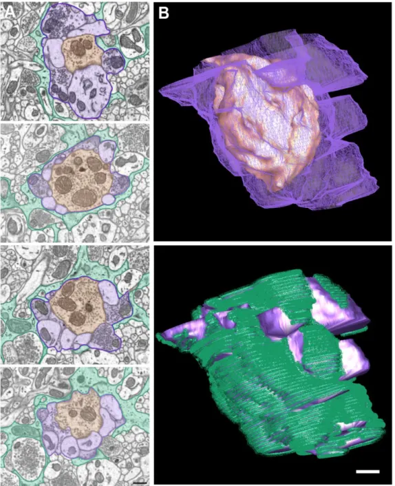
limiting neurotransmitter diffusion. On average, a single cortical astrocyte domain surrounds four neuronal cell bodies and hundreds of dendrites (Halassa, Fellin et al. 2007). In the CA1 region of the hippocampus, it has been estimated that a single astrocyte domain engulfs 140,000 synapses (Bushong, Martone et al. 2002). Thus, an astrocyte has the potential to interact with many nearby neurons. This architecture has led to the development of the tripartite synapse model. Structurally, it is composed of the presynaptic terminal, the postsynaptic bouton and the ensheathing astrocyte process. The percentage of contacted synapses as well as the degree of ensheathment varies dramatically from brain region to brain region. In the cerebellum, nearly all of the parallel and climbing fiber synapses are contacted by astrocyte processes (Spacek 1985). Of contacted climbing fiber synapses the median ensheathment by astrocytes is 94% of the synapse perimeter. In the parallel fiber synapses population, the median ensheathment dropped to 67% (Xu-Friedman, Harris et al. 2001). In the hippocampus, only 57% of synapses are contacted by astrocytes. Of these synapses, the astrocyte processes only ensheathed an average of 43% of the synaptic interface (Ventura and Harris 1999). At the synaptic glomeruli of the olfactory bulb and thalamus, astrocytes processes form multi-lamellar sheets surrounding the entire poly-synaptic unit (Spacek and Lieberman 1974; Chao, Kasa et al. 1997). This nonuniform distribution of astrocyte
processes raises the question of whether the distribution is random or whether processes are growing toward specific synapses.
In cell culture models, astrocytes extend processes toward synapse related molecules, including glutamate (Hatten 1985; Cornell-Bell, Thomas et al. 1990; Matsutani and
Yamamoto 1997). Using organotypic slice culture, astrocyte process interaction with
retracting (Haber and Murai 2006). This movement is neuronal activity-dependent, where neuronal activation encourages astrocyte encroachment of the synaptic zone (Genoud,
Quairiaux et al. 2006). Ultrastructural studies in the mouse whisker barrel cortex have shown that 24 hours of whisker stimulation causes a significant increase in the astrocytic
envelopment of excitatory synapses on dendritic spines (Genoud, Quairiaux et al. 2006). This work suggests that the amount of glutamate in the synapse is directly correlated to the degree of astrocyte coverage. The fine astrocytic processes covering a synapse can be very narrow, less than 50 nm wide, and below the resolution of light microscopy. The determination of the degree of synaptic ensheathment by an astrocytes process requires serial section electron microscopy (EM). Release probability at individual synapses is largely regulated by the size of the readily releasable vesicle pool, which can be determined at the EM level (Harris and Sultan 1995; Murthy, Schikorski et al. 2001; Dobrunz 2002; Branco, Marra et al. 2010). Though no published work has directly correlated the number of docked synaptic vesicles to the degree of astrocyte ensheathment at individual synapses, analysis of the average
percentage of astrocyte process coverage at specific varieties of synapses is correlated to the relative number of docked vesicles or the known release probability of a given variety of synapse.
neurotransmitters from the synaptic cleft. Furthermore, astrocytes may release
gliotransmitters including glutamate, D-serine and ATP to directly participate in neuronal signaling. The molecular pathways that allow astrocytes to communicate with synaptic compartments are only beginning to be understood.
Table 1. The relationship between release probability and astrocyte synapse coverage.
2.2.SYNAPSE FORMATION AND STRENGTH
Astrocytes are involved in the development, maturation and stabilization of synapses. During synaptogenesis, astrocytes can release cholesterol and thrombospondins to increase synapse number and promote synapse maturation (Ullian, Sapperstein et al. 2001;
Christopherson, Ullian et al. 2005). Conversely, the interaction of astrocytic ephrin A3 with EphA4 receptors on dendritic spines leads to spine retraction, reducing spine number and size (Murai, Nguyen et al. 2003; Bourgin, Murai et al. 2007; Fu, Chen et al. 2007) (Zhou,
Martinez et al. 2007). Synaptic strength can be modulated both by ephrin A3-induced modifications of glutamate transport and TNFα induced changes in AMPA receptor trafficking on the post-synaptic membrane.
Synapse Release Probability
Average % Astrocyte
Coverage
Reference
Cerebellar Climbing Fibers High 87% Xu-Friedman, 2001 Cerebellar Parallel Fibers Low 67% Xu-Friedman, 2001 Hippocampal
2.2.1.Thrombospondins
Thrombospondins are a family of five large extracellular matrix proteins that can mediate cell-cell and cell-matrix interactions through communication with membrane receptors, other extracellular matrix proteins and cytokines (Adams and Lawler 2004). Thrombospondins 1 and 2 are the most studied members of this family. They are secreted by astrocytes during early postnatal ages when the majority of excitatory synapses are forming (Christopherson, Ullian et al. 2005), as well as after ischemic injury (Lin, Kim et al. 2003; Liauw, Hoang et al. 2008) and after spinal cord injury (Wang, Chen et al. 2009). After ischemic stroke, thrombospondin-1/2-deficient mice show deficits in synaptic plasticity and functional recovery (Liauw, Hoang et al. 2008). In culture, thrombospondin-induced synapse formation produces ultrastructurally normal synapses; however, these synapses are
postsynaptically silent due to a lack of surface AMPA receptors (Christopherson, Ullian et al. 2005). Both in early post-natal stages and after injury, astrocytes appear to take on an
immature form. Thrombospondins are thought to work together with another yet unidentified molecule secreted from astrocytes in order to produce functional synapses.
antisense oligonucleotides blocks the upregulation of α2δ-1 and decreases mechanical allodynia (Li, Song et al. 2004; Li, Zhang et al. 2006). Post neuropathic injury,
thrombospondin 4 is upregulated in dorsal root ganglion neurons (Valder, Liu et al. 2003). Thrombospondin 4 can also bind α2δ-1 through its EGF-like domain and is secreted by astrocytes; however, its role in the pathophysiology of pain has yet to be studied. 2.2.2.Ephrins
Binding of the Eph receptor and ephrin from distinct cell surface membranes induces a bidirectional signaling cascade that alters cell adhesion and provokes actin cytoskeletal rearrangement. Ephrins A and B, as well as Eph receptors, are broadly expressed by neurons in the developing and mature central nervous system (Murai and Pasquale 2011). Ephrin-A3 becomes enriched in astrocyte processes within the first few weeks of post-natal development (Stein, Savaskan et al. 1999; Carmona, Murai et al. 2009; Galimberti, Bednarek et al. 2010). Ephrin-A3 activates EphA4 on dendritic spines and regulates the functional properties of astrocytes. The activation of EphA4 leads to retraction of the dendritic spine and to a reduction in their number and size (Murai, Nguyen et al. 2003; Bourgin, Murai et al. 2007; Fu, Chen et al. 2007; Zhou, Martinez et al. 2007). While other ephrin and Eph receptor
signaling pairs may be involved, most research has focused on ephrin-A3 and EphA4 pairing. In ephrin-A3 knockout mice, EphA4 tyrosine phosphorylation (an indication of its level of activity) is greatly reduced in the hippocampus and elevated in transgenic mice overexpressing ephrin-A3 in astrocytes (Filosa, Paixao et al. 2009). Upon activation, EphA4 phosphorylates and complexes with Cdk5 to bind and activate the guanine nucleotide
exchange factor Ephexin1. Ephexin1 activates RhoA, which is required to decrease spine size and synapse number. EphA4 tyrosine phosphorylation also signals through
produce inositol 1,4,5-triphosphate (IP3) and diacylglycerol. These mobilize calcium signaling and promote protein kinase C (PKC) activation. EphA4 signaling reduces the membrane association of F-actin severing and depolymerization factor cofilin (Zhou,
Martinez et al. 2007). Coflin is downstream of both RhoA signaling (Bernstein and Bamburg 2010) and the cdk5-Ephexin1 PLC1 pathways. These pathways may concomitantly regulate the activity of cofilin to modify the architecture of actin filaments in spines (Murai and Pasquale 2011).
EphA4 signaling also regulates adhesion receptors critical for dendritic spine maintenance. This EphA4 signaling pathway disrupts β1 integrin function in neurons,
promoting spine retraction and loss (Bourgin, Murai et al. 2007). This cell contact-dependent communication between astrocyte processes and neurons may maintain the architecture of the synapse by restricting the dendritic spine’s ability to change shape (Murai, Nguyen et al. 2003).
Forward EphA4 signaling in dendritic spines activates pathways that promote the modification of the actin cytoskeleton and adhesion molecules important for the maintenance of spine density and morphology. Additionally, forward EphA4 signaling could modulate synaptic strength. In cultured neurons, EphA4 modulates the cell surface expression of GluR1-containing AMPA receptors (Fu, Hung et al. 2011). Stimulation of EphA4 promotes its interaction with the anaphase-promoting complex (APC), enabling Cdh1 to bind and polyubiquinate GluR1. Polyubiquitination and degradation of GluR1 decreases synaptic efficacy.
importance of Ephrin-A3 reverse signaling in astrocytes has been demonstrated in vivo.
Transgenic mice overexpressing ephrin A3 under the astrocytic promoter GFAP have reduced GLT-1 and GLAST immunolabeling (Filosa, Paixao et al. 2009). Ephrin A3 knockout mice have increased immunolabeling for glial glutamate transporters GLT1 (EAAT2) and GLAST (EAAT1) in the hippocampus and cortex and an increase in glial glutamate transporter currents measured by patch-clamp in acute hippocampal slices (Carmona, Murai et al. 2009; Filosa, Paixao et al. 2009). Treatment of wild-type, but not ephrin-A3 knockout, slices with EphA2 FC fusion protein to promote reverse ephrinA-A3 signaling also reduces glutamate uptake. Interestingly, both ephrin A3 and EphA4 knockout mice exhibit a reduction in long-term potentiation (LTP) following theta-burst stimulation but not tetanic stimulation (Filosa, Paixao et al. 2009). The reduction of LTP following theta-burst stimulation can be rescued by the pharmacological blockade of glutamate transporters, arguing that the increased levels of glutamate transporters observed in ephrin A3 and EphA4 knockout mice are clearing glutamate too efficiently from the synapse to allow for the
development of LTP. Tetanic stimulation, which results in higher levels of glutamate release, may overwhelm even the increased population of glutamate transporters. However, rhythmic bursting, similar to theta bursting, of CA1 neurons is observed when animals explore novel environments (Winson 1978; Otto, Eichenbaum et al. 1991; O'Keefe 1993).
Synaptic activity may promote the trafficking of EphA4 to the membrane, allowing EphA4 activation and the downregulation of synaptic GluR1, as well as the mobilization of the cytoskeleton and adhesion molecules, which acts as a homeostatic mechanism to stabilize activity following bursts of neuronal network activity. Alternatively, synaptic activity also drives the cleavage of EphA4, which could lead to an increase in the number of dendritic spines. While the efficacy of the extracellular fragment of EphA4 in binding to ephrin-A3 has not been determined, it would be expected to inhibit ephrin reverse signaling, thereby enhancing glutamate transport via GLT1 and GLAST transporters and decreasing synaptic activity (Murai and Pasquale 2011). In vivo, prolonged synaptic activity has been shown to increase glial glutamate transporter levels as well as synaptic coverage by astrocyte processes (Genoud, Quairiaux et al. 2006).
Ephrin A3 signaling has not been studied in the dorsal horn in the context of chronic pain; however, spinal cord injury, which is also associated with chronic pain, causes an increase in EphA receptor expression on both oligodendrocytes and astrocytes (Willson, Irizarry-Ramirez et al. 2002). The remodeling of synapses in the dorsal horn post injury is likely to require astrocyte process movement and dendritic spine remodeling, making ephrins another good target for study using neuropathic pain models.
2.2.3.TNFα
that a drop in neuronal activity, as induced by a prolonged activity blockade by TTX, triggers the release of TNFα from glial cells. TNFα signaling released from glia can induce the trafficking of AMPA receptors to postsynaptic sites to increase excitatory synaptic strength (Beattie 2002). Conversely, blocking TNFα has the opposite effect (Beattie 2002). Soluble TNFα acts to compensate for the decrease in activity by increasing the level of cell-surface AMPA receptors (Stellwagen and Malenka 2006). Glial-derived TNFα containing media was prepared using mixed wild-type glial and TNFα KO neuronal cell cultures in which activity was blocked using TTX. This media was then applied to hippocampal slices prepared for electrophysiological recording. Interestingly, medium from cultures treated with TTX for 24 hours did not increase synaptic strength; it required 48 hours of conditioning before the effect was produced. This finding raises the possibility that TNFα is a factor in long-term synaptic scaling and that there may be other factors involved at shorter time-scales.
Several lines of evidence suggest a role for TNFα in the production of central sensitization in the dorsal horn as well. TNFα is induced in spinal cord glial cells after neuropathic lesions (DeLeo, Colburn et al. 1997; Xu, Xin et al. 2006; Hao, Mata et al. 2007). Intrathecal injection of TNFα induces thermal hyperalgesia and mechanical allodynia (Kwon, Shim et al. 2005; Kawasaki, Zhang et al. 2008; Gao, Zhang et al. 2009). Intrathecal injection
of a TNFα inhibitor inhibits behavioral sensitivity following peripheral nerve injury
(Sommer, Schafers et al. 2001; Schafers, Svensson et al. 2003; Marchand, Tsantoulas et al.
2009). Additionally, the application of TNFα to spinal cord slices increases spontaneous EPSC frequency but not EPSC amplitude (Kawasaki, Zhang et al. 2008; Youn, Wang et al.
2008; Li, Xie et al. 2009; Zhang, Berta et al. 2011). While this may argue for a presynaptic
by the cells being recorded from. More detailed studies will be needed to determine the role
of TNFα in the dorsal horn, particularly after neuropathic lesions.
2.3.NEUROTRANSMITTER UPTAKE
In vitro and in situ astrocytes have been shown to express a wide variety of neurotransmitter transporters including glutamate, GABA, adenosine and dopamine.
Glutamate transporters, or excitatory amino acid transporters (EAATs), are among the most studied family of neurotransmitter uptake transporters. Glia express EAAT1 and EAAT2, neurons express EAAT 3 and EAAT4, and EAAT5 is found exclusively in the retina. In rodents, the orthologs for EAAT1-3 are named glutamate aspartate transporter (GLAST), glutamate transporter 1 (Glt-1) and excitatory amino acid carrier 1 , whereas the titles of EAAT4 and 5 are conserved. While both neurons and astrocytes express glutamate transporters, the astrocytic glutamate transporters, particularly Glt-1, are the predominant transporters in most brain areas (Danbolt 2001).
little effect on the decay of evoked EPSCs (Isaacson and Nicoll 1993; Ventura and Harris 1999). In contrast, in the synapses between the parallel and climbing fibers on Purkinje cells in the cerebellum, glutamate diffusion is limited by astrocyte ensheathment, and the
inhibition of glutamate transport increases the duration of glutamatergic transmission to the postsynaptic neuron (Overstreet, Kinney et al. 1999).
Examination of BAC-generated Glt-1-eGFP/GLAST-DsRed double transgenic mice shows that in most areas of the brain, Glt-1 and GLAST are coexpressed in astrocytes, whereas the spinal cord has largely segregated populations of Glt-1- and GLAST-positive astrocytes (Regan, Huang et al. 2007). In neurons of the dorsal horn, glutamate uptake by Glt-1 contributes to the termination of EPSCs (Weng, Chen et al. 2007; Xin, Weng et al. 2009; Zhang, Xin et al. 2009). Immunohistochemical and western blot studies have shown an initial increase followed by a long-term decrease in glutamate transporters after nerve injury (Xin, Weng et al. 2009). This timeline correlates with astrocyte hypertrophy and the chronic phase of pain observed in neuropathic pain models (Vega-Avelaira, Moss et al. 2007). However, the decrease in transporter protein may be associated with the decrease in primary afferent innervations and may not generate a lack of synaptic glutamate clearance at intact primary afferent fibers.
2.4.CALCIUM MEDIATED RELEASE OF GLIOTRANSMITTERS FROM ASTROCYTES
is the canonical G alpha q-coupled pathway that elicits increases in intracellular calcium. Upon activation, Gαq activates phospholipase C (PLC). PLC then hydrolyzes the membrane lipid phosphatidylinositol 4,5 bisphosphate, cleaving it into diacylglycerol (DAG) and inositol 1,4,5-triphosphate (IP3). DAG remains bound to the membrane, whereas IP3 is released into the cytosol where it can activate the IP3 receptor on the endoplasmic reticulum. Activation of IP3 receptors causes the release of calcium from intracellular stores. Of the three subtypes of IP3 receptors known, astrocytes are only known to possess IP3 receptor subtype 2 (IP3R2). Conditional knockout of IP3R2 under the GFAP promoter abolished astrocyte calcium oscillations and increases in response to Gq GPCR agonists or neuronal stimulation (Petravicz, Fiacco et al. 2008; Agulhon, Fiacco et al. 2010).
The Gq-IP3 receptor pathway elicitation of calcium increases is thought the most prevalent physiological method of increasing intracellular calcium in astrocytes. Cultured astrocytes express voltage-gated calcium channels and P2X7 receptors (Panenka, Jijon et al. 2001; Latour, Hamid et al. 2003; D'Ascenzo, Vairano et al. 2004); however, there is little functional evidence of these channels in situ. Specific subpopulations of astrocytes, such as Bergmann glia and complex cells, have been found to display voltage-gated calcium channels as well as AMPA receptors; however, these subpopulations seem to be the exception rather than the rule (Burnashev, Khodorova et al. 1992; Hamilton, Vayro et al. 2010). In vitro, in situ and in vivo astrocytes respond to neuronal stimulation with calcium elevations attributed to the activation of their wide range of Gq-coupled GPCRs (Agulhon, Petravicz et al. 2008).
of gliotransmitters. In neuronal cells, voltage-gated calcium channels at the presynaptic membrane open in response to voltage changes, triggering an influx of calcium over a relatively small microdomain. This calcium elevation causes the release of neurotransmitter-filled vesicles into the synaptic cleft via the soluble NSF attachment protein receptor
(SNARE) complex. In ultrastructural studies, astrocytes have been found to contain sparsely distributed vesicles complete with vesicular glutamate transporters 1 or 2 as well as the vesicular SNARE proteins cellubrevin, SNAP-25 and syntaxin (Bezzi, Gundersen et al. 2004; Wilhelm, Volknandt et al. 2004; Zhang, Pangrsic et al. 2004; Montana, Malarkey et al. 2006). However, much of this work was completed in cultured astrocytes. The presence of vesicular machinery provides the basis for many studies supporting the role of astrocytes in the active modulation of neuronal transmission via gliotransmission.
selectively in astrocytes reduced long-term potentiation in the hippocampus and interfered with the formation of heterosynaptic depression (Pascual, Casper et al. 2005; Serrano, Haddjeri et al. 2006). Astrocytes release D-serine in culture (Schell, Molliver et al. 1995; Mothet, Pollegioni et al. 2005) and are thought to be the sole source of D-serine in the brain (Schell, Brady et al. 1997; Oliet and Mothet 2006). In the supraoptic nucleus, D-serine levels change as a function of the astrocytic coverage of neuronal synapses and affect both LTP and LTD (Panatier, Theodosis et al. 2006).
Astrocytes display the same receptor milieu of Gq GPCRs as neurons; therefore, the selective stimulation of astrocyte signaling, while not perturbing neuronal function, is a major hurdle for all studies on gliotransmission. The selective stimulation of astrocyte signaling has been achieved using methods as crude as mechanical stimulation of the cell, which has a high potential of damaging the cell membrane, to the more commonly used photolysis of caged calcium or IP3 molecules delivered by patch pipette. Although these techniques have been used in numerous studies and increase calcium selectively in
observed with other methods, such as calcium or IP3 uncaging (Fiacco, Agulhon et al. 2007). This study therefore does not support the current model of gliotransmission.
The Gq GPCR-mediated release of gliotransmitters relies on the activation of IP3R2 to release calcium from internal stores. IP3R2 knockout mice show abolished intrinsic calcium oscillations as well as agonist- or neuronal stimulation-evoked calcium increases in astrocytes, while leaving neuronal calcium responses intact (Petravicz, Fiacco et al. 2008; Agulhon, Fiacco et al. 2010). Contrary to the current model of gliotransmission, the lack of calcium signaling in astrocytes had no effect on basal excitatory neurotransmission or on short- or long-term potentiation in the hippocampus (Petravicz, Fiacco et al. 2008; Agulhon, Fiacco et al. 2010). This series of papers does not support the current model of
gliotransmission. More work will need to be done in the field in order to resolve these differences.
2.5.THE IMPACT OF THE PHYSIOLOGICAL ROLES ASTROCYTES ON NEUROPATHIC PAIN
Astrogliosis has been considered a marker for stress in the central nervous system since the early 1900s (Ramon y Cajal 1928). Astrocyte activation, or astrogliosis, is seen visualized by hypertrophy of the cell body and initial large processes using
immunohistochemical staining for GFAP and S100beta. These morphological changes have been linked to numerous diseases and insults, including Alzheimer’s disease, Parkinson’s disease, amyotrophic lateral sclerosis, alcoholism and physical trauma. Astrocyte
Under conditions of chronic neuropathic pain, astrogliosis begins as early as 7 days post-neuropathic injury and remains for the duration of behavioral allodynia, through 4 weeks (Tanga, Raghavendra et al. 2004). Astrogliosis may result in added roles of astrocytes, such as the release of cytokines and chemokines, which can further sensitize surrounding neurons. Alternately, astrogliosis may perturb of the existing roles of astrocytes within a neuronal network. Studies have shown that the inhibition of these changes alleviates the behavioral sensitization that occurs in chronic pain (Sweitzer, Colburn et al. 1999).
Transgenic inhibition of inflammatory pathways selectively in astrocytes lessens behavioral sensitization following formalin administration and chronic constriction injury (Fu, Zhang et al. 2007; Fu, Zhang et al. 2010). Decreases in glutamate transporter expression following the development of astrogliosis (Sung, Lim et al. 2003; Binns, Huang et al. 2005) may account for the increase in glutamate levels in the spinal cord during chronic pain and may produce the AMPA and NMDA receptor activation underlying the hyperexcitability of secondary pain transmission neurons (Cavaliere, Cirillo et al. 2007). Furthermore, the administration of glial metabolic inhibitors decreases behavioral hypersensitivity after the induction of
neuropathic pain (Tawfik, Nutile-McMenemy et al. 2007).
First, we examined the tripartite synapse structure of primary afferents in the dorsal horn to determine whether astrocytes were in a position to regulate excitatory neuronal transmission in the dorsal horn. Astrocyte interactions with synapses can affect their
formation, stability and strength through cell contact and secreted factors. Astrocytes can also modulate neuronal signaling through neurotransmitter transporters and the release of
nociceptive synapses is an important first step for the field in determining the possible astrocyte contribution to pain processing. There is considerable variability between brain regions in synaptic coverage by astrocyte processes, and this variability even extends to the synapse type within a single region. As blockade of the astrocyte glutamate transporter Glt-1 affects dorsal horn EPSCs, I hypothesize that astrocyte processes will contact synapses in the dorsal horn.
Next, we chose to investigate the role of gliotransmission in the dorsal horn through the use of transgenic mouse models blocking the calcium-dependent and/or