A DORSAL, BUT NOT VENTRAL, HIPPOCAMPAL CIRCUIT IS REQUIRED FOR EXPRESSION OF HEROIN’S CONTEXTUALLY CONDITIONED IMMUNE EFFECTS
Christina L. Lebonville
A dissertation submitted to the faculty at the University of North Carolina at Chapel Hill in partial fulfillment of the requirements for the degree of Doctor of Philosophy in the Department of Psychology & Neuroscience (Behavioral and Integrative Neuroscience) in the College of Arts
and Sciences.
Chapel Hill 2019
ii © 2019
iii ABSTRACT
Christina L. Lebonville: A dorsal, but not ventral, hippocampal circuit is required for expression of heroin’s contextually conditioned immune effects.
(Under the direction of Donald T. Lysle)
Drugs of abuse, like opioids, cause a diverse array of physiological effects. These effects can become conditioned to occur to any stimulus, for example an environmental context, that becomes associated with drug use. In terms of drug reward conditioning, exposure to drug-paired contexts can elicit craving and re-engagement in drug seeking behaviors, promoting relapse to drug use. Similarly, the immune modulating effects of opioids can be conditioned to occur with exposure to an opioid-paired context. Therefore, exposure to drug-paired contexts can
iv
behaviors by manipulating this region’s predominant outgoing projections prior to expression of heroin contextually conditioned immune modulation. The hippocampus is not a homogeneous structure, and the dorsal and ventral aspects of the hippocampus connect anatomically to distinct groups of brain regions. Therefore, understanding the relative importance of the dorsal and ventral outputs from the hippocampus will give a clearer understanding of how the hippocampus relays information about context to other brain regions. The chemogenetic technique, designer receptors exclusively activated by designer drugs (DREADDs), lends itself well to inhibiting particular neurons in hippocampal output regions and building toward an understanding of hippocampal circuits. An experiment in Chapter 2 tested whether output from the dorsal hippocampus is required for the expression of heroin contextually conditioned immune modulation. Chapter 3’s experiment tested whether output from the ventral hippocampus is required for the expression of heroin contextually conditioned immune modulation.
Chemogenetic inhibition of the dorsal hippocampus output, but not inhibition of the ventral hippocampus output, attenuated the expression of heroin conditioned immune modulation. Thus, Chapter 4’s experiment tested whether a specific anatomical connection between the dorsal hippocampus and retrosplenial cortex is required for expression of conditioned immune
modulation. Chemogenetic inhibition of the specific projection from the dorsal hippocampus to the retrosplenial cortex did not attenuate the expression of heroin conditioned immune
v
To my partner Steven Lebonville,
whose never-ending support and encouragement drive me to persist through the challenges of academic life. Thank you for being my family, anchoring me to what is most important, and
vi
ACKNOWLEDGEMENTS
There are so many who have contributed to this dissertation, even without contributing to the words on the page, because they have supported my growth and persistence as a scientist. This is
a very short list of some of those people who have been instrumental along the way. To my laboratory colleagues, Meghan E. Jones, Jacqueline E. Paniccia, and Shveta V. Parekh, you are the ones I always turn to for guidance and encouragement when things become overwhelming. From our lab social bonding moments, to deep discussions about science, I am never alone and find such joy in sharing graduate life with you. When things get tough between us, as life in the trenches often encourages, you teach me how to be a better friend and coworker. Your perspectives enrich my ability to do good science and be a leader and professional. I hope that I have been a fraction as valuable to you as you have been to me. Go Lysle Lab Ladies!
To my brilliant undergraduate research assistants, Sarah Yaghoubi, Tara M. O’Connell, and Lynde M. Wangler, I could not have done this research without you. You taught me how to lead others and to keep them passionate about the big picture. You made me into a better teacher and mentor. You became dear friends of mine and are still my most passionate supporters. I can’t wait to see how each of you changes the world for the better. I’m in awe of you.
vii
Don, you have a special place in my heart for trusting me with an enormous amount of independence, knowing that I would figure it all out and be more resilient and capable as a result. Thank you for being there alongside us during long experiment days, for having the best stories to tell, and for your delightful sense of humor.
viii
TABLE OF CONTENTS
LIST OF FIGURES ... xii
LIST OF ABBREVIATIONS ... xiii
CHAPTER 1: GENERAL INTRODUCTION ... 1
The Role of Context in Drug Behaviors ... 1
The Hippocampus and Recall of Context-Drug Associations ... 3
Hippocampal Function and Connectivity ... 5
Specific Hippocampal Connections and Contextually Conditioned Drug Behaviors ... 6
DREADDs Have the Power to Reveal Hippocampal Neurocircuitry ... 9
Examining the Diverse Hippocampus in Opioid Contextually Conditioned Immune Effects.. 11
CHAPTER 2: DORSAL HIPPOCAMPAL OUTPUT REGIONS ARE REQUIRED FOR THE EXPRESSION OF HEROIN’S CONTEXTUALLY CONDITIONED IMMUNE EFFECTS ... 14
Introduction ... 14
Materials & Methods ... 16
Animals ... 16
Drugs and Delivery ... 16
Surgical Procedure ... 17
ix
Conditioning & Testing Procedure ... 19
Blood and Spleen Tissue Collection ... 20
Brain Histology and DREADD Expression Analysis ... 21
Nitrate/nitrite Assay ... 21
RNA Extraction & RT-qPCR ... 22
Protein Extraction & iNOS ELISA ... 24
Statistical Analysis ... 25
Results ... 25
Discussion ... 29
CHAPTER 3: VENTRAL HIPPOCAMPAL OUTPUT REGIONS ARE NOT REQUIRED FOR THE EXPRESSION OF HEROIN’S CONTEXTUALLY CONDITIONED IMMUNE EFFECTS ... 34
Introduction ... 34
Materials & Methods ... 36
Animals ... 36
Drugs and Delivery ... 37
Surgical Procedure ... 38
DREADD Virus and Incubation ... 38
Conditioning & Testing Procedure ... 39
Blood and Spleen Tissue Collection ... 40
x
Nitrate/nitrite Assay ... 42
RNA Extraction & RT-qPCR ... 42
Protein Extraction & iNOS ELISA ... 44
Statistical Analysis ... 45
Results ... 46
Discussion ... 50
CHAPTER 4: A PROJECTION FROM THE DORSAL HIPPOCAMPUS TO THE RETROSPLENIAL CORTEX IS NOT REQUIRED FOR THE EXPRESSION OF HEROIN’S CONTEXTUALLY CONDITIONED IMMUNE EFFECTS ... 53
Introduction ... 53
Materials & Methods ... 55
Animals ... 55
Drugs and Delivery ... 55
Surgical Procedure ... 56
DREADD Virus and Incubation ... 57
Conditioning & Testing Procedure ... 58
Blood and Spleen Tissue Collection ... 59
Brain Histology and Injector/DREADD Expression Analysis ... 60
Nitrate/nitrite Assay ... 60
RNA Extraction & RT-qPCR ... 61
xi
Statistical Analysis ... 64
Results ... 64
Discussion ... 68
CHAPTER 5: GENERAL DISCUSSION ... 72
Primary Findings ... 72
A Deeper Look at Dorsal Hippocampal Projection Targets ... 73
Retrosplenial and Medial Entorhinal Cortices ... 73
Nucleus Accumbens Shell ... 75
Lateral Septum ... 77
Limitations & Technical Concerns ... 81
Insertion of DREADDs into Cellular Membranes ... 81
Non-Specific CNO Action... 82
Functionality of DREADD ... 84
Other Limitations ... 85
Broader Implications of Results ... 87
Conclusion ... 90
xii
LIST OF FIGURES Figure 1 – Inactivation of dorsal hippocampal output regions, including dorsal subiculum and CA1, disrupts expression of heroin
contextually conditioned suppression of nitric oxide...26 Figure 2 – Inactivation of ventral hippocampal output regions, including
ventral subiculum and CA1, does not disrupt expression of heroin contextually
conditioned suppression of nitric oxide...46 Figure 3 – Inactivation of dorsal hippocampal projections to the retrosplenial
cortex does not disrupt expression of heroin contextually conditioned
xiii
LIST OF ABBREVIATIONS
°C degrees Celsius
ΔΔCt delta delta threshold cycle (qPCR)
AAV adeno-associated virus
AAV5 adeno-associated virus serotype 5
ANOVA analysis of variance
AP anterior-posterior
BLA basolateral amygdala
CA1 Cornu Ammonis region 1 of the hippocampus
CA3 Cornu Ammonis region 3 of the hippocampus
CAMKIIα calmodulin-dependent protein kinase II alpha
cAMP cyclic adenosine monophosphate
CNO clozapine-N-oxide
CS conditioned stimulus
dCA1 dorsal Cornu Ammonis region 1 of the hippocampus
dCA3 dorsal Cornu Ammonis region 3 of the hippocampus
dDG dorsal dentate gyrus of the hippocampus
xiv dHpc dorsal hippocampus
DMSO dimethyl sulfoxide
DREADD designer receptors exclusively activated by designer drugs
dSub dorsal subiculum of the hippocampus
DV dorsal-ventral
EGFP enhanced green fluorescent protein
ELISA enzyme-linked immunosorbent assay
FAD flavine adenine dinucleotide
g gram
g/rcf gravitational force/relative centrifugal force
G-protein guanine nucleotide-binding protein
GABA gamma-aminobutyric acid
HC home cage
HOV homogeneity of variance
HPA hypothalamic-pituitary-adrenal
HSD Honestly Significant Difference
xv
IACUC Institutional Animal Care and Use Committee
iNOS inducible nitric oxide
kg killogram
KORD κ-opioid receptor derived DREADD (Gi-coupled)
LPS lipopolysaccharide
LS lateral septum
LTP long-term potentiation
µL microliter
µm micrometer
mEC medial entorhinal cortex
mg milligram
min minute
ML medial-lateral
mL milliliter
mm millimeter
mM millimolar
xvi NAc nucleus accumbens
NADPH nicotinamide adenine dinucleotide phosphate hydrogen
NIDA National Institute on Drug Abuse
NO nitric oxide
PAG periaqueductal gray nucleus
PCR polymerase chain reaction
RNA ribonucleic acid
RSC retrosplenial cortex
RSCgb retrosplenial cortex granular area b/c
RT-qPCR reverse transcription quantitative real-time polymerase chain reaction
qPCR quantitative real-time polymerase chain reaction
vCA1 ventral Cornu Ammonis region 1 of the hippocampus
vCA3 ventral Cornu Ammonis region 3 of the hippocampus
vDG ventral dentate gyrus of the hippocampus
vHpc ventral hippocampus
vSub ventral subiculum of the hippocampus
1
CHAPTER 1: GENERAL INTRODUCTION The Role of Context in Drug Behaviors
Learning about events that occur in a particular environmental context allows us to use our past experiences to guide future behavior in that context. Generally, learning associations between contexts and important events confers an advantage, allowing for better prediction and adaptive responses to these events (Maren et al., 2013). However, some contextual associations contribute to maladaptive behavior, like drug addiction. Drug-associated contexts serve an important role in maintaining addiction by increasing the likelihood of relapse (Caprioli et al., 2007; Crombag et al., 2008; Janak and Chaudhri, 2010). For example, entering an environment where past drug experiences have occurred can increase motivation for seeking and taking a wide range of drugs including alcohol (Janak and Chaudhri, 2010), cocaine (Fuchs et al., 2008), nicotine (Diergaarde et al., 2008), and heroin (Bossert and Stern, 2014) in an animal model of relapse called “context-induced reinstatement of drug seeking”. It is thought that being in a drug-associated context prompts the expression of learned behaviors and sensations surrounding drug use (ex. approach, seeking, craving). This might be due to the context becoming a Pavlovian conditioned stimulus (CS) for the rewarding/reinforcing effects of drugs or by the context acting as an occasion setter for drug seeking and taking behaviors that have led to obtaining drug reward previously (Crombag et al., 2008).
In the study of addiction, a predominant focus on relapse behaviors makes sense.
2
potently modulate the immune system (Fecho et al., 1993; Fecho and Lysle, 2000; Fecho et al., 2000; Lysle et al., 1993; Lysle and How, 2000; Nelson et al., 2000). It has been shown that stimuli associated with immune altering substances can influence immune function by themselves through Pavlovian conditioning (Ader and Cohen, 1975). When the immune modulating effects of opioids become associated with a particular context, exposure to these contexts even in the absence of opioids can modulate the immune system to a similar degree (Coussons et al., 1992; Lysle and Ijames, 2002). Therefore, opioid contextually conditioned immune effects can add to the societal cost of addiction by contributing to the increased incidence of infection seen in opioid users (Govitrapong et al., 1998; Horsburgh et al., 1989; Louria et al., 1967; Risdahl et al., 1998). By understanding the neural mechanisms behind the recall or expression of contextually conditioned immune effects, we might be able to develop behavioral or pharmacological interventions to help ameliorate these costs.
The potent ability of a drug-associated context to not only increase the probability of relapse to abuse behavior but to also exert health consequences beyond those of drug use itself, makes it vital to understand the neural mechanisms of context-induced effects more broadly. If governed by a shared mechanism, both contextually conditioned immune and reward effects may be able to be blocked simultaneously with a single treatment. However, if each are governed by diverging mechanisms, it means that treatments blocking the abuse promoting effects of contexts will not address the immune compromising effects of contexts and vice versa. Ultimately, the study of contextually-elicited immune effects with drugs like opioids provides a unique perspective from which to study the general role of context in drug behaviors.
3
mechanisms of opioid contextually conditioned immune effects with the known mechanisms of the context-induced reinstatement of drug seeking model. The two animal models show complete overlap in required brain regions [for review see (Bossert et al., 2013; Szczytkowski et al.,
2011)], and both share a dependency on the neural representation of a context. An additional benefit of comparing these two models is that they both use standard operant chambers as the behavior-producing context. Additional neural mechanisms engaged with larger or more complex environmental contexts could confound investigations of the neural mechanisms of context alone. For example, a frequently used animal model of contextual drug reward behaviors, conditioned place preference, involves movement between two or more connected chambers. As a result, in this and other models of drug reward, navigational and more complex spatial
processing could be engaged. Furthermore, this processing might obscure mechanisms of contextual representation per se because both navigational and contextual representation occurs within the same brain region – the hippocampus [see (Riaz et al., 2017)].
The Hippocampus and Recall of Context-Drug Associations
The hippocampus has long been seen as the processor of context and context-relevant memories (Maren et al., 2013; Smith and Mizumori, 2006). The notorious case of Henry Molaison in the 1950’s led to the hippocampus being accepted as a brain region important for retrieval of episodic memory (Scoville and Milner, 1957). In the 1970’s, the hippocampus became considered a processor of physical space (spatial context) due to the discovery of
4
Mizumori, 2006). Recently, significant technological advances have led to the view that ensembles of cells (including place cells) are how the hippocampus represents context (Smith and Bulkin, 2014). Sparse cell ensembles that become active during exposure to a relevant context can be studied using specialized transgenic mice models with activity-dependent cell tagging techniques. Such studies have shown that a distinct set of cells form an ensemble with repeated exposure to a specific context, and this ensemble is preferentially active during re-exposure to that same context. It is proposed that activity of these ensembles allow for identification of the current context and retrieval of context-specific memories and therefore behaviors. This ability to represent unique contexts and their meaning with different ensembles of cells, may explain the hippocampus’ role in controlling contextually motivated behaviors with drugs, leading to an increased likelihood of relapse.
Accordingly, addiction research has applied this foundational work from the learning and memory field in understanding the hippocampus’ role in the ability of context to influence responses to drugs of abuse. A theoretical model has been proposed where the hippocampus encodes the context around the experiences with drugs and is involved in the recall of contextual memories that promote drug taking and seeking behavior (Everitt and Robbins, 2005; Robbins et al., 2008). A context-specific hippocampal ensemble has been shown to be sensitive to drug exposure that occurs in that context, to be particularly active around times when drug-seeking behavior occurs, and to be necessary for expression of context-specific drug behaviors (Trouche et al., 2016; Xia et al., 2017).
heroin-5
paired context prevented the context’s ability to modulate the immune system (Szczytkowski et al., 2013). Since both contextually conditioned immune and reward drug responses rely on a functional hippocampus, this region may be of particular interest for pharmacological or behavioral interventions hoping to ameliorate both relapse behaviors and negative health outcomes associated with drug abuse. However, before such a treatment can be developed, we must understand how hippocampal signaling ultimately leads to these drug behaviors. Once the context is identified by increased activity in context-specific ensembles, the hippocampus is thought to promote drug behaviors by engaging reward and motivational brain areas. These connections between the hippocampus and the canonical “reward circuitry” are actually quite complex and frequently misunderstood. A close study of these connections reveals important unanswered questions about how the hippocampus processes context.
Hippocampal Function and Connectivity
6
basolateral amygdala, hypothalamus, and infralimbic cortex (Amaral and Witter, 1989; Bienkowski et al., 2018; Naber and Witter, 1998; Witter, 2006).
Since dHpc and vHpc connections are incredibly distinct from one another and non-overlapping, it may be that by virtue of engaging one set of targets versus the other, different functions of the dHpc and vHpc arise. In fact, experiments have shown that the dHpc and vHpc have dissociable roles in different types of behavior [see (Fanselow and Dong, 2010)]. The dHpc is thought to primarily support spatial, navigational, and episodic memory, while the vHpc is thought to be involved in contextually motivated fear and reward memory (“emotional memory”), although not all evidence supports this clear distinction (see Chapter 5: General Discussion). Despite these functional distinctions, inactivation of neural signaling in either the dHpc (Fuchs et al., 2005; Ge et al., 2017; Xie et al., 2010) or vHpc (Lasseter et al., 2010) attenuates the expression of context-induced reinstatement with cocaine or heroin. Likewise, inactivation of neural signaling in the dHpc attenuates expression of heroin contextually
conditioned immune modulation (Szczytkowski et al., 2013), but the role of the vHpc has yet to be tested. There is reason to believe that the dHpc, and perhaps also the vHpc, is involved in the expression of contextually conditioned drug behaviors, generally. What follows is a summary of which connections are theoretically important to contextually driven drug behaviors and which regions of the hippocampus, the dHpc or vHpc, might make these connections.
Specific Hippocampal Connections and Contextually Conditioned Drug Behaviors Connections between the hippocampus and ventral striatum have been proposed to support the expression of contextually conditioned reward behaviors (van der Meer and Redish, 2011). Regions in the ventral striatum are well-known to modulate motivational states,
7
hippocampus might represent the context and then signal to the NAc which would be able to promote the necessary reward-motivated behaviors (Lansink et al., 2009; van der Meer and Redish, 2011).The hypothesis that the hippocampus initiates retrieval of contextually conditioned drug behaviors via the NAc is supported by the fact that in recall (replay) of contextual drug reward associations, activity the hippocampus precedes activity in the NAc (Lansink et al., 2009). Thus, the activation of a contextual ensemble for a drug-paired context is theoretically able to contribute to expression of drug-related behavior in that context by reactivating relevant experiential and motivational states mediated by the NAc (Destexhe et al., 2015).
There is support for the importance of this connection between the hippocampus and NAc to contextual initiation of drug behaviors (Sjulson et al., 2018), but it is unclear whether this connection arises from the dHpc or vHpc. Their respective roles likely depend on which
8
one study, lateral NAc shell also mediated context-induced reinstatement of heroin seeking (Bossert et al., 2007), which could suggest a role for dHpc communication to the NAc shell. As a result, either the dHpc, vHpc, or both regions of the hippocampus could mediate context-induced drug behaviors by engaging with different regions of the NAc shell.
Connections between the hippocampus and other reward/motivational regions have also been implicated in the expression of contextually conditioned behaviors with drugs of abuse. Specifically, serial information processing between the dHpc and basolateral amygdala (BLA) has been shown to be required for expression of context-induced reinstatement of cocaine seeking (Fuchs et al., 2007b). Similarly, the BLA has also been implicated in contextually conditioned immune modulation with opioids (Szczytkowski and Lysle, 2008, 2010). A hippocampal projection to the BLA arises only from the vHpc, which has direct, reciprocal connections with the BLA (Pikkarainen et al., 1999; Pitkänen et al., 2000). This means that despite the fact that functional disconnection of the dHpc and BLA interferes with expression of contextually conditioned reward behaviors, there is no direct connection between these regions. Thus, the dHpc must interact with the BLA indirectly through some area(s) with connectivity to both regions, whereas the vHpc is well-positioned to mediate contextually conditioned drug behaviors along with the BLA. To our knowledge, no one has determined how the dHpc and BLA can interact even though this interaction is implicated in not only expression of
9
In summary, there are important gaps in understanding the roles of connections
originating from the dHpc and vHpc in contextually conditioned behaviors with drugs, especially with conditioned immune modulation. The evidence indicates that both the dHpc and vHpc may be necessary, but approaching their role at the circuit level highlights outstanding questions that need to be answered. First, how is the dHpc initiating immune modulating or reward behavior to a context, especially if it needs to interact with the BLA to do so? Second, is the vHpc important to contextually conditioned immune modulation as it is in conditioned reward? A powerful chemogenetic tool allows us to begin to answer these important questions.
DREADDs Have the Power to Reveal Hippocampal Neurocircuitry
Looking only at hippocampal ensembles does not reveal any information about how the hippocampus is engaging behavior through communication with other brain regions.
Hippocampal outputs to other areas might convey the net processing from such activated ensembles and may be a simpler way to investigate hippocampal contextual initiation of
behavior. If we could inhibit these outputs specifically, then we would be able to investigate the respective roles of the dHpc and vHpc. This is where designer receptors exclusively activated by designer drugs (DREADDs) are particularly useful.
10
after a 3-4 week-long incubation period. As modified guanine nucleotide-binding protein (G-protein) coupled receptors, several different types of DREADDs have been produced that couple to different types of G-proteins. The family of G-protein coupled receptors produces different intracellular signaling cascades and effects upon activation. A DREADD coupled with a Gi protein and expressed in neurons, when activated by systemically delivered CNO, will inhibit adenylyl cyclase, decreasing levels of cyclic adenosine monophosphate (cAMP) and preventing numerous cellular functions. The activated Gi-protein also activates inwardly-rectifying
potassium channels which hyperpolarize the neuron and prevent action potentials. Thus, a Gi -coupled DREADD inhibits neuronal function upon activation with CNO (Armbruster et al., 2007; Roth, 2016).
Whereas GABA agonists can be used for localized cellular inhibition of hippocampal output regions, DREADD technology allows for specific cells and their projections to be inhibited. For example, by including promotor elements in the genetic construct, you can limit expression of DREADDs to cells expressing particular genes (e.g. calmodulin-dependent kinase II alpha, CAMKIIα). Furthermore, once you determine that a population of cells is important for an effect, you can then take advantage of the fact that DREADDs are expressed throughout the cell, including efferent projections to other brain regions. By delivering CNO specifically to where the projections terminate in these regions, specific projections arising from your
11
of hippocampal regions/projections in contextually conditioned immune effects, it is important to have a model that captures what impact opioid-associated contexts might have on the health consequences of opioid use.
Examining the Diverse Hippocampus in Opioid Contextually Conditioned Immune Effects Exposure to opioid-associated contexts in animals suppresses several peripheral measures of immunity including lymphocyte proliferation, natural killer cell activity, the production of proinflammatory cytokines, and the production of nitric oxide [NO, (Coussons et al., 1992; Lysle and Ijames, 2002; Saurer et al., 2008; Szczytkowski and Lysle, 2008)]. NO production has been particularly useful as an in vivo measure of immune function after exposure to opioids. NO is released by multiple immune cells, greatly aids resistance to infections, and serves as a regulator of immune function (Bogdan, 2001; Lewis et al., 2010; MacMicking et al., 1995; Nathan and Shiloh, 2000; Uehara et al., 2015). By strongly inducing NO production with lipopolysaccharide (LPS), an immunogenic component of gram-negative bacterial walls, you can measure NO by looking at levels of splenic inducible nitric oxide (iNOS), the enzyme responsible for producing NO, and plasma nitrate/nitrite, byproducts of NO degradation. Using this model of immune challenge, numerous studies have sought to characterize the neural mechanisms of heroin contextually conditioned suppression of NO (Lysle and Ijames, 2002; Paniccia et al., 2018; Szczytkowski et al., 2011; Szczytkowski et al., 2013; Szczytkowski and Lysle, 2007, 2008, 2010). It is through these experiments that the hippocampus was found to be important for the expression of this contextually conditioned immune effect.
12
more broadly and to complement the work that has been done so far with drug reward. The goals of following experiments are to 1) delineate the specific roles of dHpc and vHpc outputs in heroin contextually conditioned suppression of NO and 2) to build on this information to probe functionally relevant connections between the hippocampus and other structures.
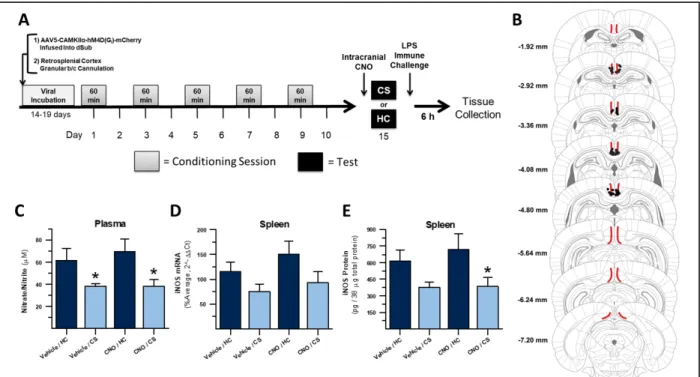
It is unclear how the dHpc contributes to heroin conditioned immune modulation. The dHpc has also been implicated in conditioned drug-reward behavior to a context (Fuchs et al., 2005) and disconnection of the dHpc and the BLA attenuates context-induced reinstatement of cocaine seeking, despite the two areas not having direct connectivity (Fuchs et al., 2007b). Therefore, understanding how the dHpc communicates with these reward structures would be valuable for understanding both context-immune and context-reward associations with drugs of abuse. Chapter 2 of this dissertation describes an experiment employing DREADD-mediated inhibition of dHpc output regions during expression of heroin contextually conditioned suppression of NO. This experiment validates the involvement of dHpc outputs and identifies specific dHpc projection targets that could support hippocampal initiation of contextually conditioned drug behaviors. In Chapter 4, the dHpc output to one of these targets, the
retrosplenial cortex, is specifically inhibited using a combination of dHpc DREADD expression and intra-retrosplenial CNO administration during expression of heroin contextually conditioned suppression of NO.
13
14
CHAPTER 2: DORSAL HIPPOCAMPAL OUTPUT REGIONS ARE REQUIRED FOR THE EXPRESSION OF HEROIN’S
CONTEXTUALLY CONDITIONED IMMUNE EFFECTS Introduction
The dorsal hippocampus (dHpc), like the rest of the hippocampus, is thought to process information mostly in a serial, unidirectional fashion. Sensory information from cortex arrives at the dorsal dentate gyrus (dDG) via the perforant path, is then transmitted from the dDG to the dorsal CA3 (dCA3) subregion via the mossy fiber path, and is conveyed to the dorsal CA1 subregion (dCA1) via Schaffer Collaterals. Information from this tri-synaptic circuit is then finally routed to the dorsal subiculum (dSub) by dCA1 (Knierim, 2015; O'Mara, 2005). Information processed by the dHpc in this way is then relayed to other brain regions
predominantly through projections directly from dCA1 and dSub, which are considered the main output regions of the dHpc (Witter, 2006). Fitting with the vital role of the dHpc in processing contextual information, these regions have been shown to be important in
contextually-influenced behaviors with drugs of abuse. Xia et al showed that dCA1 neurons encode
associations between nicotine reward and context in a conditioned place preference paradigm (Xia et al., 2017). Trouche et al similarly showed that more dCA1 neurons active during context-cocaine association, were re-activated during re-exposure to the context-cocaine-paired context than to a saline-paired context. Repeated inhibited of these cocaine-context representing neurons
15
Much less direct evidence exists to evaluate the likely importance of dSub neurons to recall of a drug-associated context. One study showed that exposure to a cocaine-associated context increased activity in dSub neurons, although this did not differ significantly from unpaired controls where the context was not associated with cocaine (Franklin and Druhan, 2000). Another study showed that inactivation of the dSub disrupted forming a context-cocaine reward association as measured by cocaine-seeking behavior in a subsequent re-exposure to the associated context (Martin-Fardon et al., 2007), but this study manipulated the learning, not the expression of the conditioned behavior. Despite the scarcity of supporting evidence, if we accept the dSub as a predominant output of dHpc information, the evidence implicating dCA1 in
context-drug associations and behavior would predict that dSub would be equally important to such processing.
Furthermore, outgoing projections from these output regions of the dHpc have been indirectly implicated in contextual models of drug behaviors from circuit-level analyses. For example, Fuchs et al have shown that inhibition of the dHpc prevents context-induced reinstatement of cocaine seeking (Fuchs et al., 2005) and that functional disconnection of the dHpc and the BLA attenuates context-induced reinstatement of cocaine seeking through interfering with context-cocaine memory reconsolidation (Fuchs et al., 2007b; Wells et al., 2011).
16
of information within the hippocampus would lead to the prediction that dCA1 and dSub would be critically important for the expression of heroin contextually conditioned immune modulation, but there is a possibility that this information might be relayed in some manner other than
through these classical output regions (see Chapter 5: General Discussion). Additionally, it is unclear which projection neurons in these heterogeneous regions would mediate the relay of this information. Knowing which neuronal activity in the dHpc is required for the expression of heroin conditioned immune modulation will be an important step toward understanding how the dHpc might relay contextual information in this phenomenon.
The current experiment aimed to chemogenetically inhibit neurons in dCA1 and dSub just prior to re-exposure to a heroin-paired context in order to test their hypothesized role in mediating the expression of heroin contextually conditioned immune modulation.
Materials & Methods
Animals
Adult, male Lewis rats weighing initially 225-250 g (N = 36) were purchased from Charles River Laboratories (Kingston, NY, USA). Rats were housed individually on a reversed, 12-h light-dark cycle and all experimental procedures took place during the animals’ active dark period (7 am – 7 pm). Food and water were provided ad libitum in home cages and animals were handled regularly. All experimental procedures were conducted in accordance with federal guidelines and with approval from the University of North Carolina at Chapel Hill Institutional Animal Care and Use Committee (IACUC).
Drugs and Delivery
17
mg/mL solution, and stored at 4°C. During each conditioning session, rats were administered 1.0 mg/kg heroin subcutaneously. This dosage was based on our experiments showing that it reliably alters measures of nitric oxide (NO) in spleen and blood plasma following endotoxin immune challenge (Lysle and How, 2000; Lysle and Ijames, 2002; Szczytkowski and Lysle, 2007). Lipopolysaccharide (LPS, derived from E. coli, serotype O55:B5, Cat# L2880, MilliporeSigma, St. Louis, MO, USA) was dissolved in sterile 0.9% saline the day before use to produce a 1 mg/mL solution, which was then stored at 4°C. Following the test session, LPS was injected subcutaneously at a dose of 1.0 mg/kg which produces sickness behavior and production of NO measures. We have used this particular LPS serotype to previously investigate heroin-and conditioning-induced changes in immune response. Replications of these experiments should employ the same serotype, if possible, as activity between serotypes can vary (Caroff et al., 2002). The synthetic DREADD agonist clozapine-N-oxide (CNO, Cat# C0832, MilliporeSigma) was prepared as a solution on the same day as it was used while also protecting this
light-sensitive reagent from light. CNO first dissolved in 100% dimethyl sulfoxide (DMSO) and then diluted with sterile 0.9% saline to a final concentration of 3.0 mg/mL CNO and 0.5% DMSO. CNO was injected subcutaneously at a dose of 3.0 mg/kg. All injected reagents stored at 4°C were allowed to come to room temperature just before use.
Surgical Procedure
To deliver a designer receptors exclusively activated by designer drugs (DREADD) containing virus in vivo to the dorsal hippocampus (dHpc) output regions, rats underwent
18
incision was made in the skin covering the skull. Holes were drilled into the exposed skull at bilateral coordinates for dorsal subiculum (dSub). Coordinates relative to bregma were AP -6.0 mm, ML ±2.8 mm, DV -3.5 mm (Paxinos and Watson, 2007). Injectors (33 gauge, Plastics One, Roanoke, VA, USA) containing virus and connected with tubing to syringes and a microinfusion pump (PHD 2000, Harvard Apparatus, Holliston, MA, USA) were lowered at a 0° lateral angle to the appropriate DV coordinate but were raised 0.1 mm DV to create a pocket before virus infusion. The virus was infused at the received titer of 4.4x10¹² GC/mL, volume of 0.7 µL per hemisphere, and a rate of 0.05 µL/min. Injectors were then left in place for 10 min to allow for diffusion away from the injection site. Injectors were raised slowly (over 1-2 min) to limit spread of the virus up the injection tract. All animals received DREADD virus. The incision was closed using 4-0 nylon monofilament non-absorbable suture. Animals were given at least two weeks to recover prior to undergoing conditioning.
DREADD Virus and Incubation
DREADDs are delivered in vivo using viruses. In these experiments, the DREADD plasmid CAMKIIα-hM4D(Gi)-mCherry, a gift from Bryan Roth, was purchased pre-packaged in
an adeno-associated virus serotype 5 (AAV5) from Addgene (Viral prep 50477-AAV5; http://n2t.net/addgene:50477; RRID:Addgene_50477, Cambridge, MA, USA). The
Ca2+/calmodulin-dependent protein kinase II alpha (CAMKIIα) promoter element was chosen based on pilot studies showing that virus with this promoter exhibited stronger expression and less dorsal-ventral spread relative to virus with the human synapsin 1 (hSyn) promoter
(unpublished data). Furthermore, the CAMKIIα promoter element may allow for the DREADD to be expressed preferentially in excitatory neurons, especially CA1 pyramidal neurons
19
al., 1996), which is one of our main target populations. The mCherry reporter element produced fluorescent signal in transduced cells that allowed for localization of DREADD-expressing cells postmortem. Following virus delivery, the virus was allowed to incubate to promote DREADD expression for at least two weeks prior to the initiation of any training. By the time animals were treated with CNO, total incubation time was at least four weeks.
Conditioning & Testing Procedure
All rats in these studies were Pavlovian conditioned using five, 60-min sessions every 48 hours where they received an injection of heroin, the unconditioned stimulus (US), and were immediately placed in a distinct context, the conditioned stimulus (CS). This training regimen has repeatedly produced a conditioned immunomodulatory response to the heroin-paired context alone in our laboratory (Lebonville et al., 2016; Paniccia et al., 2018; Szczytkowski et al., 2011; Szczytkowski et al., 2013; Szczytkowski and Lysle, 2010). The CS was a standard operant chamber (BRS/LVE, Laurel, MD, USA; W 30.5 cm x H 26.7 cm x D 24.1 cm) that was enclosed by a sound and light attenuating outer chamber (W 50.8 cm x H 36.8 cm x D 34.3 cm). To distinguish these chambers from any home cage stimuli, the conditioning chambers were housed in a separate room from the vivarium and contained distinct auditory (noise-masking house fan), tactile (metal footshock bar floor), visual (metal side walls), and olfactory (cedar bedding) cues. Between animals, the chambers were thoroughly cleaned with Roccal-D Plus (Zoetis,
Kalamazoo, MI).
20
immune response to LPS, another group of animals remained in home cage instead of being re-exposed to the CS before LPS challenge. Prior work in our laboratory has demonstrated that the LPS response of these heroin-conditioned home-cage control animals are not different than unmanipulated animals, saline conditioned controls, or animals that received heroin and CS-exposure in an unpaired manner (Lysle and Ijames, 2002). Collectively, these results indicate not only that immunomodulation to a heroin-paired context is a conditioned response to the CS and not ancillary effects of conditioning procedures or heroin dosing, but also that the use of only one of these equivalent control groups in future experiments is valid. We believe the heroin-conditioned home-cage control to be the most important and thus it is used here.
Blood and Spleen Tissue Collection
Immediately after CS (or home cage control) exposure, all animals received an LPS immune challenge and were sacrificed by cervical dislocation without anesthesia 6 h later for brain, blood, and spleen collection. This time point is optimized to detect measures of NO production in spleen and blood plasma. Blood was collected in heparinized syringes, transferred to tubes, and spun at 2000 g/rcf and 4°C for 20 min. Plasma was collected and stored at -80°C. Spleen tissue has shown robust expression of iNOS in response to LPS in multiple immune cell types (Bandaletova et al., 1993) and has reliably demonstrated opioid-conditioned
21
Brain Histology and DREADD Expression Analysis
Whole brains were extracted and post-fixed in 4% paraformaldehyde at 4°C for 48 hours with agitation. Then brains were cryoprotected at 4°C in 30% sucrose containing 0.1% sodium azide until sunk (from 6-8 days). Brains were next embedded in frozen section compound
(VWR, Radnor, PA, USA), frozen in a -23 to -25°C freezing microtome, covered with aluminum foil, and stored at -80°C. Brains were allowed to warm to -20 to -21°C before being sliced into 40 µm coronal sections on a cryostat (Leica CM 3050 S, Leica Microsystems, Buffalo Grove, IL, USA). Free-floating slices were stored in a cold solution of ethylene glycol and
polyvinylpyrrolidone at -20°C. Desired sections were slide mounted onto charged glass slides (FisherBrand Superfrost, ThermoFisher Scientific, Waltham, MA, USA), allowed to air dry under dark conditions, and then coverslip mounted using HardSet VECTASHIELD mounting medium with DAPI (Vector Laboratories, Burlingame, CA, USA). Fluorescent microscopy (Leica DM6000 B widefield light microscope, Leica Microsystems, Buffalo Grove, IL, USA) was used to verify positive bilateral DREADD expression in the hippocampal region of interest though localization of the mCherry fluorescent tag. Any animals that did not show bilateral mCherry expression in dSub were removed from all subsequent analyses.
Nitrate/nitrite Assay
22
acid and 0.1% N-(1-Naphthyl)ethylenediamine dihydrochloride in dH2O) was added and color allowed to develop for 10 min at room temperature. Bubbles were popped using a syringe needle and the bottom of the plate was wiped clean before measuring absorbance at 550 nm in a
spectrophotometer. Total nitrate/nitrite concentration was determined from a concurrently run known standard dilution series with a 4-parameter logistic curve fit.
RNA Extraction & RT-qPCR
Reverse transcription quantitative real-time polymerase chain reaction (RT-qPCR) was performed on spleen samples to measure iNOS messenger ribonucleic acid (mRNA) expression. First, spleen tissue free of residual RNAlater was homogenized in 1 mL TRI-Reagent (Molecular Research Center, Cincinnati, OH, USA) using a bead mill homogenizer (Precellys Evolution, Bertin Instruments, Montigny-le-Bretonneux, France) and the following parameters: 7500 rpm, 30 s, 30 s pause, 6 cycles, cooling samples on ice between every three cycles. RNA was purified using TRI-Reagent’s manufacturer protocols with the following modifications: performed optional homogenization step to remove debris, added 100 µL RNAse Free H2O in conjunction with 100 µL of BCP to reduce the density of the homogenate and aid phase separation in PhaseLock Gel Tubes (Heavy formulation, 5Prime/Quantabio, Beverly, MA, USA), and conducted three RNA pellet washes with 75% ethanol. The purified RNA pellet was dissolved by 55-60°C incubation in 150 µL of RNase Free H2O.
23
Technologies, Inc., Santa Clara, CA, USA). A minimum RNA integrity number (RIN) of 8.0 was considered indicative of high quality, intact RNA. All samples demonstrated A260 nm/280 nm values close to 2.1, indicating high purity.
cDNA synthesis was performed on a Veriti 96 Well Fast Thermal Cycler (Applied Biosystems, ThermoFisher Scientific) using the Advantage RT-for-PCR Kit, according to the manufacturer protocol (Clontech/Takara Bio, Mountain View, CA, USA). Priming for the RT reaction was carried out using Oligo(dT) primers. Input RNA concentration was equalized across samples (1 µg). Undiluted cDNA from each sample was pooled into a single sample of which five serial 1:10 dilutions were made to evaluate qPCR efficiency. Remaining cDNA from each sample was then diluted 1:5 in PCR-grade H2O.
24
efficiency of cDNA pools were at least 92% and roughly equivalent between the two genes, two important prerequisites for relative qPCR analysis. The comparative delta delta Ct method (ΔΔCt) was used for data analysis (Livak and Schmittgen, 2001; Schmittgen and Livak, 2008).
Any Ct value in a triplicate that differed by 0.5 or more from the other two was removed from analysis. The validity of L13A as a reference gene was verified using a 2 x 2 ANOVA to show that L13A expression did not differ in any way by group (F(3,28) = .178, p = .910). The first normalization (ΔCt) was to the reference gene, L13A, while the second normalization was to an
average of iNOS ΔCt for the experiment, since the experimental design did not have a single
control group. ΔΔCt values were linearly transformed into 2-ΔΔCt values for graphical
representation.
Protein Extraction & iNOS ELISA
Spleen tissue was thawed and homogenized on ice in sterile, glass, Dounce grinders. Cells were lysed using two freeze-thaw cycles, where the protein was released into supernatant (protease inhibitor buffer). The homogenate was then centrifuged and the supernatant containing isolated protein collected. Total protein was quantified by Bradford Assay as previously
described (Lebonville et al., 2016). To quantify iNOS protein, 38 µg of total protein from each sample was run in triplicate in a rat iNOS sandwich ELISA (Cat #: abx256135, Abbexa Ltd., Cambridge, UK) following the manufacturer’s protocol. An automatic plate washer (EL 403, BioTek Instruments Inc., Winooski, VT, USA) was used to uniformly wash the plate.
25
Statistical Analysis
A 2 x 2 analysis of variance (ANOVA) was performed on all data sets using statistical software (SPSS Statistics 24 and 25, IBM, Armonk, NY, USA). For all tests, the level of
significance was set to p ≤ .05. The validity of using an ANOVA with the current data was tested
using Shapiro-Wilk Test for normality and Levene’s Test for homogeneity of variance (HOV). All assumptions were supported for this experimental data. Planned contrasts were performed with the CS and HC groups within each drug treatment (vehicle or CNO) to test for a
conditioned effect with exposure to the CS. Any ancillary effects were probed using Tukey’s Honestly Significant Difference (HSD) post-hoc test. The presence of statistical outliers was probed using Grubb’s test. Statistical outliers were removed from final analysis. For RT-qPCR data, statistical analysis was performed with ΔΔCt values (without linear transformation) because
these values tend to better meet the assumptions of an ANOVA in our experience. Results
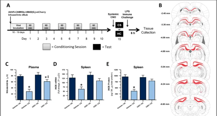
These studies tested the effect of DREADD-mediated inhibition of dorsal hippocampal (dHpc) outputs on the expression of heroin contextually conditioned suppression of NO. The experimental design is displayed in Figure 1A. Virally-transduced cells, as determined by mCherry staining, were present in multiple areas of the dorsal hippocampus (dCA1, dSub, dDG, and dCA2), post-subiculum, overlying proximal retrosplenial granular and dysgranular cortex, and deep layers of visual cortex (Figure 1B).
26
due to insufficient plasma collection (final n = 7-8). No subjects were dropped from iNOS qPCR and ELISA analyses (final n = 8).
Plasma nitrate/nitrite concentration during the test for the expression of heroin
contextually conditioned suppression of NO following chemogenetic inhibition of dHpc outputs is shown in Figure 1C. A 2 x 2 ANOVA revealed significant differences in plasma nitrate/nitrite concentration between treatment groups (F(3,27) = 14.264, p < .001). There was a significant interaction between exposure regimen (CS or HC) and injection (CNO or Veh) on plasma nitrate/nitrite (F(1,27) = 4.725, p = .039). There were also significant main effects of exposure (F(1,27) = 29.321, p < .001) and injection (F(1,27) = 8.002, p = .009) on this measure. In
vehicle-Figure 1. Inactivation of dHpc output regions, including dSub and dCA1, disrupts expression of heroin contextually conditioned suppression of NO. Experimental timeline (A). DREADD
27
treated animals, a planned comparison revealed that CS-exposed animals demonstrated
significantly less nitrate/nitrite in response to LPS than animals that remained in their home cage (p < .001). This comparison shows that exposure to the CS dampened the production of NO to LPS stimulation, confirming expression of the conditioned effect. In CNO-treated animals, a planned comparison also revealed that CS-exposed animals demonstrated significantly less nitrate/nitrite in response to LPS than animals that remained in their home cage (p = .027). This result would seem to indicate that in CNO-treated animals, there was still significant conditioned suppression of NO, yet post-hoc analysis showed that CNO-treated CS-exposed animals showed significantly higher nitrate/nitrite concentration than vehicle-treated CS-exposed animals (p < .001) which we interpret to be indicative of partial attenuation of conditioned suppression of nitrate/nitrite when dHpc output inhibition preceded CS exposure.
28
Furthermore, post-hoc analysis showed that CNO-treated CS-exposed animals showed significantly higher iNOS mRNA expression than vehicle-treated CS-exposed animals (p = .018). These results indicate that there was no significant conditioned suppression of NO when dHpc output inhibition preceded CS exposure.
Splenic iNOS protein expression during the test for the expression of heroin contextually conditioned suppression of NO is shown in Figure 1E. A 2 x 2 ANOVA revealed significant differences in iNOS protein expression between treatment groups (F(3,28) = 13.838, p < .001). There was a significant interaction between exposure regimen (CS or HC) and injection (CNO or Veh) on iNOS protein expression (F(1,28) = 9.145, p = .005). There were also significant main effects of exposure (F(1,28) = 25.081, p < .001) and injection (F(1,28) = 7.288, p = .012) on this measure. In vehicle-treated animals, a planned comparison revealed that CS-exposed animals demonstrated significantly less iNOS protein expression in response to LPS than animals that remained in their home cage (p < .001). This comparison shows that exposure to the CS dampened the production of NO to LPS stimulation, confirming expression of the conditioned effect in vehicle-treated animals. In CNO-treated animals, a planned comparison revealed that CS-exposed animals showed similar iNOS protein expression in response to LPS compared to animals that remained in their home cage (p = .172). Furthermore, post-hoc analysis showed that CNO-treated CS-exposed animals showed significantly higher iNOS mRNA expression than vehicle-treated CS-exposed animals (p = .002). These results indicate that there was no
29
of conditioned suppression of measures of NO production after exposure to a heroin-paired context.
Discussion
Research has shown that one role of the dorsal hippocampus (dHpc) is to process contextual information that can help guide future behavior. In addiction, context reliably influences and prompts drug taking and seeking behavior, but context can also prompt other changes in behavior unrelated to the rewarding aspects of drug use. With heroin, since it is a potent modulator of immune function, these effects can be recapitulated through exposure to a heroin-paired context. Our laboratory has repeatedly shown that these conditioned immune responses to a heroin-paired context require the dHpc. However, little is known about how the contextual processes of the dHpc ultimately lead to changes in behavior.
The study described in this chapter represents the first step to understanding dHpc processing at both the local and circuit level during the expression of heroin contextually conditioned immune modulation. Chemogenetic inhibition of dorsal subiculum (dSub) and dorsal CA1 (dCA1), two of the main output regions of the dHpc, disrupted the expression of heroin contextually conditioned immune suppression, as determined by measures of nitric oxide production in response to LPS. This experiment is the first to manipulate specific populations of dHpc neurons in this paradigm and provide empirical evidence that this chemogenetic technique can be used to ask questions about the broader circuitry of contextually influenced behaviors that require the dHpc.
These results complement substantial evidence that Ca2+/calmodulin-dependent protein kinase II alpha (CAMKIIα) expressing neurons in the dHpc are crucial to formation of
30
study long-term potentiation (LTP), a cellular mechanism of learning and memory through synaptic plasticity (Takeuchi et al., 2014), an abundance of evidence exists to demonstrate the importance of dHpc CAMKIIα to both pre-synaptic (Hojjati et al., 2007) and post-synaptic (Incontro et al., 2018; Lisman et al., 2012) plasticity. Perturbations in CAMKIIα and subsequent
interference with LTP has also been linked with deficits in contextual and spatial memory. Mice with a disrupting mutation or deletion of the CAMKIIα gene from birth or at the time of training show impaired dCA1 LTP as well as impaired spatial and contextual learning (Achterberg et al., 2014; Giese et al., 1998; Silva et al., 1992a; Silva et al., 1992b).
Furthermore, there is evidence that CAMKIIα mechanisms are important to drug
contextual memory. Chronic opiate administration was shown to elevate CAMKIIα mRNA expression in dCA1 (Chen et al., 2008), and given it’s important role in synaptic plasticity, this may represent encoding of drug associations. CAMKII inhibition in the dHpc impaired
acquisition and drug-primed reinstatement of morphine conditioned place preference (Lu et al., 2000). Interestingly, these studies also implicated dHpc CAMKII in the development of
morphine dependence as well, suggesting that the dHpc may be involved in drug synaptic plasticity beyond simply encoding contextual associations. Less evidence supports the role of CAMKIIα expressing neurons in contextual memory recall, that is, during expression of a
previously learned hippocampal-dependent contextual task. This is likely due to the fact that few investigate the role of CAMKIIα expressing neurons themselves and are instead looking at the
role of CAMKIIα expression by these neurons. In the case of CAMKIIα, this gene seems to have
more of a reconsolidation or extinction (new learning) role at the time of memory recall than a role in memory retrieval itself (Vigil and Giese, 2018). Cao et al found that transient CAMKIIα
31
memories (Cao et al., 2008). Thus, the distinction between CAMKIIα expression and CAMKIIα expressing neurons is important in studies like this one which investigates the global role of the neurons themselves, without necessarily looking at that gene’s expression by those neurons. In the context of this chapter, the overwhelming evidence of CAMKIIα’s importance to dHpc
function and memory formation merely provides evidence that these neurons might be sites of important previous plasticity that is relied on during expression of heroin contextually
conditioned immune modulation.
This experiment targeted prominent hippocampal output regions with the aim of beginning to elucidate important Hpc projections. However, it is altogether possible that
inhibiting CAMKIIα expressing neurons perturbed local hippocampal signaling and this resulted
in impaired expression instead of the intended inhibition of outgoing projections. This possibility could be ruled out by specifically inhibiting these CAMKIIα-DREADD expressing neurons at
their projection terminals in areas outside of the hippocampus. A site-directed CNO infusion at an area receiving projections from CAMKIIα-DREADD expressing neurons would demonstrate
conclusively that signals from dHpc outputs are required for the expression of heroin
contextually conditioned immune modulation. Chapter 4 will describe the results from such an experiment. Additionally, concerns of CNO action non-specifically through
DREADD-independent mechanisms have gained much attention recently (Gomez et al., 2017; Mahler and Aston-Jones, 2018). These concerns are not specific to Chapter 2’s experiment and so will be discussed at length in Chapter 5: General Discussion.
This experiment targeted prominent hippocampal output region, dSub, but virus
32
2006) so this spread is not unique to our DREADD delivery system. Additionally, dCA1 is also considered an output region of the dHpc and so spread to this region does not interfere with testing the question about dHpc outputs and connectivity. However, DREADD expression was also seen in other regions other than dCA1 and dSub within and outside of the hippocampus, and this represents a major limitation of this study. Deep layers of cortex, including retrosplenial and visual cortex, and post-subiculum also consistently expressed the mCherry tag. The contributions of these areas to the observed effect could be ruled out using a site-directed CNO infusion in these areas. However, given the close proximity of these areas to the intended dHpc targets, which is likely why DREADD expression spread to these areas in the first place, it would be challenging to ensure that CNO action was restricted to these areas in such an experiment. The same limitation would also apply to a site-directed CNO infusion directly into the dHpc. Given these issues, a more promising way to determine specificity of the effect to inhibition of dHpc outputs is to specifically target dHpc projection areas that do not receive inputs from these overlying extra-hippocampal regions in follow-up experiments. Not only will experiments such as these confirm specificity of the effects seen here to inhibition of dHpc output regions, but also will help to understand the larger circuitry of heroin contextually conditioned immune
modulation.
Our previous work has implicated the dHpc in the expression of heroin contextually conditioned immune modulation. The current results build on past data to implicate a specific population of neurons, those expressing CAMKIIα in the dSub and dCA1 regions of the dHpc.
33
core. The potential role of each of these projections in heroin contextually conditioned immune modulation is discussed in more detail in Chapters 4 and 5. This experiment demonstrates that CAMKIIα-DREADDs can be successfully used to investigate how the dHpc might relay
34
CHAPTER 3: VENTRAL HIPPOCAMPAL OUTPUT REGIONS ARE NOT REQUIRED FOR THE EXPRESSION OF HEROIN’S
CONTEXTUALLY CONDITIONED IMMUNE EFFECTS Introduction
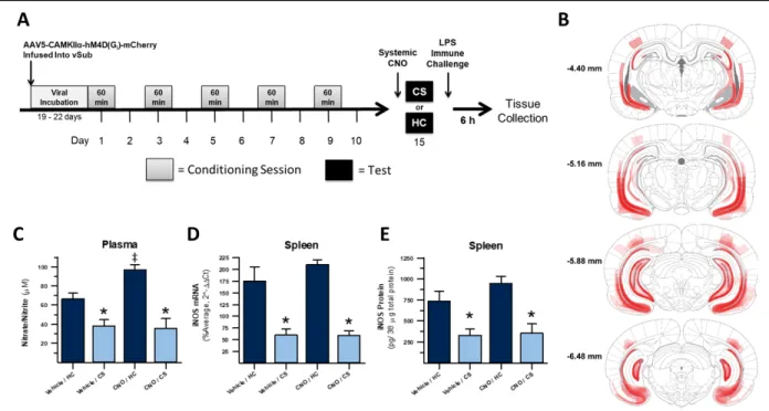
Chapter 2 focused on the role of the dorsal hippocampus (dHpc) in the expression of heroin contextually conditioned immune modulation. This chapter will focus on the role of the ventral hippocampus (vHpc) in this paradigm. Like the dHpc, the vHpc also processes
information serially. Serial pathways route information ultimately to ventral subiculum (vSub) and ventral CA1 (vCA1) (Knierim, 2015; O'Mara, 2005) which then send projections to other regions of the brain. Both vSub and vCA1, mirroring their dorsal counterparts, are considered the main output regions from the vHpc (Witter, 2006). Unlike the role of the dHpc in processing mostly spatial and contextual information, the vHpc has been thought to process mostly emotional (i.e. stress and reward) information. Most relevant to this dissertation is the fact that the vHpc has direct connections with all of the brain regions implicated in both conditioned immune and conditioned reward behaviors including the medial nucleus accumbens shell
(medial NAc shell), ventral tegmental area (VTA), and basolateral amygdala (BLA). Anatomical connectivity with important brain regions alone would not necessarily imply a role of the vHpc outputs in heroin contextually conditioned immune modulation. Yet, more questions than answers arise when reviewing relevant behavioral evidence.
35
Comparatively, there is more evidence implicating the vSub in drug-context associations and behaviors. Several studies have shown that exposure to a cocaine-associated context increases activity in vCA1 and vSub neurons (Franklin and Druhan, 2000; Neisewander et al., 2000), although some of these effects were modest and did not differ significantly from unpaired controls where the context was not associated with cocaine. Reversible lidocaine-mediated lesions of vCA1/vSub has been shown to block context-induced reinstatement of cocaine seeking (Atkins et al., 2008). GABAergic inhibition of the vSub, but surprisingly not vCA1, reduced context-induced reinstatement of heroin seeking (Bossert and Stern, 2014). The vSub was also shown to be important for context-induced reinstatement of ethanol seeking (Marchant et al., 2016a). Moreover, the role of these regions seems to be due to their projections to the nucleus accumbens. For example, stimulating the vSub alone is enough to both increase dopamine release in the nucleus accumbens and increase reinstatement of drug-seeking behavior with cocaine (Vorel et al., 2001) and d-amphetamine (Taepavarapruk and Phillips, 2003). Pathway specific inhibition of projections from the vSub to nucleus accumbens also reduces context-induced reinstatement of heroin seeking (Bossert et al., 2016). Overall, these data indicate that the vHpc, especially vSub, is required across different drugs of abuse for contextual influences on drug behaviors and that this role is likely due in part to its modulation of dopamine signaling in the nucleus accumbens.
36
less involved in these tasks (Vann et al., 2000). The vCA1 possesses place cells that could be capable of contextual processing, though they are fewer in number and have much lower spatial resolution (e.x. larger place fields) than cells in dCA1 (Jung et al., 1994). Floresco and
colleagues found that inhibition of the vCA1 and vSub disrupted acquisition of escape behavior in the Morris Water Maze, a spatial memory task (Floresco et al., 1996). Notably this
manipulation did not affect performance during a subsequent test after temporary inhibition of vCA1/vSub had passed, indicating that the spatial information necessary to perform the task was encoded independent of these vHpc outputs. However, spatial memory in these tasks is more navigational than contextual. More convincingly, Atkins and colleagues demonstrated that inhibition of vCA1/vSub causes a deficit in the ability to discriminate between saline- and cocaine-paired contexts (Atkins et al., 2008). This study also showed that this manipulation blocked context-induced, but not discrete cue-induced reinstatement of cocaine seeking, which would seem to indicate a role for the vHpc output areas in contextually influenced drug
behaviors.
No study to date has investigated the role of the vHpc, much less specific output subregions, in heroin contextually conditioned immune modulation. The current experiment aimed to chemogenetically inhibit neurons in vSub and vCA1 just prior to re-exposure to a heroin-paired context in order to test their hypothesized role in mediating the expression of heroin contextually conditioned immune modulation.
Materials & Methods
Animals
37
12-h light-dark cycle and all experimental procedures took place during the animals’ active dark period (7 am – 7 pm). Food and water were provided ad libitum in home cages and animals were handled regularly. All experimental procedures were conducted in accordance with federal guidelines and with approval from the University of North Carolina at Chapel Hill Institutional Animal Care and Use Committee (IACUC).
Drugs and Delivery
Heroin (diacetylmorphine hydrochloride) was procured from the National Institute on Drug Abuse (NIDA) drug supply program, dissolved in sterile 0.9% saline to produce a 1.0 mg/mL solution, and stored at 4°C. During each conditioning session, rats were administered 1.0 mg/kg heroin subcutaneously. This dosage was based on our experiments showing that it reliably alters measures of nitric oxide (NO) in spleen and blood plasma following endotoxin immune challenge (Lysle and How, 2000; Lysle and Ijames, 2002; Szczytkowski and Lysle, 2007). Lipopolysaccharide (LPS, derived from E. coli, serotype O55:B5, Cat# L2880, MilliporeSigma, St. Louis, MO, USA) was dissolved in sterile 0.9% saline the day before use to produce a 1 mg/mL solution, which was then stored at 4°C. Following the test session, LPS was injected subcutaneously at a dose of 1.0 mg/kg which produces sickness behavior and production of NO measures. We have used this particular LPS serotype to previously investigate heroin-and conditioning-induced changes in immune response. Replications of these experiments should employ the same serotype, if possible, as activity between serotypes can vary (Caroff et al., 2002). The synthetic DREADD agonist clozapine-N-oxide (CNO, NOCD-135, NIDA Drug Supply Program, Bethesda, MD, USA) was prepared as a solution on the same day as it was used while also protecting this light-sensitive reagent from light. CNO first dissolved in 100%
38
3.0 mg/mL CNO and 0.5% DMSO. CNO was injected subcutaneously at a dose of 3.0 mg/kg. All injected reagents stored at 4°C were allowed to come to room temperature just before use.
Surgical Procedure
To deliver a designer receptors exclusively activated by designer drugs (DREADD) containing virus in vivo to ventral hippocampus (vHpc), rats underwent intracranial surgery. Rats were anesthetized with 1.0 mL/kg of a 9:1 (vol:vol) mixture of ketamine hydrochloride (100 mg/mL) and xylazine (100 mg/mL) injected intraperitoneally. Head-shaven animals were placed in a stereotaxic apparatus, the surgical site sterilized, and an incision was made in the skin covering the skull. Holes were drilled into the exposed skull at bilateral coordinates for ventral subiculum (vSub). Coordinates relative to bregma for vSub were AP -6.0 mm, ML ±4.6 mm, DV -8.5 mm (Paxinos and Watson, 2007). Injectors (33 gauge, Plastics One, Roanoke, VA, USA) containing virus and connected with tubing to syringes and a microinfusion pump (PHD 2000, Harvard Apparatus, Holliston, MA, USA) were lowered at a 0° lateral angle to the appropriate DV coordinate but were raised 0.1 mm DV to create a pocket before virus infusion. The virus was infused at the received titer of 4.4x10¹² GC/mL, volume of 0.7 µL per hemisphere, and a rate of 0.05 µL/min. Injectors were then left in place for 10 min to allow for diffusion away from the injection site. Injectors were raised slowly (over 1-2 min) to limit spread of the virus up the injection tract. All animals received DREADD virus. The incision was closed using 4-0 nylon monofilament non-absorbable suture. Animals were given at least two weeks to recover prior to undergoing conditioning.
DREADD Virus and Incubation
39
an adeno-associated virus serotype 5 (AAV5) from Addgene (Viral prep 50477-AAV5; http://n2t.net/addgene:50477; RRID:Addgene_50477, Cambridge, MA, USA). The
Ca2+/calmodulin-dependent protein kinase II alpha (CAMKIIα) promoter element was chosen based on pilot studies showing that virus with this promoter exhibited stronger expression and less dorsal-ventral spread relative to virus with the human synapsin 1 (hSyn) promoter
(unpublished data). Furthermore, the CAMKIIα promoter element may allow for the DREADD to be expressed preferentially in excitatory neurons, especially CA1 pyramidal neurons
(Achterberg et al., 2014; Guo et al., 2010; Johansen et al., 2010; Liu and Jones, 1996; Tsien et al., 1996), which is one of our main target populations. The mCherry reporter element produced fluorescent signal in transduced cells that allowed for localization of DREADD-expressing cells postmortem. Following virus delivery, the virus was allowed to incubate to promote DREADD expression for at least two weeks prior to the initiation of any training. By the time animals were treated with CNO, total incubation time was at least four weeks.
Conditioning & Testing Procedure
40
in a separate room from the vivarium and contained distinct auditory (noise-masking house fan), tactile (metal footshock bar floor), visual (metal side walls), and olfactory (cedar bedding) cues. Between animals, the chambers were thoroughly cleaned with Roccal-D Plus (Zoetis,
Kalamazoo, MI).
Six days after the final conditioning session (day 15), animals were tested for the expression of a conditioned immune response by being re-exposed to the CS (conditioning chamber) for 60 min. Thirty-minutes before CS re-exposure, animals received either an injection of CNO (experimental) or vehicle (control). As a behavioral control representing a typical immune response to LPS, another group of animals remained in home cage instead of being re-exposed to the CS before LPS challenge. Prior work in our laboratory has demonstrated that the LPS response of these heroin-conditioned home-cage control animals are not different than unmanipulated animals, saline conditioned controls, or animals that received heroin and CS-exposure in an unpaired manner (Lysle and Ijames, 2002). Collectively, these results indicate not only that immunomodulation to a heroin-paired context is a conditioned response to the CS and not ancillary effects of conditioning procedures or heroin dosing, but also that the use of only one of these equivalent control groups in future experiments is valid. We believe the heroin-conditioned home-cage control to be the most important and thus it is used here.
Blood and Spleen Tissue Collection