RESTRICTION OF APOPTOSIS IN MATURE NEURONS BY MULTIPLE REDUNDANT BRAKES
Ryan Patrick Annis
A dissertation submitted to the faculty of the University of North Carolina at Chapel Hill in partial fulfillment of the requirements for the degree of Doctor of Philosophy in the Curriculum of
Neurobiology.
Chapel Hill 2016
Approved by:
Mohanish Deshmukh Benjamin Philpot Jay Brenman
Kathleen Caron
iii ABSTRACT
Ryan Patrick Annis: Restriction of Apoptosis in Mature Neurons by Multiple Redundant Brakes
(Under the Direction of Mohanish Deshmukh)
Apoptotic cell death is a key part of normal nervous system development, as a process that allows for the controlled removal of developing neurons that fail to properly integrate into the nervous system. However, once the nervous system is established, the production of new neurons is halted in most areas of the nervous system, meaning that neurons remaining after the establishment of the nervous system must last for the lifetime of the organism. Aberrant or accidental activation of the apoptotic pathway would be deleterious for this long-term neuronal survival, and neurons have been found to restrict the apoptotic pathway as they mature. However, while the resistance of mature neurons to apoptosis is well documented, the precise molecular mechanisms underlying this resistance have remained unclear.
iv
miR-29 family of miRNAs was the only brake restricting Bax activation in mature neurons, or if other brakes on Bax activation exist.
The work presented in this dissertation addresses this question by examining the status of the apoptotic pathway in neurons from mice in which all three members of the miR-29 family have been deleted. I found that in neurons lacking miR-29 expression, the apoptotic pathway remains restricted. I identified miR-24 as a microRNA that is also upregulated with neuronal maturation, which can act redundantly with miR-29 by
v
vi
ACKNOWLEDGEMENTS
vii
TABLE OF CONTENTS
TABLE OF CONTENTS ... vii
LIST OF FIGURES ... xi
LIST OF TABLES ... xiii
LIST OF ABBREVIATIONS... xiv
CHAPTER 1: INTRODUCTION ... 1
1.1 Apoptosis: A Brief History ... 1
The BCL-2 Family ... 3
Caspases ... 5
1.2 Apoptosis in Development and Disease ... 6
1.3 Apoptosis in Neurons ... 7
1.4 Apoptosis in Nervous System Pathologies ... 12
Alzheimer’s Disease ... 13
Parkinson’s Disease ... 15
Amyotrophic Lateral Sclerosis ... 17
1.5: Introduction to microRNAs ... 19
1.6 MicroRNAs in Nervous System Development and Disease ... 20
1.7 Increasing Resistance to Apoptosis with Neuronal Maturation... 22
viii
Post-Mitochondrial Apoptotic Brakes ... 26
Pre-Mitochondrial Apoptotic Brakes ... 30
1.10 Introduction to miR-29 ... 33
miR-29 miRNAs as Regulators of Neuronal Apoptosis ... 34
1.11 Figures and Legends ... 38
1.14 Tables ... 52
CHAPTER 2: MATURE NEURONS DYNAMICALLY RESTRICT APOPTOSIS VIA REDUNDANT PRE-MITOCHONDRIAL BRAKES ... 53
2.1: Introduction ... 53
2.2 Results... 56
Maturing neurons simultaneously restrict apoptosis both pre- and post- mitochondria... 56
Loss of miR-29 is not sufficient to re-sensitize mature neurons to apoptosis ... 58
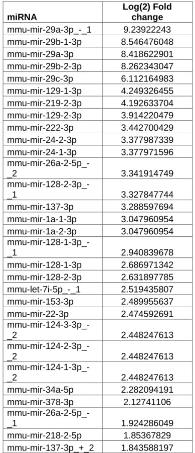
Other microRNAs with predicted targets in the apoptotic pathway are upregulated during neuronal maturation ... 59
Overexpression of miR-24 in Young Neurons Inhibits Cyt c Release and Cell Death ... 60
miR-24 Can Inhibit Bim and Puma Induction in NGF-Deprived Neurons ... 61
2.3: Discussion ... 63
2.4 Materials and Methods ... 68
2.5 Figures and Figure Legends ... 74
CHAPTER 3: SPATIAL AND TEMPORAL REGULATION OF BAX ACTIVATION IN NEURONS ... 94
3.1 Introduction ... 94
ix
3.2: Materials and Methods ... 100
3.3 Results... 102
Treatment with ABT-737 Causes Selective Degeneration of Axons in Young Neurons... 102
ABT-737 Induces Global Cytochrome c Release in Young Sympathetic Neurons ... 102
ABT-737 Induces Axon-Specific Cytochrome c Release in Mature Neurons ... 103
NGF Deprivation Sensitizes Mature Neuron Somas to ABT-737-Induced Cytochrome c Release ... 105
ABT-737 Treatment Results in Elevated Bim Levels in Young, but not Mature Neurons ... 106
Direct Stimulation of Bax with BH3-Peptides as an Alternative Approach to Explore Bax Activation in Neurons ... 107
3.4: Figures and Figure Legends ... 114
CHAPTER 4: DISCUSSION ... 126
4.1: Summary of Findings ... 126
Mature Neurons Dynamically Regulate Apoptosis Via Redundant Pre-Mitochondrial Brakes ... 127
Spatial and Temporal Regulation of Bax Activation in Neurons ... 128
4.2: Clinical Relevance ... 129
Overexpression of miRNAs for Therapeutic Purposes ... 129
x
4.3: Non-Apoptotic Roles for Apoptotic Proteins ... 134
Bax and Caspases in Synaptic Plasticity and Synapse Remodeling ... 135
Concluding Remarks ... 138
xi
LIST OF FIGURES
Figure 1.1: The BCL-2 Family of Proteins ... 39
Figure 1.2: Effector and Initiator Caspases ... 41
Figure 1.3: Timecourse of Developmental Milestones in Sympathetic Neuron Development ... 43
Figure 1.4: Apoptosis is Essential for Proper Brain Development ... 45
Figure 1.5: The Intrinsic Apoptotic Pathway in Sympathetic Neurons ... 47
Figure 1.6: Overview of microRNA Biogenesis ... 49
Figure 1.7: Resistance of Sympathetic Ganglia to Apoptosis with Maturation ... 51
Figure 2.1: Neuronal maturation is associated with progressive resistance to neuronal apoptosis at both pre and post-mitochondrial checkpoints ... 75
Figure 2.2: Mature miR-29 deficient neurons remain resistant to NGF-deprivation induced cyt c release. ... 77
Figure 2.3: Other miRNAs predicted to regulate the apoptotic pathway are also induced with neuronal maturation... 79
Figure 2.4: Overexpression of miR-29 or miR-24 is sufficient to inhibit cyt c release and cell death in young, NGF deprived neurons. ... 81
Figure 2.5: Overexpression of miR-29 or miR-24 is sufficient to inhibit the induction of Bim and Puma in young sympathetic neurons. ... 83
Figure 2.6: Inhibition of miR-24 Function in miR-29 Knockout Neurons Leads to Partial Restoration of Apoptotic Response ... 85
Figure 3.1: Indirect vs Direct Activation Models for Bax Activation ... 115
Figure 3.2: Treatment of Young Sympathetic Neurons with ABT-737 Induces Axon Degeneration ... 117
xii
Figure 3.4: ABT-737 Upregulates Bim Expression in Sympathetic Neurons ... 121
Figure 3.5 Direct Injection of BH3-only Domain Peptides Is Sufficient
to Cause Cytochrome c Release in Young, but not Mature SCGs... 123
Figure 3.6 Optimization of Digitonin Concentration for Permeablizing
xiii
LIST OF TABLES
Table 1.1: miRNAs in Neurodegeneration and Stroke ... 52
xiv
LIST OF ABBREVIATIONS
Aβ: Amyloid Beta
AD: Alzheimer’s Disease
Ago: Argonaute
ALS: Amyotrophic Lateral Sclerosis
APAF-1: Apoptosis Associated Factor 1
APP: Amyloid Precursor Protein
BACE1: β-Site APP-Cleaving Enzyme 1
BH: BCL-2 Homology
BCL: B-Cell Lymphoma
CARD: Caspase Activation and Recruitment Domain
C. elegans: Caenorhabditis elegans
CNS: Central Nervous System
Cyt c: Cytochrome c
dATP: Deoxyadenosine Triphosphate
DED: Death Effector Domain
xv DLK: Dual Leucine Zipper Kinase
DRG: Dorsal Root Ganglia
GABA: Gamma-Aminobutyric Acid
HD: Huntington’s Disease
HSP: Heat Schock Protein
FOXO: Forkhead Box
IAP: Inhibitor of Apoptosis Protein
JNK: c-Jun N-Terminal Kinase
LNA: Locked Nucleic Acid
MCAO: Middle Cerebral Artery Occlusion
MCL- Mantle Cell Lymphoma
miR/miRNA: microRNA
MLK: Mixed Lineage Kinase
MOMP: Mitochondrial Outer Membrane Permeablization
MPP+: 1-methyl-4-phenylpyridinium
MPTP: 1-methyl-4-phenyl-1,2,3,6-tetrahydropyridine
xvi NFT: Neurofibrillary Tangle
NGF: Nerve Growth Factor
NMDA: N-Methyl-D-Aspartate
P: Postnatal Day
Pol II: RNA Polymerase II
PD: Parkinson’s Disease
PI3K: Phosphoinositide 3-kinase
PNS: Peripheral Nervous System
qRT-PCR: quantitative RT-PCR
RISC: RNA-Induced Silencing Complex
RNA: Ribonucleic Acid
SNc: Substantia Nigra
SOD1: Superoxide Dismutase 1
UTR: Untranslated Region
XAF1: XIAP Associated Factor 1
1
CHAPTER 1: INTRODUCTION
1.1 Apoptosis: A Brief History
Death is an unavoidable and important aspect of life, but death is something that all organisms, including humans, instinctually avoid in most cases. However, in most eukaryotes, decisions about life and death are constantly taking place at a cellular level, and are crucially important for both the development and health of complex organisms like mammals. The work of many researchers throughout the years has uncovered a process of voluntary, genetically programmed self-destruction that takes place in mammalian cells, called apoptosis.
The term apoptosis was first coined by the researchers Kerr, Wyllie, and Curry in 1972. They described a process of “controlled cell deletion” that opposed the process of mitosis, or cell division, in mammalian cells for the purposes of regulating the size of cell populations (Kerr, Wyllie et al. 1972). Early observations in developing chick embryos found that the same populations of cells would undergo cell death at the same time-point during embryonic development, suggesting that cell death might be a normal, voluntary, and perhaps genetically programmed part of development (Hinchliffe and Johnson, 1980).
2
elegans). C. elegans was an ideal model system to explore programmed cell death, because every worm consists of exactly 1090 cells, of which 131 are lost during development in a highly reproducible fashion (Sulston and Horvitz 1977). Using a forward genetic screen in these worms, Horvitz identified the first “death genes”, called ced-3 and ced-4. Horvitz showed that functional ced-3 and ced-4 genes were necessary for the execution of the cell death program (Ellis and Horvitz 1986). In 1988,
researchers identified the B-Cell Lymphoma 2 (BCL-2) oncogene, which promoted survival in mammalian B-Cells (Vaux, Cory et al. 1988). A gene with similar function, ced-9, was discovered in C. elegans in 1992 (Hengartner, Ellis et al. 1992), and later that same year, it was discovered that expression of human BCL-2 in C.elegans was able to inhibit cell death, providing strong evidence that BCL-2 performed an
evolutionarily conserved function of opposing cell death in both worm and mammalian cells (Vaux, Weissman et al. 1992). Indeed, later work would find that BCL-2 and ced-9 are actually the same evolutionarily conserved gene (Hengartner and Horvitz 1994).
The evidence that an evolutionarily conserved programmed cell death pathway existed between worms and humans set researchers to searching for other homologues conserved between the two organisms. In 1993, a mammalian homologue of ced-3 was discovered, which would become the first member of the family of proteins known as cysteine aspartic proteases, or caspases, now known to be essential for the execution of mammalian apoptosis (Yuan, Shaham et al. 1993). In 1997, a mammalian
3
Horvitz 1998). In 1996, the central role played by the mitochondria in apoptosis was established by the discovery that translocation of the electron transport chain protein cytochrome c (cyt c) was essential for the activation of the apoptotic program. (Liu, Kim et al. 1996).
The connection between cyt c release from the mitochondria and BCL-2 was established by the finding that BCL-2 overexpression is able to inhibit apoptosis by preventing cyt c release (Kluck, Bossy et al. 1997). It was subsequently discovered that cyt c release is governed by complex interactions between multiple members of what came to be known as the BCL-2 family of proteins. Named after the founding member of the family (described above), the BCL-2 family is now known to consist of subgroups of multiple anti- and pro-apoptotic members that serve redundant functions(Danial and Korsmeyer 2004). The first pro-apoptotic member of the family, Bax, was discovered in 1993 based on its interaction with BCL-2 (Oltvai, Milliman et al. 1993). It was
subsequently found that Bax and BCL-2 have opposing functions in the regulation of apoptosis, with Bax promoting the release of cyt c from the mitochondria and BCL-2 acting to inhibit this process (Jurgensmeier, Xie et al. 1998; Rosse, Olivier et al. 1998).
The BCL-2 Family
4
These members are characterized by conservation of BH1-4 domains and act to inhibit cell death.
The second group, known as multidomain proapoptotic BCL-2 family proteins, consists of Bax and Bak. These members exist as inactive monomers in the cytosol, and undergo conformational changes that result in their translocation to the
mitochondria to promote the release of cyt c and subsequently apoptosis. Bax and Bak exhibit conserved expression of BH1-3 domains and serve as key gatekeepers on the apoptotic pathway, as loss of Bax and Bak expression is sufficient to render cells
resistant to all known intrinsic apoptotic stimuli (Lindsten, Ross et al. 2000; Wei, Zong et al. 2001).
The third and largest group of BCL-2 family proteins consists of the pro-apoptotic BH3-only domain proteins, which bear strong structural homology with the pro-death c. elegans protein EGL-1(Conradt and Horvitz 1998). This family promotes Bax activation and apoptosis by acting upstream of the mitochondria, and is regulated at both the transcriptional level and by post-translational modification (Lomonosova and Chinnadurai 2008). BH3-only domain proteins promote the activation of Bax and apoptosis, but there is some controversy over the precise mechanism by which this is accomplished. Two non-mutually exclusive models have been proposed, called the direct and indirect activation models.
5
activators to Bax itself is essential for apoptosis to proceed. In the indirect activation model, Bax and Bak are constitutively active and held in check by binding to the anti-apoptotic BCL-2 family proteins. In this model, the BH3-only domain proteins promote apoptosis by liberating active Bax from BCL-2 family proteins and permitting the translocation of Bax to the mitochondria, where it promotes apoptosis (Shamas-Din, Brahmbhatt et al. 2011). These models are not mutually exclusive and requirements for the induction of apoptosis may vary based on the endogenous expression levels of the various BCL-2 family genes in different cell types.
Caspases
The ultimate execution of the apoptotic pathway in mammalian cells is carried out by caspases (Figure 1.2). Caspases are a family of cysteine proteases that can be divided into two functional groups: effectors and initiators. Effector caspases, a group comprised of Caspases-3, -6, and -7, exist as inactive zymogens in the cytosol of mammalian cells, and remain inactive until cleaved by initiator caspases, a group
comprised of Caspase-2, -9, -8, and -10. Caspase-8 and-10 act in the extrinsic pathway of apoptosis, and contain a Death Effector Domain (DED), while Caspase-2 and -9 act in the intrinsic apoptosis pathway, and contain Caspase Activation Recruitment
Domains (CARDs) (Riedl and Shi 2004).
6
cleavage and activation of Caspase-9. Caspase-9 then cleaves and activates executioner caspases such as Caspase-3/7, which cleave a variety of downstream substrates, resulting in the final death of the cell (Li, Nijhawan et al. 1997; Zou, Henzel et al. 1997).
1.2 Apoptosis in Development and Disease
Defects in the apoptotic program are associated with a wide range of disorders, most notably cancer (Hanahan and Weinberg 2000; Hanahan and Weinberg 2011). Indeed, the anti-apoptotic BCL-2 gene was discovered based on its overexpression in follicular lymphoma (Vaux, Cory et al. 1988). Since this discovery, abundant evidence has accumulated that many cancer types exhibit mutations or changes in gene
expression that interfere with the proper function of the apoptotic pathway. For example, the well-known tumor suppressor P53 is mutated in 50% of human cancers, and is known to induce apoptosis through transcriptional upregulation of the BH3-only domain proteins Puma and Noxa (Oda, Ohki et al. 2000; Nakano and Vousden 2001). Mutations in P53 that interfere with its function prevent the induction of the cell death program and allow cancer cells to continue to survive and proliferate.
The apoptotic pathway also plays a key role in the development of many
7
this study, however, will focus on neurons, a population of cells unique in many ways from others in the body, not least of which is their ability to precisely spatially and temporally regulate the apoptotic pathway.
1.3 Apoptosis in Neurons
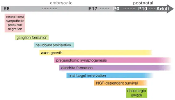
Neurons are the building blocks and key functional units of the nervous system. They are unique among cells in their ability to form connections, called synapses, with muscles, glands, and other neurons, through which they transmit signals via the release of chemicals known as neurotransmitters. The development of the nervous system is governed by a variety of complex and overlapping processes. Neurons differentiate from neuronal precursor cells, and must migrate to the appropriate location and form connections with the correct targets. These processes often occur simultaneously at the cellular level during development (Figure 1.3). Regulation of these processes in various populations of neurons is the subject of intensive ongoing research that is beyond the scope of this dissertation. Instead, we will focus on the apoptotic pathway in neurons, which is employed in the developing nervous system to remove neurons that fail to integrate into the nervous system properly as a result of defects in differentiation, migration, or target innervation.
8
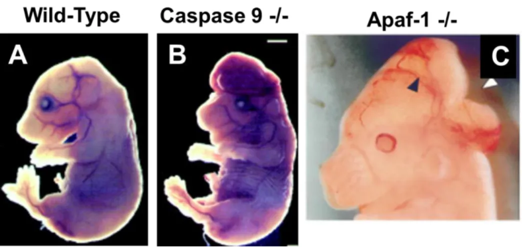
if lost, cannot easily be replaced. One might expect, given the requirement that neurons survive for the lifetime of an organism, that these cells would not possess an active apoptotic pathway. On the contrary, however, apoptosis plays a key role in nervous system development and function. Indeed, mice deficient for key proteins in the apoptotic pathway exhibit craniofacial defects and early embryonic lethality stemming from overproliferation of neuronal precursors (Figure 1.4).
The observation that neurons undergo cell death as a part of normal
development was first made by Dr. Erich Kallius in 1926 (Ernst 1926). Many of the key early observations in the neuronal cell death field, however, were made by Dr. Rita Levi Montalcini and Dr. Viktor Hamburger beginning in 1949. In 1960, Dr. Montalcini injected neonatal mice with a neutralizing antibody to the newly-discovered protein Nerve
Growth Factor, or NGF, and observed almost total degeneration in the mouse sympathetic nervous system (Levi-Montalcini and Booker 1960). This observation represented both a key discovery in the role of cell death in neuronal development, as well as the birth of the sympathetic neuron model of neuronal apoptosis, which has proven to be one of the most informative models of neuronal cell death to date.
Sympathetic neurons are a population of neurons in the peripheral nervous system (PNS) that are perhaps the best understood model of neuronal apoptosis. Mouse sympathetic neurons undergo extensive cell death as a part of normal
9
sympathetic neuron terminals to promote cell survival. Neurons that properly innervate their targets and obtain NGF survive and mature, while neurons that fail to innervate their targets are eliminated by apoptosis during the first postnatal week (Glebova and Ginty 2005).
Sympathetic neurons are an attractive model for the study of apoptosis because they can be isolated from neonatal mice and maintained in culture by adding exogenous NGF to the cell culture media. Switching cells to NGF-free media results in neuronal death within 48-72 hours, and early studies noted that this death features the hallmarks of apoptosis, including chromatin condensation, DNA fragmentation, declines in RNA and protein synthesis, and membrane disruption. This death was also found to depend on protein synthesis, as pharmacological inhibition of protein synthesis is able to
significantly delay cell death. This requirement for active protein synthesis highlights the voluntary “self-destructive” nature of apoptosis in neurons (Martin, Schmidt et al. 1988; Martin, Ito et al. 1992; Deckwerth and Johnson 1993; Edwards and Tolkovsky 1994).
NGF deprivation has been found to induce the intrinsic apoptosis pathway in sympathetic neurons (Figure 1.5). In this pathway, an apoptotic stimulus such as NGF deprivation leads to both the upregulation of the transcription factor c-jun and its
10
overexpression of BCL2 inhibits cell death and cyt c release in NGF-deprived
sympathetic neurons, and neurons from BCL2- deficient animals die faster than their wild-type counterparts (Garcia, Martinou et al. 1992; Greenlund, Korsmeyer et al. 1995). Similarly, overexpression of BCL-xL and MCL-1 have been found to promote neuronal resistance to apoptosis (Gonzalez-Garcia, Garcia et al. 1995; Arbour, Vanderluit et al. 2008).
Since the BH3-only proteins govern the activation of Bax and the release of cyt c from the mitochondria, their roles in neurons have been the subject of considerable study. It is now known that NGF-deprivation in sympathetic neurons leads to the transcriptional upregulation of four BH3-only proteins: Bim, Puma, BMF, and HRK (Kristiansen, Menghi et al. 2011). Overexpression of these genes is sufficient to induce or accelerate cell death in neurons; however, knockout of any of these genes on their own is at best only partially protective from apoptosis in neurons, suggesting that these proteins may have overlapping functions (Whitfield, Neame et al. 2001; Wyttenbach and Tolkovsky 2006; Coultas, Terzano et al. 2007; Pfeiffer, Anilkumar et al. 2014). Indeed, it has been found that knockout of Bim and Puma together provides significantly
enhanced protection from apoptosis in the cerebellar granule neuron model of apoptosis compared to individual knockout of either gene alone, and additional deletion of Bid to generate Bim/Puma/Bid knockout neurons phenocopies knockout of Bax/Bak in the cerebellum (Ren, Tu et al. 2010).
11
promoter of the BH3-only gene Bim and participate in its transcriptional upregulation (Whitfield, Neame et al. 2001). It has subsequently been discovered that the activation of Bim in NGF-deprived sympathetic neurons requires the participation of a number of other factors in addition to c-jun, including the Myb and Forkhead Box (FOXO)
transcription factors, which bind to the Bim promoter and first intron (Gilley, Coffer et al. 2003; Biswas, Shi et al. 2007). More recent studies discovered binding sites for the transcription factor NF-Y in the Bim promoter, and found that NF-Y forms a complex with FOXO3A and CBP/P300 proteins to promote increased Bim transcription. It has been hypothesized that CBP/P300 may integrate the contributions of various pro-survival and death signals at the Bim promoter to regulate Bim induction (Hughes, Kristiansen et al. 2011).
The various transcription factors that control Bim induction in sympathetic neurons undergoing apoptosis are regulated by several different pathways. C-jun phosphorylation is known to be regulated by the MLK/JNK pathway. During apoptosis, MLKs become active and phosphorylate JNKs, which in turn phosphorylate c-jun. Phospho-c-jun translocates to the nucleus, where it acts as a pro-death transcription factor (Davis 2000). Activity of the FOXO transcription factors is governed by their phosphorylation. It has been found that the AKT pathway, which is stimulated by NGF, phosphorylates FOXO transcription factors, resulting in their sequestration in the cytoplasm by 14-3-3 proteins. In response to NGF deprivation, the activity of the PI3K-AKT pathway declines, leading to the dephosphorylation of FOXOs, which then
translocate from the cytoplasm to the nucleus to stimulate pro-apoptotic gene
12
pathway stimulated by NGF, the Raf-MEK-ERK pathway, is thought to promote survival by phosphorylating and activating the transcription factor RSK, which promotes survival by phosphorylating and activating the pro-survival factor CREB (Riccio, Ahn et al. 1999). More recently, ERK has been found to directly repress Bim gene induction by direct binding to the 3’UTR of Bim RNA, although to date the mechanism remains unclear (Hughes, Gilley et al. 2011).
Other pro-death transcription factors, such as the Myb proteins, are thought to be regulated by cell cycle induction. It has been found that neurons stimulated to undergo apoptosis upregulate cell-cycle genes like Cyclin D1 and reenter the cell cycle, and that pharmacological inhibitors of cell cycle progression, such as the CDK inhibitors
Roscovitine and Flavopiridol, are potent inhibitors of neuronal apoptosis (Freeman, Estus et al. 1994; Liu and Greene 2001).
1.4 Apoptosis in Nervous System Pathologies
13 Alzheimer’s Disease
Alzheimer’s Disease (AD) is a progressive neurodegenerative disorder
characterized by memory problems, dementia, and progressive neuronal death, which was estimated to afflict 5.1 million Americans as of 2013. The chief risk factor for AD has been found to be age, and the number of patients is expected to grow rapidly as the population ages, according to the US centers for Disease Control.
The pathological hallmarks of AD are neurofibrillary tangles composed of the cytoskeletal associated protein Tau and extracellular plaques composed of the protein Amyloid-β (Aβ), as well as progressive loss of neurons and synapses (Serrano-Pozo, Frosch et al. 2011). The extent to which apoptosis contributes to the loss of neurons and synapses during AD is controversial. Some studies have reported widespread expression of apoptotic markers, while others have reported small or absent changes in expression of apoptotic markers between diseased brains and age-matched controls (Su, Anderson et al. 1994; Troncoso, Sukhov et al. 1996; Selznick, Holtzman et al. 1999; Su, Zhao et al. 2001; Woodhouse, Dickson et al. 2006). Characterizing cell death in post-mortem samples from degenerating human brains is challenging, mainly
14
Interestingly, while the evidence that apoptosis contributes to neuron loss in AD patient brains is controversial, substantial evidence exists that Beta-Amyloid (Aβ), one of the proteins that make up the hallmark lesions found in the AD brain, has a pro-apoptotic effect on neurons. Incubation of cortical neurons with purified Aβ has been found to induce robust cell death (Morishima, Gotoh et al. 2001). Aβ-induced death has been found to be Bax dependent and dying neurons were found to exhibit cleaved caspase 3, but pharmacological inhibition or genetic knockout of caspase 3 failed to prevent cell death of Aβ treated telencephalic neurons. This suggests that arrest of apoptosis after the point of cyt c release is not able to prevent neurons from dying in reponse to Aβ, but inhibition prior to the release of cyt c can promote survival (Selznick, Zheng et al. 2000). More recently, it has been found that exposure to Aβ causes
increases in the levels of Bim and active Bax, while decreasing the levels of BCL-2, in organotypic hippocampal slice cultures or in the hippocampi of 2-3 month old mice injected with Aβ intrahippocampally (Kudo, Lee et al. 2012). Aβ was subsequently found to upregulate Bim via a Foxo3a-dependent mechanism in cultured cortical neurons (Sanphui and Biswas 2013).
While the evidence for Aβ toxicity is substantial, the role that Tau, plays in AD pathology is less clear. Tau has been well established to exist in a
15
no longer associates with microtubules, and accumulates in the cytosol in the form of neurofibrillary tangles (NFTs).
It is thought that a combination of loss of endogenous Tau function as well as pathological gain-of-function in hyperphosphorylated Tau contributes to neuronal dysfunction in AD (Ballatore, Lee et al. 2007). For example, in mouse models of
tauopathy, neurons exhibit defects in axonal transport that can be rescued by treatment with microtubule stabilizing drugs (Zhang, Maiti et al. 2005). Interestingly,
hyperphosphorylation of Tau has been found in several studies to be neuroprotective and to inhibit apoptosis, suggesting that the toxic effects of Tau may be due to indirect effects or toxic intermediates rather than a direct toxic effect of the hyperphosphorylated Tau present in NFTs (Li, Wang et al. 2007; Liu, Liao et al. 2010; Duan, Chai et al.
2013). A recent finding also found that Tau expression is important for the clearance of Aβ plaques in cultured hippocampal neurons and mouse brain, suggesting that there may be crosstalk between Tau and Aβ during AD pathogenesis (Lonskaya, Hebron et al. 2014)
Parkinson’s Disease
Parkinson’s Disease (PD) is the second most common neurodegenerative
16
The reason for the classical motor symptoms of PD is the progressive loss of dompaminergic neurons in the Substantia Nigra (SNc) region of the brain. PD is also associated with the presence of insoluble protein aggregates known as Lewy Bodies, which are made up of aggregated α-synuclein protein, among other proteins. In addition to the Nigro-Striatal pathway, other brain nuclei have been found to degenerate with Lewy Body pathology. The degeneration of these nuclei is believed to contribute to many of the non-motor symptoms of PD (Jellinger 2012; Dexter and Jenner 2013).
Similar to AD, the contribution of apoptosis to the progression of PD is controversial. Evidence from animal and cell culture models of PD suggested that apoptosis played a prominent role in PD. PD has been modeled in vitro and in mouse models using inhibitors of mitochondrial complex I, namely the toxins MPP+, MPTP, and rotenone. These toxins cause energy failure and oxidative stress in cells, and their injection into mice causes a Parkinson’s-like pathology (Dauer and Przedborski 2003). Injection of these compounds into mice was found to cause increased apoptosis in the Substantia Nigra (SNc) region of the brain (Tatton and Kish 1997; Viswanath, Wu et al. 2001).
17
found to be protective in mouse models of PD (Vila, Jackson-Lewis et al. 2001; Perier, Bove et al. 2007). Overexpression of BCL-2 was also found to be protective in both in vitro and in vivo models of PD, although the degree of protection varies depending on the dosage regimen of MPTP (Offen, Beart et al. 1998; Yang, Matthews et al. 1998; Vila and Przedborski 2003).
The abundance of evidence for the involvement of apoptosis in preclinical models of PD led to the initiation of clinical trials examining whether a drug that inhibits neuronal apoptosis could improve patient outcomes in PD. The drug tested, CEP-1347, is a Mixed Lineage Kinase Inhibitor, which inhibits apoptosis at an early point in the pathway by preventing MLK-JNK activation. CEP-1347 was found to inhibit neuronal apoptosis in multiple models of developmental and pathological cell death (Wang, Ma et al. 2004). Unfortunately, the CEP-1347 trial was stopped early when an interim analysis of the data determined that it would be “futile” to continue the trial due to the drug’s lack of efficacy (Parkinson Study Group PRECEPT Investigators, 2007). The reason for the failure of promising preclinical data to translate to the clinic is unclear, but may reflect inadequacy of the current animal and cellular models to accurately depict human disease, or the presence of other “back-up” cell death pathways, such as necrotic or autophagic cell death, that are engaged when apoptosis is inhibited (Venderova and Park 2012).
Amyotrophic Lateral Sclerosis
18
include muscle weakness, atrophy, and spasticity. Approximately 5-10% of ALS cases are familial, and have been found to be caused by specific mutations, most notably a toxic gain-of-function mutation in the Superoxide Dismutase 1 (SOD1) gene. Familial ALS can also be caused by hexanucleotide repeat expansion of the C9ORF72 gene, and mutations in the RNA processing gene TARDBP.
The pathology of ALS involves progressive loss of motor neurons in the anterior horns of the spinal cord and in the motor nuclei of the brainstem. Denervation of
muscles after motor neuron death leads to muscle atrophy. Most ALS patients die within 2-3 years of the onset of symptoms, usually of respiratory muscle paralysis.
19
As the research summarized above demonstrates, while there is evidence that apoptosis contributes to the progression of some neurodegenerative diseases, it has been difficult to translate even the most promising preclinical data into successful treatments in humans. While improved animal models of neurodegeneration would no doubt aid in this process, it appears at this point that inhibition of apoptosis will not serve as a “magic bullet” in the treatment of neurodegenerative disorders.
1.5: Introduction to microRNAs
MicroRNAs (miRNAs) are small (~22 nucleotide) single-stranded RNAs that act as regulators of gene expression. miRNAs are typically transcribed from genomic DNA by RNA Polymerase II (Pol II) to produce a primary transcript, or miRNA. These pri-miRNAs include stem-loop structures that will eventually be processed into mature miRNAs, as well as varying amounts of surrounding RNA. In the next step of miRNA biogenesis, these stem-loop structures are cleaved from the surrounding RNA by the microprocessor complex, which includes the proteins DGCR8 and Drosha. The product of this cleavage event is a ~65 nucleotide hairpin structure known as a pre-miRNA. Following this cleavage, pre-miRNAs are exported from the nucleus to the cytoplasm via the nuclear pore complex. The most important protein for this export process is the nuclear transport receptor Exportin-5 (Ha and Kim 2014).
20
RNA-Induced Silencing Complex (RISC), at which point one strand, the passenger strand, is removed by cleavage or unwinding. The strand remaining in the RISC, known as the guide strand, is able to participate in RNA silencing at this point, while the
removed passenger strand is typically degraded rapidly. Argonaute proteins, especially Argonaute 2 (Ago2), are critical for miRNA silencing activity (Huntziner et al., 2011). miRNAs bind primarily to the 3’ untranslated regions (3’UTRs) of target mRNAs in a sequence-dependent manner and mediate their silencing through translational inhibition or RNA degradation (Ha and Kim 2014). A schematic of this process is shown in Figure 1.6. Target selection is mediated by a short sequence within each miRNA known as the seed sequence, with miRNAs within families (e.g. miR-29a, b, and c) sharing conserved seed sequences.
1.6 MicroRNAs in Nervous System Development and Disease
21
miRNAs have also been found to play crucial roles in neuronal development and disease. For example, deletion of Dicer using the cerebral cortex-specific Emx1-Cre line results in a range of defects, including decreased survival and differentiation (De Pietri Tonelli, Pulvers et al. 2008; Kawase-Koga, Otaegi et al. 2009). Deletion of Dicer using the CAMKII promoter, which is specific to postmitotic neurons of the cortex and
hippocampus, results in decreased cortical size, enhanced neuronal cell death, defects in axon pathfinding, and impaired dendritic branching (Davis, Cuellar et al. 2008). Certain aspects of these Dicer-deletion phenotypes have been attributed to specific miRNAs. miRNAs, including miR-124, miR-9, and Let-7 have been found to promote neuronal differentiation, neurite outgrowth, and synaptogenesis (Bian and Sun 2011; McNeill and Van Vactor 2012). miR-128 was found to be highly expressed in adult neurons and to regulate neuronal excitability and mouse motor behavior. Knockdown of miR-128 in the brain was found to lead to neuronal hyperexcitability and fatal epilepsy, while overexpression of miR-128 was found to suppress motor activity and alleviate motor abnormalities associated with Parkinson’s-like disorders in mice (Tan, Plotkin et al. 2013).
microRNAs have also been implicated in various neurodegenerative disease states and in cases of acute neuronal injury, such as stroke. Selective ablation of Dicer in adult forebrain neurons was found to result in cellular shrinkage, neurodegeneration, and abnormal phosphorylation of the cytoskeletal protein Tau, which is a hallmark of Alzheimer’s Disease (Hébert, Papadopoulou et al. 2010).
22
may play a role in the etiology of neurodegenerative disorders (Table 1.1). While the candidates summarized in Table 1.1 were selected for analysis based primarily on their ability to target disease-specific genes, such as α-synuclein in Parkinson’s disease or β-Site APP-Cleaving Enzyme 1 (BACE1) in Alzheimer’s Disease. However, disruption of other miRNA functions, such as the ability of miR-29 to regulate apoptosis, could also play an important role in the progression or onset of neurodegenerative disease, and merits further investigation (Kole, Swahari et al. 2011). The effects of these
non-disease-specific functions of miRNAs will likely become clearer as more miRNA-specific knockout organisms are generated. For example, in Drosophila, knockout of miR-34 has been shown to lead to late-onset brain degeneration, decreased lifespan, and a gene-expression profile consistent with accelerated aging. Conversely, overgene-expression of miR-34 extended lifespan and mitigated neurodegeneration induced by overexpression of a pathogenic (poly-glutamine) ataxin-3 protein (Liu, Landreh et al. 2012).
1.7 Increasing Resistance to Apoptosis with Neuronal Maturation
23
system did undergo changes in response to inhibited NGF signaling, including decreases in metabolism and catecholamine synthesis, these changes were largely reversible once NGF signaling was restored (Figure 1.5) (Angeletti, Levi-Montalcini et al. 1971; Goedert, Otten et al. 1978; Otten, Goedert et al. 1979; Gorin and Johnson 1980).
Interestingly, this phenomenon of increased neuronal resistance to apoptosis with maturation can be recapitulated in vitro. Neurons isolated from neonatal mice and cultured in the presence of NGF for 5 days will undergo robust apoptosis in response to NGF deprivation, with virtually 100% of cells dying by 72 hours post-NGF-deprivation. In contrast, neurons maintained in culture for 28 days are remarkably resistant to NGF deprivation, with >90% of cells surviving for an extended period of time in the absence of NGF. These mature neurons were found to undergo some of the initial steps of the apoptosis pathway in response to NGF-deprivation, including phosphorylation of the transcription factor c-jun, but never progressed to the point of Bax activation and mitochondrial outer membrane permeablization (MOMP) (Lazarus, Bradshaw et al. 1976; Chun and Patterson 1977; Deshmukh and Johnson 1997; Easton, Deckwerth et al. 1997). These findings were also recapitulated in another peripheral neuron
population, the Dorsal Root Ganglia (DRG) neurons, which, similar to sympathetic neurons, are dependent on NGF for survival early in development and lose this dependence as they mature (Lindsay 1988; Kimpinski, Campenot et al. 1997;
24
during a critical early postnatal period, but these neurons persist if the cochlea is
removed later in development (Tierney, Russell et al. 1997; Mostafapour, Cochran et al. 2000; Mostafapour, Del Puerto et al. 2002; Harris and Rubel 2006).
In addition to developmental cues, enhanced survival of mature neurons has also been observed following pathological or traumatic stimuli. For example, nerve
transection has been found to cause widespread cell death in multiple neuronal
populations in young mice, but significantly reduced cell death in neurons of older mice subjected to the same insult (Rich, Luszczynski et al. 1987; Yan and Johnson 1988; Yu 1988; Snider and Thanedar 1989; Sendtner, Kreutzberg et al. 1990; Kuzis, Coffin et al. 1999; McKernan, Caplis et al. 2006).
In the case of hypoxic-ischemic brain injury, it has been found that widespread caspase-dependent death occurs in the neonatal brain in vivo when mice are subjected to hypoxic ischemic insult. This death exhibits classical hallmarks of apoptosis, including DNA fragmentation, TUNEL staining, and the presence of apoptotic morphology
detected by electron microscopy (Cheng, Deshmukh et al. 1998). In contrast, older mice subjected to the same insult show marked decreases in the number of cells exhibiting apoptotic morphology, as well as decreased Caspase 3 activation (Hu, Liu et al. 2000).
25
2004). While the outcome of a viral infection depends on many factors, such as immune response, changes in cell-surface receptors, development of anatomical barriers, etc., there is evidence that increased neuronal resistance to apoptosis may also play a role in this acquired resistance to viral infection with increasing age. For example, infection of 1-day old mice with Sindbis virus leads to death of 100% of the infected mice within 8 days, and widespread neuronal cell death in the CNS as determined by histology and TUNEL staining. In contrast, virus-infected 4-week old mice displayed no mortality or clinically apparent disease, and no increase in CNS cell death (Labrada, Liang et al. 2002). Importantly, overexpression of the anti-apoptotic protein BCL-2 in the brains of neonatal mice infected with Sindbis virus significantly increased the survival of the infected mice (7.8% mortality vs. 72.1 and 78.1% mortality in mice infected with control viruses) and decreased apoptosis in infected brains (Levine, Goldman et al. 1996).
The observation that older neurons are more resistant to apoptosis may also have clinical relevance in the field of anesthesiology. Studies have found that commonly used anesthetic agents cause widespread apoptosis in the brains of neonatal mice, and that this leads to behavioral deficits in these mice later in life (Jevtovic-Todorovic,
Hartman et al. 2003; Zhou and Ma 2014) Correlative studies examining learning and behavioral problems in cohorts of human patients have produced mixed results,
26
The precise mechanism through which anesthetics cause apoptosis in the young brain is has not been definitively determined. It is thought that the ability of most
anesthetics to antagonize excitatory N-Methyl-D-Aspartate (NMDA) receptors and potentiate inhibitory Gamma-Aminobutyric Acid (GABA) receptor activity in the
developing brain plays a role (Chiao and Zuo 2014). Indeed, neurons in the young brain have been found to be acutely sensitive to either excitation or inhibition of NMDA
receptor activity. Excitation of NMDA receptors through direct injection of NMDA, or inhibition of NMDA receptors by direct injection of the NMDA antagonist MK-801 into the brains of developing mice were found to cause widespread apoptosis in neonatal rodent brains, which was decreased with increasing age (McDonald, Silverstein et al. 1988; Ikonomidou, Bosch et al. 1999). It is likely that changes in the relative levels of NMDA and GABA receptors in the brain, as well as changes in the cellular response to manipulation of GABA/NMDA receptor activity, play a role in this response. However, developmental changes in the apoptotic machinery likely also have an effect.
1.8 Molecular Mechanisms Underlying Increased Neuronal Resistance to Apoptosis
Post-Mitochondrial Apoptotic Brakes
Maturing neurons across the nervous system have been found to restrict cell death by regulating the apoptotic pathway at various points before and after
27
complex. This complex then cleaves and activates executioner caspases, most notably Caspase 3/7, which cleave a large number of substrates, resulting in the ultimate death of the cell.
Evidence that apoptosis is restricted has been definitively demonstrated in the sympathetic neuronal model of apoptosis. As previously described, young neurons have been found to undergo robust cell death in response to apoptotic stimuli, while mature neurons have been found to be markedly resistant to apoptosis induction. To investigate whether mature neurons restrict apoptosis at a checkpoint after cyt c release,
experiments were conducted utilizing microinjection of purified cyt c directly into the cytosol of sympathetic neurons. While cyt c injection is insufficient to induce death in young sympathetic neurons maintained in NGF, young neurons can be induced to develop so-called “competence to die” by depriving cells of NGF in the absence of Bax or the presence of inhibitors of protein synthesis, which arrest the apoptotic pathway prior to the point of cyt c release (Deshmukh and Johnson 1998). Development of competence depends on the inactivation and/or degradation of X-Linked Inhibitor of Apoptosis Protein (XIAP). XIAP is known to be able to bind to Caspase 3 and Caspase 9 and inhibit their activity (Riedl, Renatus et al. 2001; Shiozaki, Chai et al. 2003). NGF deprivation leads to downregulation of XIAP levels, and neurons from XIAP-deficient animals are acutely sensitive to cytosolic injection of cyt c (Potts, Singh et al. 2003).
In contrast to young sympathetic neurons, mature neurons are not sensitive to cyt c microinjection, even when subjected to the same treatments that induce
28
expression of Apaf-1 at the chromatin level, preventing apoptosome formation and subsequent caspase activation to arrest the cell death pathway (Wright, Smith et al. 2007). Interestingly, Apaf-1 levels have been found to decline during development in multiple neuronal tissues, including the cerebral cortex, cerebellum, and retina (Yakovlev, Ota et al. 2001; Donovan and Cotter 2002; Ota, Yakovlev et al. 2002; Johnson, Huang et al. 2007). This suggests that restriction of Apaf-1 expression is a conserved mechanism to restrict apoptosis during the maturation of multiple neuronal populations.
While maturing sympathetic neurons and dorsal root ganglion neurons have been found to specifically downregulate Apaf-1 while maintaining the levels of Caspase 3 and Caspase 9, other neuronal populations have been found to restrict apoptosis by downregulating the expression of caspases as well. Decreases in Caspase 3
expression with maturation have been observed in cortex, cerebellum, photoreceptors, and neurons of the AVCN in the brainstem (Yakovlev, Ota et al. 2001; Donovan and Cotter 2002; Harris, Hardie et al. 2005; Johnson, Huang et al. 2007). In the motor
neurons of the spinal cord, it was found that the ratio of XIAP to XIAP Associated Factor 1 (XAF1) increases with maturation. XAF1 is a protein which acts to promote apoptosis by restricting the ability of XIAP to inhibit caspases, and increasing the XIAP/XAF1 ratio enhanced the ability of XIAP to inhibit cell death and contributed to the enhanced
29
Heat shock protein 27, or HSP27, has been found to be a critical determinant in the survival of sensory and motor neurons in response to axotomy. In young mice subjected to nerve transection, it was observed that the majority of neurons in the transected ganglia die, but a subset survive, and the surviving neurons were found to express HSP27. In adult mice subjected to the same insult, a much greater percentage of neurons survive, and 100% of neurons were found to express HSP27, suggesting that the enhanced survival of DRG or motor neurons in response to axotomy is due to the increased ability of adult neurons to upregulate HSP27 in response to this insult (Costigan, Mannion et al. 1998). Overexpression of HSP27 was found to promote survival of young DRG neurons when delivered in vivo, while knockdown of HSP27 in mature DRGs was found to sensitize them to death in response to axotomy (Benn, Perrelet et al. 2002).
Interestingly, HSP27 may promote survival via different mechanisms in different cell types. In neurons, it was found that HSP27 prevents caspase 3 activation but not cyt c release (Benn, Perrelet et al. 2002). Consistent with this observation, it has been found that HSP27 can directly bind to and sequester cyt c and caspase 3 (Bruey, Ducasse et al. 2000; Concannon, Orrenius et al. 2001). However, a separate study in cancer cells found that HSP27 was able to indirectly prevent Bax activation by
30 Pre-Mitochondrial Apoptotic Brakes
Similar to other aspects of apoptosis regulation during neuronal maturation, the characterization of pre-mitochondrial apoptotic brakes has been most thoroughly
performed in the model of mouse sympathetic neurons. The early stages of apoptosis in mouse sympathetic neurons deprived of NGF involve changes in the phosphorylation status of its receptor, TRKA. In response to NGF stimulation, the TRKA receptor becomes phosphorylated, and exerts pro-survival and trophic effects through
stimulation of a several downstream pathways, most notably the Ras-Raf-Mek-ERK and PI3-Kinase pathways (Kaplan and Miller 2000).
Upon NGF deprivation, levels of phospho-TRKA decline, leading to diminished stimulation of downstream pro-survival pathways and increased stimulation of pro-death pathways, such as the MLK-JNK pathway described above. Interestingly, while
TRKA levels decline rapidly in young neurons deprived of NGF, phospho-TRKA levels have been found to decline much more slowly in mature neurons. It has been suggested that this increased persistence of TRKA signaling in the absence of NGF could contribute to the resistance to apoptosis observed with neuronal maturation (Tsui-Pierchala and Ginty 1999).
31
early events associated with NGF deprivation still proceeded as they did in young neurons. For example, both young and mature neurons exhibit a decrease in soma diameter and protein synthesis in response to NGF deprivation and both young and mature neurons display decreases in their rates of glucose uptake, RNA synthesis, and protein synthesis within the first 24 hours of NGF deprivation (Easton, Deckwerth et al. 1997).
When examining the components of the apoptotic pathway more directly, it was found that c-jun phosphorylation, known to be a key event in the induction of apoptosis in sympathetic neurons, still occurred within 6 hours in mature neurons deprived of NGF, and persisted out to 48 hours in culture. Furthermore, induction of several genes found to be induced with NGF deprivation in young neurons, including cyclophilin, c-jun, and c-fos, still occurred in mature neurons deprived of NGF. Taken together, these results suggested that the early events of the apoptotic pathway in mature neurons were able to proceed as in young neurons, but neurons failed to die (Easton, Deckwerth et al. 1997). A subsequent study examined the status of Bax and cyt c in mature
32
Bax-knockout neurons are capable of surviving long-term in the absence of any trophic stimulation (Deckwerth, Elliott et al. 1996).
In contrast to sympathetic neurons, some other neuronal populations appear to repress Bax activation as they mature. Bax expression has been found to decline during the maturation of cerebellum, dorsal root ganglia, and forebrain neurons (Vekrellis, McCarthy et al. 1997; Vogelbaum, Tong et al. 1998; Polster, Robertson et al. 2003). Interestingly, in the case of forebrain neurons, it was found that a percentage of Bax is present at the mitochondrial outer membrane of purified mitochondria in the absence of any apoptotic stimulus, and that the amount of Bax present at the outer membrane declined with maturation. This correlated strongly with decreasing ability of purified mitochondria to release cyt c in response to incubation with purified Bax-BH3-only peptide, and suggests that Bax may exist at the mitochondria of young neurons in a “primed” state that increases the sensitivity of cells to apoptosis.
The findings summarized above highlight the increasing resistance of diverse populations of neuronal cells to apoptosis as they mature. The fact that different populations of neurons restrict the pathway in different ways (e.g. repressing Bax vs expressing Bax with maturation) suggests that apoptotic proteins may perform other functions. Indeed, it has been found recently that activation of caspase 3 in a limited fashion is required for Long-Term Depression (LTD) and AMPA receptor internalization in hippocampal neurons (Li, Jo et al. 2010) . Thus, different populations of neurons may restrict apoptosis in different ways depending on their specific stresses and
33 1.10 Introduction to miR-29
The miR-29 family of miRNAs are of particular interest to the study of neuronal maturation and apoptosi. The 29 family is composed of 3 members: 29a, miR-29b, and miR-29c. These family members are transcribed from two separate
chromosomes in humans and mice. miR-29a and miR-29b1 are cotranscribed from chromosome 7, while miR-29b2 and miR-29c are cotranscribed from chromosome 1. miR-29b1 and b2 are identical in sequence and are collectively referred to as miR-29b in their mature form. The three family members share a conserved seed sequence, which is thought to be important for target selectivity and specificity.
The miR-29 family is widely expressed and is one of the more extensively studied microRNAs to date. It has been identified as playing a role in regulating a
diverse range of cellular pathways, including innate/adaptive immune response (Ma, Xu et al. 2011) (Ma et al., 2011), lipid metabolism (Kurtz, Peck et al. 2014), and the
regulation of various extracellular matrix components, such as collagen proteins, in the fibrosis of various tissues such as lung (Xiao, Meng et al. 2012; Montgomery, Yu et al. 2014)(Montgomery et al., 2014; Xiao et al., 2012), kidney (Qin, Chung et al. 2011), and heart (van Rooij, Sutherland et al. 2008). Of particular interest to this dissertation, however, is the role that miR-29 has been found to play in the regulation of apoptosis.
34
miR-29 in malignant cells was found to reduce MCL-1 protein levels and sensitize these cells to Tumor Necrosis Factor-induced apoptosis, while transfection of non-malignant cells with a Locked-Nucleic Acid (LNA) designed to inhibit miR-29 activity resulted in increased MCL-1 levels and decreased sensitivity to apoptosis (Mott, Kobayashi et al. 2007). Consistent with a pro-apoptotic role for miR-29 in cancer, miR-29 has been found to be suppressed or deleted in a number of cancer types, including Acute Myeloid Leukemia (Eyholzer, Schmid et al. 2010), Mantle Cell Lymphoma (Zhang, Zhao et al. 2012), and B-Cell Chronic Lymphocytic Leukemia (Pekarsky, Santanam et al. 2006). miR-29 was also found to promote p53 expression and apoptosis in HeLa cells by downregulating P85-alpha and CDC42, which are two proteins that negatively regulate p53 (Park, Lee et al. 2009).
miR-29 miRNAs as Regulators of Neuronal Apoptosis
Recently, it has been found that mature sympathetic neurons fail to upregulate BH3-only domain genes in response to NGF deprivation (Kole, Swahari et al. 2011). The same study found that maturing neurons of multiple neuronal populations
upregulate members of the 29 family of microRNAs, 29a, 29b, and miR-29c. It was found that each of the BH3-only genes that are typically upregulated with NGF deprivation in sympathetic neurons, namely Bim, BMF, Puma, and HRK,
35
miR-29b in young neurons was able to provide significant protection for those neurons against diverse apoptotic insults, including NGF deprivation, DNA damage, and
Endoplasmic Reticulum stress (Kole, Swahari et al. 2011). microRNAs, with their ability to simultaneously target multiple different genes in the same pathway, provide an attractive candidate for regulation of the redundant members of the BH3-only domain proteins in maturing neurons.
Studies performed in other labs further strengthened the case for the miR-29 family as promoters of neuronal survival. miR-29b was found to be decreased in the infarct area of mice subjected to brain ischemia by middle cerebral artery occlusion (MCAO), and exogenous expression of miR-29b mimics resulted in a significant
36
miR-29 may also regulate neuronal apoptosis through mechanisms independent of its ability to target BH3-only genes. A recent study examining the effects of ethanol on cell death in the developing cerebellum found that mir-29 levels were decreased in neurons exposed to ethanol, and that exogenous expression of miR-29 could dramatically decrease cell of cerebellar granule neurons treated with ethanol. Interestingly, the authors noted that they observed no effect of miR-29 mimic expression on BH3-only gene levels in cerebellar granule neurons, and that this protection instead was mediated by inhibition of an SP1-RAX-PKR cascade by miR-29 (Qi, Zhang et al. 2014)(Qi and Zhang et al., 2014). Additionally, a recent study identified Aquaporin 4 as another potential target of miR-29 that at least partially mediates its neuroprotective effects during ischemia (Wang, Huang et al. 2015). The apparently opposite effects of miR-29 upregulation on apoptosis in cancer cells and neurons may be due to differential
dependence of these different cell types on MCL-1 for survival. They may also be due to the effect of miR-29 on downregulating cell cycle gene expression, such as CDK6
(Zhao, Lin et al. 2010). Inhibition of cell-cycle progression can lead to cell-cycle arrest and apoptosis in mitotic cells, but has been found to be protective in neurons.
The findings summarized above raise many interesting questions. Firstly, miR-29 upregulation is the only brake identified so far that neurons can employ to block
37
38 1.11 Figures and Legends
Figure 1.1: The BCL-2 Family of Proteins
The BCL-2 family can be broken down into three subgroups based on structure and function. The anti-apoptotic members of the family contain BH1-BH4 domains. The pro-apoptotic members can be further subdivided into the multidomain pro-pro-apoptotic
proteins and the BH3-only domain proteins. The multidomain pro-apoptotic proteins Bax and Bak lack BH4 domains and are essential for the permeablization of the
39
Figure 1.1: The BCL-2 Family of Proteins
40 Figure 1.2: Effector and Initiator Caspases
Domain structure of effector caspases (Caspase 3, 6, 7) and initiator caspases (Caspase 2, 8, 9, 10). Cleavage sites are indicated by black arrows, while alternate cleavage sites, thought to be important for caspase regulation, are indicated with gray arrows. Catalytic cysteines are indicated in red. Death Effector Domains (DED) are important for participation in the extrinsic apoptotic pathway, while Caspase Activation and Recruitment Domains (CARD) are important for participation in the intrinsic
41
Figure 1.2: Effector and Initiator Caspases
42
Figure 1.3: Timecourse of Developmental Milestones in Sympathetic Neuron Development
Figure 1.3 shows the timecourse of the key events in the formation of the sympathetic nervous system. Proper formation of the nervous system requires neurons to properly migrate, differentiate, and make connections with the proper targets, often
43
44
Figure 1.4: Apoptosis is Essential for Proper Brain Development
45
Figure 1.4: Apoptosis is Essential for Proper Brain Development
46
Figure 1.5: The Intrinsic Apoptotic Pathway in Sympathetic Neurons
In sympathetic neurons, apoptotic stimuli, such as growth factor deprivation, lead to the activation of Mixed Lineage Kinases (MLKs). MLKs then phosphorylate and activate members of the c-Jun N-Terminal Kinase (JNK) family of proteins, which phosphorylate the transcription factor c-Jun. Upon phosphorylation, c-Jun translocates from the
cytoplasm to the nucleus, where it cooperates with other transcription factors to upregulate multiple redundant members of the pro-death BH3-only domain family of proteins. Increased levels of BH3-only proteins promote apoptosis both by directly activating Bax and neutralizing the anti-apoptotic members of the BCL-2 family (BCL-2, BCL-XL, BCL-w, and MCL-1) which oppose Bax activation. Once activated, Bax
undergoes conformational changes, resulting in its oligomerization and translocation to the mitochondria, where it mediates the permablization of the outer mitochondrial membrane and the release of the electron transport chain protein cytochrome c (cyt c) into the cytoplasm. Once in the cytoplasm, cyt c forms the apoptosome complex with Apaf-1 and procaspase 9, resulting in procaspase 9 cleavage and activation. Active Caspase 9 then cleaves Caspase 3, which in turn cleaves a large number of
47
48
Figure 1.6: Overview of miRNA Biogenesis and Function
49 Figure 1.6: Overview of microRNA Biogenesis
50
51
Figure 1.7: Resistance of Sympathetic Ganglia to Apoptosis with Maturation
52 1.14 Tables
Table 1.1: miRNAs in Neurodegeneration and Stroke
Disorder miRNA Status Mechanism Citation
Alzheimer's
Disease miR-107 Decreased
Targets BACE1 to inhibit Beta-Amyloid
Production Wang et al., 2008
Targets Cofilin Yao et al., 2010
miR-29 Decreased
Targets BACE1 to inhibit Beta-Amyloid
Production Hebert et al., 2008
miR-298
Decreased (Mouse)
Targets BACE1 to inhibit Beta-Amyloid Production Boissonneault et al., 2009 miR-328 Decreased (Mouse)
Targets BACE1 to inhibit Beta-Amyloid Production
Boissonneault et al., 2009
miR-15a Decreased
Targets ERK1, which is
can phosphorylate Tau. Hebert et al., 2010
miR-153 Decreased
Represses expression
of APP. Long et al., 2012
Huntington's Disease
miR-9/9* Decreased
Represses expression
of REST/CoREST. Packer et al., 2008 Parkinson's
Disease miR-7 Decreased Targets α-Synuclein Junn et al., 2009
miR-34 Decreased
Deletion leads to neurodegeneration in Drosophila.
Minones-Moyano et al., 2011
miR-433
Target Mutated
Increased FGF20 Expression leads to increased α-Synuclein
expression. Wang et al., 2008
ALS miR-206 Increased
Slows ALS progression by inhibiting HDAC4 and FGF Pathways.
Williams et al., 2009
Stroke miR-21 Increased
Targets and
downregulates FASLG. Buller et al., 2010
miR-124a Increased
Mimic delivery decreases infarct area
and apoptosis Sun et al., 2013
miR-29 Decreased
miR-29 mimic delivery decreases infarct size and improves
53
CHAPTER 2: MATURE NEURONS DYNAMICALLY RESTRICT APOPTOSIS VIA REDUNDANT PRE-MITOCHONDRIAL BRAKES
2.1: Introduction
In recent years, it has become increasingly clear that the threshold to undergo apoptosis can be markedly different in different cell types. For example, primary mitotic cells are sensitive to apoptotic insults, whereas postmitotic cells such as neurons, cardiomyocytes and myotubes have acquired mechanisms for restricting apoptosis (Benn, Perrelet et al. 2002; Potts, Singh et al. 2003; Sanchis, Mayorga et al. 2003; Mayorga, Bahi et al. 2004; Nam, Mani et al. 2004; Wright, Linhoff et al. 2004; Potts, Vaughn et al. 2005; Wright, Smith et al. 2007; Vaughn and Deshmukh 2008; Smith, Huang et al. 2009; Kole, Swahari et al. 2011; Xiao, Ferry et al. 2011; Gama, Swahari et al. 2014; Oláh, Szczesny et al. 2015). Such differences in the regulation of apoptosis are physiologically important because while mitotic cells are at continual risk of becoming cancerous and need to maintain their ability to die rapidly, this risk is significantly lower in terminally differentiated postmitotic cells. Indeed, the ability of organisms to maintain the long‐term survival of postmitotic cells such as neurons is critical for normal physiological functions (Wright et al., 2002).
54
highly restricted as young neurons become mature (Kole, Annis et al. 2013). Apoptosis plays an important role in the developing nervous system, where it is estimated that more than 50% of neurons that are initially produced will die by apoptosis as a part of normal neuronal development (Oppenheim 1991). However, once the nervous system is fully formed and neurons are appropriately wired, it is physiologically important for these neurons to survive long term. Indeed, the apoptotic pathway becomes highly restricted with neuronal maturation, but the molecular details are not completely understood.
55
We and others have previously shown that one mechanism by which mature neurons become resistant to apoptosis is via the epigenetic silencing of Apaf-1(Wright et al., 2007; Donovan and Cotter, 2002; Madden et al., 2007; Yakovlev et al., 2001). However, mature neurons induced to undergo apoptosis with NGF deprivation fail to release cyt c, (a process that is unaffected by Apaf-1 levels), despite maintaining Bax levels (Easton et al., 1997; Putcha et al., 2000). Interestingly, mature neurons still undergo the initial steps in the apoptotic pathway, such as c-jun phosphorylation, after NGF deprivation (Easton et al., 1997). This suggested the presence of one or more brakes upstream of cyt c release but downstream of c-jun phosphorylation in mature neurons. Indeed, mature neurons were found to have markedly elevated levels of the microRNA (miRNA) miR-29, which targets and represses multiple redundant members of the BH3-only family of proteins (Kole et al., 2011). A neuroprotective role for miR-29 is further supported by the results of in vivo models of neuronal insult, which have found that overexpression of miR-29 is able to reduce cell death in ischemic stroke (Khanna, Rink et al. 2013; Ouyang, Xu et al. 2013; Pandi, Nakka et al. 2013), spinal cord injury (Liu et al., 2015), and ethanol-induced toxicity (Qi et al., 2014). However, it is unclear at this point whether miR-29 induction is the only brake employed to inhibit cyt c release in mature neurons, or if other redundant brakes also exist.
56
genes. Our results highlight the ability of mature neurons to engage multiple, redundant mechanisms to restrict the apoptotic pathway and help ensure their long-term survival.
2.2 Results
Maturing neurons simultaneously restrict apoptosis both pre- and post-
mitochondria
In previous studies of neuronal maturation, the mechanisms by which mature neurons become resistant to apoptosis have been investigated in post-natal day 28 neurons. At this time-point, neurons engage multiple mechanisms to restrict apoptosis both up-stream and down-stream of mitochondrial permeablization. However, the exact timing for when these brakes are initiated during the maturation process is not known. Therefore, to investigate the time course and mechanisms by which neurons become resistant to apoptosis with maturation, we matured neurons for increasing lengths of time and assessed their susceptibility to apoptotic stimuli. Sympathetic neurons were isolated from neonatal (postnatal day 0-1) mouse pups and maintained in culture for 5-25 days. Apoptosis was then induced by deprivation of Nerve Growth Factor (NGF). While neurons maintained in culture for 5-7 days remained vulnerable to NGF
deprivation-induced apoptosis, marked resistance to apoptosis was observed by as early as 9 days in vitro (DIV) (Figure 2.1A, B).