34 Vol. 64, No. 1
© 2016 The Pharmaceutical Society of Japan Chem. Pharm. Bull.64, 34–41 (2016)
Regular Article
Synthesis and Optimization of New 3,6-Disubstitutedindole Derivatives
and Their Evaluation as Anticancer Agents Targeting the MDM2/MDM
x
Complex
Mohamed Salah Rezk,
a,bMohammad Abdel-Halim,
aAdam Keeton,
cDerek Franklin,
d,eMatthias Bauer,
fFrank Michael Boeckler,
fMatthias Engel,
bRolf Wolfgang Hartmann,
bYanping Zhang,
d,eGary Anthony Piazza,
cand Ashraf Hassan Abadi*
,aa Department of Pharmaceutical Chemistry, Faculty of Pharmacy and Biotechnology, German University in Cairo;
Cairo 11835, Egypt: b Department of Pharmaceutical and Medicinal Chemistry, Saarland University; Saarbrücken
66123, Germany: c Department of Oncologic Sciences and Pharmacology, Drug Discovery Research Center,
Mitchell Cancer Institute, University of South Alabama; Mobile, AL 36604, U.S.A.: d Department of Radiation
Oncology and Lineberger Comprehensive Cancer Center, University of North Carolina at Chapel Hill; NC 27514, U.S.A.: e Department of Pharmacology, University of North Carolina at Chapel Hill; NC 27599–7365, U.S.A.: and f Department of Pharmaceutical & Medicinal Chemistry, Institute of Pharmaceutical Sciences,
Eberhard-Karls-University Tuebingen; Auf der Morgenstelle 8, Tuebingen 72076, Germany.
Received August 5, 2015; accepted October 30, 2015
Twelve derivatives of the general formula 3-substituted-6-chloroindoles were synthesized and tested for their growth inhibitory effects versus p53+/+ colorectal cancer HCT116 and its p53 knockout isogenic cells;
colorectal cancer cell p53−/− SW480; the lung cancer cell line p53−/− H1299; mouse embryonic fibroblasts
(MEF) p53+/+ and its p53 knockout isogenic cells. The compounds were also evaluated for their ability to
induce p53 nuclear translocation and binding to murine double minute 2 (MDM2) and murine double minute 4 (MDM4). Of these, compound 5a was the most active in inhibiting the growth of cells, with selectivity to-wards the p53+/+ cell lines, and it showed stronger binding to MDM4 rather than MDM2. The activity profile
of compound 5a is strongly similar to that of Nutlin-3.
Key words indole derivative; p53; murine double minute 4 (MDM4); murine double minute 2 (MDM2)
In nearly all human cancers inactivation of tumor suppres-sor p53 can be among the underlying causes of the uncon-trolled cell proliferation, that is why the reactivation of the p53 protein is considered one of the attractive approaches for cancer treatment that is currently targeted.1)
p53 plays a crucial role in the prevention of cancer develop-ment through acting as “the guardian of the genome” by ac-tivation of the transcription of a number of genes that are in-duced in response to various stress signals and are responsible for the regulation of the cell cycle check points, DNA repair, and finally induction of apoptosis.2–5)
Two major structurally related oncoproteins: murine double minute 2 (MDM2) and 4 (MDM4/x) regulate the activity of p53 through three parallel mechanisms: (1) MDM2/x binds to the transactivation domain (TAD) of p53 resulting in the inhi-bition of p53-responsive gene expression, (2) MDM2/x exports p53 from the nucleus where it can no longer activate tran-scription, and (3) by ubiquiltylating the MDM2–p53 complex causing it to undergo proteosomal degradation.6–8) MDMx is clearly involved in the first two mechanisms through forma-tion of heterodimers with MDM2.9)
In approximately half of known cancer cases, p53 activity has been nullified by mutations and other genomic alterations in the DNA-binding domain of p53.1) In most of remaining cancer cases, the p53 retains its wild-type form, but it be-comes functionally inactive by the effect of the overexpressed MDM2/x oncoprotein.6) Non-peptide small-molecules which inhibit the MDM2/x–p53 protein–protein interaction (PPI) can restore the normal activity of the wild-type p53 in MDM2/x
-overexpressing cells and lead to selective apoptosis or cell cycle arrest in cancer cells.10–14)
X-Ray crystallography has established the structural basis of p53–MDM2 interaction. The crystal structure showed that the interaction occurred between a small but deep hydropho-bic cleft in the MDM2 and a short α helix formed by residues 13–29 of p53 in which the three hydrophobic amino acids (Phe19, Trp23, Leu26) play a crucial role.15) Based on these in-teractions various classes of small-molecule MDM2 inhibitors have been reported. Nutlins, cis-imidazoline analogues, are the prototypes of this class of inhibitor.10,16,17) Subsequently, several spiro-oxindole derivatives e.g. MI-219 and 1,4-benzo-diazepine-2,5-diones e.g. TDP222669, were introduced (Fig. 1). More recently, indolyl-hydantoin derivatives (e.g. RO-2443) were reported to potently block p53 binding with both MDM2 and MDMx by inhibitor-driven homo- and/or heterodimeriza-tion of MDM2 and MDMx proteins.18,19)
In this report, we explore the scope and limitations of in-dolylhydantoin compounds by synthesizing twelve novel small organic in the context of compound RO-2443 reported by Graves et al.19) The new molecules possess a 6-chloroindole ring, the core hydantoin ring is kept or changed to the struc-turally related thiohydantoin or thiazolidinedione. The termi-nal nitrogen was substituted by different halogen-substituted benzyl derivatives or 3,4-difluorobenzoyl substituent. The compounds were tested for their effects on cell lines and mod-els with different expression state to p53 and for p53 nuclear translocation as shown hereunder.
Vol. 64, No. 1 (2016) 35
Results and Discussion
The synthetic protocol of the target compounds is depicted in Charts 1 and 2. In brief, the 6-chlorindole-3-carboxalde-hyde was prepared by reaction of 6-chloroindole with di-methylformamide and phosphorus oxychloride, the arylidenes (4a–c) were formed by reaction of the aldehyde with the commercially available hydantoin, thiahydantoin or thazol-idinedione, followed by arylation by the appropriate benzyl halide, Chart 1. To prepare the saturated, flexible analogue of 5d (compound 7), intermediate 4a was reduced using ex-cess sodium borohydride followed by benzylation using the 3,4-difluorobenzyl chloride. Also, compound 4a was benzo-ylated to obtain the oxo-analogue of 5d (compound 8), Chart 2. All the synthesized compounds were sufficiently character-ized by 1H-, 13C-NMR as well as LC-MS. The analytical data were consistent with the provided molecular structures of the
designed compounds.
As a preliminary assay, all the compounds were tested for their potential growth inhibitory activity against the wild type (p53+/+) HCT116 human colorectal carcinoma cell line. The
resulting activities of the compounds are shown in Table 1. All the tested compounds showed anticancer activity against HCT116 with IC50 values ranging from 2.83 to 24 µM, where
5a was the most potent compound with an IC50 value of 2.83 µM and 5g the most potent thiohydantoin derivative with an IC50 value of 5.10 µM. The thiazolidinedione derivative 5J was the least active among all tested compounds. In most of cases, the hydantoin derivatives were more active than their thiohydantoin and thiazolidinedione congeners e.g.5a, d and e
versus5f, h and i. In addition the hydantoins were more active than their thiazolidinedione derivative 5cversus j. This indi-cates the importance of the nature of the core ring on growth
Fig. 1. Representative Examples of Known MDM2 and MDM4 Antagonists
i) POCl3, s: DMF, 1.5 h, 0°C; 2 h, room temp. ii) Hydantoin (4a) or thiohydantoin (4b) or thiazolidinedione (4c), NaHCO3, s: H2O or ethanol, 70°C, pH=7;
HOCH2CH2NH2, 90°C; overnight, reflux. iii) Substituted benzyl chlorides, K2CO3, C: KI, s: DMF, 45 min, reflux.
36 Chem. Pharm. Bull. Vol. 64, No. 1 (2016)
inhibition of cancerous cell line HCT116.
In our attempt to test the selectivity across various cancer cell lines with various expression levels of p53; the most potent compounds 5a, b, d, g were further tested against p53-mutated cell lines; human adenocarcinoma strain (SW480) and p-53 null human non-small cell lung carcinoma cell line (H1299). The results in Table 2 showed moderate selectivity of the tested compounds towards the HCT116 cell line relative to SW480 and H1299 cell lines with selectivity index (SI) of 2.36 and 3.26 for compound 5a. Nutlin-3 was the most selective compound with SI of 8.00 and 11.11, respectively.
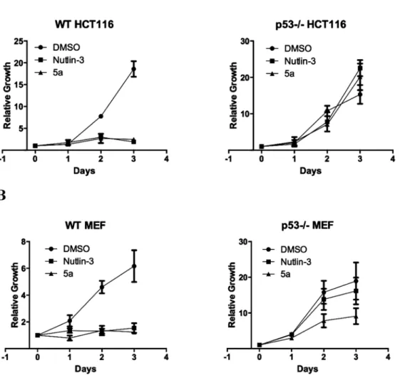
In order to elucidate the p53-dependent mode of action, the most potent and selective compound (5a) was further tested against (p53−/−) HCT116, (p53−/−) mouse embryonic
fibroblasts (MEF), wild type (p53+/+) HCT116 and wild type
(p53+/+) MEF. These isogenic cell lines are commonly used
to show the p53/MDM2-dependent mode of action.20) Indeed, compound 5a and Nutlin-3 potently inhibited the growth of HCT116 and MEF wild type p53 cell lines but showed greatly reduced activity in the same cell lines with p53 deletion as shown in Fig. 2, supporting the hypothesis that the cell growth inhibitory activity of both 5a and Nutlin-3 is p53-dependent.
Additionally, a p53 translocation assay was performed using immunocytochemical staining and high content screening. Se-lection of the tested compounds 5a, b, g were based on activ-ity against wild type (p53+/+) HCT116 cell line using Nutlin-3
as a positive control. From the results shown in Table 3, it’s clear that compounds 5a and g induced a dose-dependent in-crease in the nuclear p53 at lower concentrations which inhibit
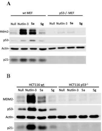
growth. For compound 5a, this effect apparently reached a plateau between 5.56 and 16.67 µM, which might be attributed to the poor solubility and high lipophilicity of the compound; however, compound 5g showed its strongest effect at 50 µM. The highest increase in nuclear p53 was observed after the treatment with 5a at 16.67 µM, which is consistent with re-sults obtained from cellular growth assays suggesting that the dominant mode of action for compound 5a might be through inhibiting p53 stability. Western blotting data supported these findings and further showed that p53 accumulation induced by
5a was accompanied by up-regulation of MDM2 and p21 pro-teins in wild type MEF and HCT116 cells (data represented in Fig. 3), all these data confirm that the mode of action of 5a is p53-dependent through inhibition of the protein–protein inter-action between p53 and its oncoproteins MDM2/x.
In an attempt to further explore the mode of action of 5a; an assessment of the binding affinity characteristics of the compound towards MDM2 was examined via competitive fluorescence polarization assay described by Vogel et al.21)
Kd values were calculated from IC50 values using equations described by Nikolovska-Coleska et al.22) Nutlin-3 was used as control, and as expected, Nutlin-3 disrupted these complexes with Kd values for MDM2 and MDM4 that were similar to growth inhibitory IC50 values for HCT116 and SW480 cells, respectively.
In contrast, 5a displayed approximately 14-fold difference between Kd values and growth inhibitory IC50 for the same pairs (MDM2 vs. HCT116 and MDM4 vs. SW480). This is consistent with the expected mechanism for this and a similar
i) NaBH4, CoCl2, CH3COOH, s: iPrOH, s: DMF, overnight, rt. ii) 3,4-Difuorobenzyl chlorides or 3,4-difluorobenzoyl chloride, K2CO3, C: KI, s: DMF, 45 min, reflux.
Chart 2
Table 1. The Growth Inhibitory Activity of the Synthesized Compounds against Wild Type HCT116 Cell Line
Compound IC50 HCT116 (µM) Compound IC50 HCT116 (µM)
5a 2.8 5g 5.1
5b 3.7 5h 9.3
5c 5.5 5i 13.2
5d 5.0 5j 19.8
5e 7.8 7 24.0
5f 7.4 8 12.2
5g 5.1 Nutlin-3 2.0
Table 2. The Activity of the Most Potent Compounds against Different Cell Lines
Compound IC50 (µM)
HCT116 SW480 H1299
5a 2.8 6.7 9.2
5b 3.7 n.d 11.3
5d 5.0 13.0 n.d
5g 5.1 5.7 16.6
Vol. 64, No. 1 (2016) 37
scaffold,19) which was shown to affect p53 stability through MDM2 and MDMx homo- and heterodimerization rather than direct binding to MDM2 (results shown in Table 4). However, we also cannot completely rule out that affinity to other tar-gets could contribute to the observed cellular profile e.g. while
5g and 5a have very similar IC50 in anti-proliferation assays,
5g barely affects the expression of MDM2 and p21.
In summary, compound 5a showed considerable growth inhibitory activity, while biochemical and biological assays showed that 5a disrupts the MDM2/x–p53 interaction and ac-tivates the p53 pathway in cells with wild-type p53 leading to cell cycle arrest in both normal and tumor cells.
In conclusion, indolylhydantoin seems to be a promising scaffold for MDM2/MDMx inhibition.
Experimental
Chemistry General Solvents and reagents were ob-tained from commercial suppliers and used as received. A Bruker DRX 300 spectrometer was used to obtain 1H- and 13C-NMR spectra. The chemical shifts are referenced to the residual protonated solvent signals or tetramethylsilane (TMS) was used as a reference. Electrospray LC-MS analysis was performed using a TSQ quantum (Thermo Electron Corpo-ration) instrument prepared with a triple quadrupole mass detector (ThermoFinnigan) and an electrospray ionization (ESI) source. All samples were inserted using an autosampler (Surveyor, ThermoFinnigan) by an injection volume of 10 µL. The MS detection was determined using a source collision induced dissociation (CID) of 10 V and carried out at a spray
Fig. 2. Compound 5a Exhibits p53 Dependent Inhibition of Cell Proliferation in Both HCT116 (A) and MEF Cells (B)
Cell proliferation in the presence of DMSO, 10 µM Nutlin-3, or 10 µM compound 5a were completed using the BioRad TC10 Automated Cell Counter. Curves are the
combination of multiple experiments MEF (n=4) and HCT116 (n=2). Relative growth was calculated as current cell number divided by Day 0 cell number. Error bars represent S.E.M.
Table 3. p53 Translocation Assay
Compound conc. Fold change in nuclear p53 staining vs. DMSO control
50.00 µM 16.67 µM 5.56 µM 1.85 µM
5a 4.24±1.63 5.56±0.87 3.85±0.40 1.64±0.24
5b 1.10±0.25 1.48±0.28 1.17±0.23 0.92±0.15
5g 4.87±2.39 2.06±0.41 1.21±0.23 1.06±0.43
38 Chem. Pharm. Bull. Vol. 64, No. 1 (2016)
voltage of 4.2 kV, a nitrogen sheath gas pressure of 4.0×10⁵ Pa, a capillary temperature of 400°C, a capillary voltage of 35 V and an auxiliary gas pressure of 1.0×10⁵ Pa. The stationary phase used was an RP C18 NUCLEODUR 100-3 (125×3 mm) column (Macherey–Nagel). The solvent system consisted of water containing 0.1% trifluoroacetic acid (TFA) (A) and 0.1% TFA in acetonitrile (B). HPLC method: flow rate 400 µL/min. The percentage of B started at an initial of 5%, was increased up to 100% during 16 min, kept at 100% for 2 min, and flushed back to 5% in 2 min.
General Procedure of Vilsmeier–Haack Reaction A so-lution of 6-chloroindole (30 mmol) in N,N-dimethylformamide (DMF) (5 mL) was added dropwise to a solution of phospho-rus oxychloride (44 mmol) in DMF (32 mL) which had been stirred for 1.5 h at 0°C. The reaction mixture was warmed to room temperature and stirred for 2 h. The solution with the precipitate formed was poured on ice and basified with solid KOH and left overnight. The precipitate was filtered and washed with a mixture of 10% ethylacetate in petroleum ben-zene several times, then left to dry to give the aldehyde.
6-Chloro-1H-indole-3-carbaldehyde (3)
Synthesized according to the general procedure of Vils-meier–Haack reaction; light brick red solid; yield 4.04 g (75%).
General Procedure of Knoevenagel Condensation Imidazolidine-2,4-dione or its appropriate sulfur-isostere (10.0 mmol) was dissolved in 10 mL of water or ethanol at 70°C with stirring. After complete dissolution, the pH of the mixture was adjusted to 7.0 with saturated NaHCO3 solution. Ethanolamine (0.9 mL) was added to the reaction mixture, and the temperature was increased to 90°C by use of an oil bath. To this, an equimolar quantity of 6-chloro-1H -indole-3-carbaldehyde (3) (10.0 mmol) solution in 10 mL of alcohol was added dropwise with continuous stirring. The temperature was raised to 120°C and kept under reflux at that temperature overnight. The mixture was cooled, the precipitate was filtered and washed with water in order to remove the soluble impuri-ties, then left to dry.
5-((6-Chloro-1H -indol-3-yl)methylene)imidazolidine-2,4-dione (4a)
Synthesized according to the general procedure of Kno-evenagel condensation using imidazolidine-2,4-dione and 6-chloro-1H-indole-3-carbaldehyde (3); yellowish white solid; yield: 1.60 g (61%); 1H-NMR (300 MHz, dimethyl sulfoxide (DMSO)) δ: 12.29 (s, 1H), 11.97 (s, 1H), 10.49 (s, 1H), 8.19 (s, 1H), 7.82 (d, J=8.6 Hz, 1H), 7.48 (d, J=1.9 Hz, 1H), 7.13 (dd, J=8.5, 1.9 Hz, 1H), 6.88 (s, 1H); 13C-NMR (75 MHz, DMSO)
δ: 164.49, 153.42, 136.18, 128.09, 127.06, 125.67, 122.56, 120.52, 119.73, 111.52, 108.46, 103.10; MS ESI: m/z=385.71 (M)+; purity: 97%.
5-((6-Chloro-1H -indol-3-yl)methylene)-2-thioxoimidazolidin-4-one (4b)
Synthesized according to the general procedure of Kno-evenagel condensation using 2-thioxoimidazolidin-4-oneand 6-chloro-1H-indole-3-carbaldehyde (3); redsolid; yield: 1.35 g (49%); 1H-NMR (300 MHz, DMSO) δ: 12.79 (s, 1H), 11.94 (s, 1H), 11.59 (s, 1H), 8.40 (s, 1H), 8.20 (d, J=8.5 Hz, 1H), 7.52 (d, J=1.9 Hz, 1H), 7.13 (dd, J=8.5, 1.9 Hz, 1H), 7.10 (s, 1H); 13C-NMR (75 MHz, DMSO) δ: 169.90, 158.62, 136.79, 135.19, 132.96, 127.05, 125.36, 121.19, 120.73, 115.72, 111.78, 111.24.
5-((6-Chloro-1H-indol-3-yl)methylene)thiazolidine-2,4-dione (4c)
Synthesized according to the general procedure of Knoeve-nagel condensation using thiazolidine-2,4-dione and 6-chlo-ro-1H-indole-3-carbaldehyde (3); yellow solid; yield: 1.25 g (45%); 1H-NMR (300 MHz, DMSO) δ: 12.44 (s, 1H), 12.23 (s, 1H), 8.18 (s, 1H), 7.93 (d, J=8.6 Hz, 1H), 7.83 (s, 1H), 7.28 (dd, J=8.0, 1.1 Hz, 1H), 7.19 (dd, J=8.5, 1.4 Hz, 1H), 13C-NMR (75 MHz, DMSO) δ: 167.56, 165.77, 137.28, 131.63, 128.26, 126.35, 126.02, 121.91, 120.60, 114.97, 112.56, 111.01.
General Procedure of Benzylation To a suspension of 5-((6-chloro-1H-indol-3-yl) methylene) imidazolidine-2,4-dione or its appropriate sulfur-isostere (4a–c) (10 mmol), K2CO3 (40 mmol) and KI (0.6 mmol) in dry DMF (20 mL), the ap-propriate benzyl chloride was added (12 mmol). The resulting mixture was refluxed for 45 min. The reaction was stopped by adding water, and then the mixture was extracted with EtOAc (3×20 mL). The combined organic phases were washed with brine, dried over anhydrous Mg2SO4, filtered and concen-trated. The crude residue was then purified by column chro-matography on silica gel.
5-((6-Chloro-1H -indol-3-yl)methylene)-3-(4-chlorobenzyl)-imidazolidine-2,4-dione (5a)
The title compound was prepared by reaction of 5-((6-chloro-1H-indol-3-yl) methylene) imidazolidine-2,4-dione
Fig. 3. Western Blotting Analysis for MDM2, p53 and p21 Proteins in Wild Type and (p53−/−) MEF (A) and HCT116 Cells (B) When Treated
with 10 µM Nutlin-3, 10 µM5a or 10 µM5g and Incubated for 24 h
Table 4. Fluorescence Polarization Assay
MDM2 MDMx
IC50 (µM) S.E. (µM) Kd (µM) IC50 (µM) S.E. (µM) Kd (µM)
5a 160 5.5 41.2 330 14.0 90.3
Vol. 64, No. 1 (2016) 39
(4a) and 1-chloro-4-(chloromethyl) benzene according to the general procedure for benzylation; yellowish white solid; yield: 1.55 g (40%); 1H-NMR (300 MHz, DMSO) δ: 11.97 (s, 1H), 10.49 (s, 1H), 8.19 (s, 1H), 7.82 (d, J=8.6 Hz, 1H), 7.48 (d, J=1.9 Hz, 1H), 7.44–7.38 (m, 2H), 7.37–7.29 (m, 2H), 7.13 (dd, J=8.5, 1.9 Hz, 1H), 6.88 (s, 1H), 4.68 (s, 2H); 13C-NMR (75 MHz, DMSO) δ: 163.59, 154.42, 136.18, 135.75, 132.08, 129.33, 128.54, 128.07, 127.06, 125.67, 122.56, 120.52, 119.73, 111.52, 108.46, 103.00, 40.58; MS (ESI): m/z=387.39 (M+H)+;
purity: 98.2%.
5-((6-Chloro-1H -indol-3-yl)methylene)-3-(3-chlorobenzyl)-imidazolidine-2,4-dione (5b)
The title compound was prepared by reaction of 5-((6-chloro-1H-indol-3-yl) methylene) imidazolidine-2,4-dione (4a) and 1-chloro-3-(chloromethyl) benzene according to the general procedure for benzylation; yellowish white solid; yield: 1.69 g (44%); 1H-NMR (300 MHz, DMSO) δ: 11.97 (s, 1H), 10.50 (s, 1H), 8.19 (s, 1H), 7.83 (d, J=8.5 Hz, 1H), 7.48 (d, J=1.8 Hz, 1H), 7.43–7.32 (m, 3H), 7.31–7.23 (m, 1H), 7.13 (dd, J=8.5, 1.8 Hz, 1H), 6.89 (s, 1H), 4.69 (s, 2H); 13C-NMR (75 MHz, DMSO) δ: 163.62, 154.40, 139.21, 136.18, 133.13, 130.53, 128.10, 127.47, 127.28, 127.07, 126.06, 125.68, 122.53, 120.53, 119.74, 111.53, 108.47, 103.11, 40.67; MS (ESI): m/z=387.68 (M+H)+; purity: 96%.
5-((6-Chloro-1H -indol-3-yl)methylene)-3-(3,4-dichlorobenzyl)-imidazolidine-2,4-dione (5c)
The title compound was prepared by reaction of 5-((6-chloro-1H-indol-3-yl) methylene) imidazolidine-2,4-dione (4a) and 1,2-dichloro-4-(chloromethyl) benzene according to the general procedure for benzylation; yellowish white solid; yield: 2.00 g (49%); 1H-NMR (300 MHz, DMSO) δ: 11.97 (s, 1H), 10.50 (s, 1H), 8.19 (s, 1H), 7.82 (d, J=8.6 Hz, 1H), 7.61 (d, J=8.3 Hz, 1H), 7.58 (d, J=2.0 Hz, 1H), 7.48 (d, J=1.9 Hz, 1H), 7.29 (dd, J=8.3, 2.0 Hz, 1H), 7.13 (dd, J=8.5, 1.9 Hz, 1H), 6.88 (s, 1H), 4.69 (s, 2H); 13C-NMR (75 MHz, DMSO) δ: 163.60, 154.34, 137.85, 136.18, 131.09, 130.80, 130.13, 129.54, 128.11, 127.76, 127.07, 125.67, 122.53, 120.53, 119.73, 111.53, 108.46, 103.15, 40.69; MS (ESI): m/z=419.50 (M−H)+; purity: 96.9%.
5-((6-Chloro-1H -indol-3-yl)methylene)-3-(3,4-difluorobenzyl)-imidazolidine-2,4-dione (5d)
The title compound was prepared by reaction of 5-((6-chloro-1H-indol-3-yl) methylene) imidazolidine-2,4-dione (4a) and 4-(chloromethyl)-1,2-difluorobenzene according to the general procedure for benzylation; faint yellow solid; yield: 1.74 g (45%); 1H-NMR (300 MHz, DMSO) δ: 11.97 (s, 1H), 10.49 (s, 1H), 8.19 (s, 1H), 7.82 (d, J=8.5 Hz, 1H), 7.48 (d, J=1.8 Hz, 1H), 7.46–7.33 (m, 2H), 7.13 (dd, J=8.5, 1.9 Hz, 2H), 6.88 (s, 1H), 4.68 (s, 2H); 13C-NMR (75 MHz, DMSO) δ: 163.60, 162.32, 154.38, 150.87, 136.18, 134.45, 128.08, 127.07, 125.67, 124.25, 122.58, 120.52, 119.73, 117.64, 116.67, 111.53, 108.47, 103.05, 40.58; MS (ESI): m/z=387.77 (M)+; purity:
98.5%.
5-((6-Chloro-1H -indol-3-yl)methylene)-3-(4-(trifluoromethyl)-benzyl)imidazolidine-2,4-dione (5e)
The title compound was prepared by reaction of 5-((6-chloro-1H-indol-3-yl) methylene) imidazolidine-2,4-dione (4a) and 1-(chloromethyl)-4-(2,2,2-trifluoroethyl)-benzeneaccording to the generalprocedure for benzylation; yellow solid; yield: 2.31 g (55%); 1H-NMR (300 MHz, DMSO-d6) δ: 11.98 (s, 1H), 10.53 (s, 1H), 8.20 (s, 1H), 7.83 (d, J=8.5 Hz, 1H), 7.72 (d, J=8.2 Hz, 2H), 7.52 (d, J=8.1 Hz,
2H), 7.48 (d, J=1.7 Hz, 1H), 7.13 (dd, J=8.5, 1.8 Hz, 1H), 6.89 (s, 1H), 4.78 (s, 2H); 13C-NMR (75 MHz, DMSO) δ: 163.63, 154.40, 141.44, 136.19, 128.08, 128.07, 127.08, 125.99, 125.67, 125.50, 122.39, 122.53, 120.53, 119.73, 111.53, 108.46, 103.14, 40.83; MS (ESI): m/z=419.68 (M)+; purity: 96%.
5-((6-Chloro-1H -indol-3-yl)methylene)-3-(4-chlorobenzyl)-2-thioxoimidazolidin-4-one (5f)
The title compound was prepared by reaction of 5-((6-chloro-1H-indol-3-yl) methylene)-2-thioxoimidazolidin-4-one (4b) and 1-chloro-4-(chloromethyl) benzene according to the general procedure for benzylation; orange solid; yield: 1.52 g (38%); 1H-NMR (300 MHz, DMSO) δ: 11.94 (s, 1H), 11.59 (s, 1H), 8.40 (s, 1H), 8.20 (d, J=8.5 Hz, 1H), 7.59–7.53 (m, 2H), 7.52 (d, J=1.9 Hz, 1H), 7.46–7.36 (m, 2H), 7.13 (dd, J=8.5,1.9 Hz, 1H), 7.10 (s, 1H), 4.59 (s, 2H); 13C-NMR (75 MHz, DMSO) δ: 169.91, 158.72, 136.77, 136.62, 135.19, 132.96, 132.00, 130.73, 128.48, 127.05, 125.36, 121.19, 120.73, 115.72, 111.78, 111.24, 32.47; MS (ESI): m/z=403.68 (M+H)+;
purity: 95%.
5-((6-Chloro-1H -indol-3-yl)methylene)-3-(3,4-dichlorobenzyl)-2-thioxoimidazolidin-4-one (5g)
The title compound was prepared by reaction of 5-((6-chloro-1H-indol-3-yl) methylene)-2-thioxoimidazolidin-4-one (4b) and 1,2-dichloro-4-(chloromethyl) benzene according to the general procedure for benzylation; dark yellow solid; yield: 1.70 g (39%); 1H-NMR (300 MHz, DMSO) δ: 11.95 (s, 1H), 11.62 (s, 1H), 8.41 (s, 1H), 8.19 (d, J=8.5 Hz, 1H), 7.84 (d, J=1.9 Hz, 1H), 7.63–7.53 (m, 2H), 7.52 (d, J=1.9 Hz, 1H), 7.15 (dd, J=8.5, 1.9 Hz, 1H), 7.08 (s, 1H), 4.59 (s, 2H); 13C-NMR (75 MHz, DMSO) δ: 169.88, 158.50, 139.01, 136.83, 135.10, 132.92, 130.94, 130.88, 130.64, 129.95, 129.20, 127.08, 125.34, 121.20, 120.77, 115.90, 111.79, 111.21, 31.91; MS (ESI): m/z=437.90 (M+H)+; purity: 98%.
5-((6-Chloro-1H -indol-3-yl)methylene)-3-(3,4-difluorobenzyl)-2-thioxoimidazolidin-4-one (5h)
The title compound was prepared by reaction of 5-((6-chloro-1H-indol-3-yl) methylene)-2-thioxoimidazolidin-4-one (4b) and 4-(chloromethyl)-1,2-difluorobenzene accord-ing to the general procedure for benzylation; orange solid; yield: 1.60g (40%); 1H-NMR (300 MHz, DMSO) δ: 11.95 (s, 1H), 11.61 (s, 1H), 8.41 (s, 1H), 8.21 (d, J=8.6 Hz, 1H), 7.67–7.57 (m, 1H), 7.52 (d, J=1.9 Hz, 1H), 7.44–7.33 (m, 2H), 7.16 (dd, J=8.5, 1.9 Hz, 1H), 7.09 (s, 1H), 4.59 (s, 2H); 13C-NMR (75 MHz, DMSO) δ: 169.91, 158.60, 151.40, 147.38, 136.82, 135.42, 135.13, 132.94, 127.08, 125.80, 125.35, 121.22, 120.75, 117.73, 117.73, 115.82, 111.79, 111.24, 32.11; MS (ESI): m/z=403.60 (M)+; purity: 96%.
5-((6-Chloro-1H -indol-3-yl)methylene)-2-thioxo-3-(4-(trifluoromethyl)benzyl)imidazolidin-4-one (5i)
40 Chem. Pharm. Bull. Vol. 64, No. 1 (2016)
5-((6-Chloro-1H -indol-3-yl)methylene)-3-(3,4-dichlorobenzyl)-thiazolidine-2,4-dione (5j)
The title compound was prepared by reaction of 5-((6-chloro-1H-indol-3-yl) methylene) thiazolidine-2,4-dione (4c) and 1,2-dichloro-4-(chloromethyl) benzene according to the general procedure for benzylation; yellow solid; yield: 1.22 g (28%); 1H-NMR (300 MHz, DMSO-d
6) δ: 12.23 (s, 1H), 8.18 (s, 1H), 7.93 (d, J=8.6 Hz, 1H), 7.83 (s, 1H), 7.60 (d, J=8.3 Hz, 2H), 7.53 (d, J=8.0, 1H), 7.28 (dd, J=8.0, 1.1 Hz, 1H), 7.19 (dd, J=8.5, 1.4 Hz, 1H), 4.82 (s, 2H); 13C-NMR (75 MHz, DMSO) δ: 167.56, 165.77, 137.28, 137.12, 131.63, 131.34, 130.93, 130.52, 130.32, 128.45, 128.26, 126.35, 126.02, 121.91, 120.60, 114.97, 112.56, 111.01, 43.86; MS (ESI): m/z=437.62 (M)+; purity: 97%.
5-((6-Chloro-1H-indol-3-yl)methyl)imidazolidine-2,4-dione (6)
Synthesized by a reaction between a solution of 5-((6-chloro-1H-indol-3-yl) methylene) imidazolidine-2,4-dione (4a) (2 mmol) in a mixed solvent of DMF–isopropanol (1 : 2, 60 mL), CoCl2 (12 mmol), acetic acid (90 mmol) and NaBH4 (120 mmol) portion wise. The mixture was stirred at room temperature overnight and then diluted with ethyl acetate (100 mL). The mixture was washed sequentially with saturated NaHCO3 (50 mL), 1 N HCl (50 mL), saturated NaCl (50 mL) and then dried over anhydrous MgSO4, filtered and concentrated. The crude residue was then purified by column chromatogra-phy on silica gel; white powder; yield: 0.13 g (25%); 1H-NMR (300 MHz, DMSO) δ: 11.11 (s, 1H), 11.01 (s, 1H), 8.37 (s, 1H), 7.51 (d, J=8.5 Hz, 1H), 7.36 (d, J=1.8 Hz, 1H), 6.92 (dd, J=8.5, 1.9 Hz, 1H), 6.35–6.27 (m, 1H), 4.46 (t, J=4.1 Hz, 1H), 3.18 (dd, J=14.9, 4.3 Hz, 1H), 3.06 (dd, J=14.9, 4.3 Hz, 1H); 13C-NMR (75 MHz, DMSO) δ: 174.02, 156.51, 136.16, 127.27, 125.59, 122.93, 120.09, 118.68, 110.85, 107.58, 57.18, 25.96.
5-((6-Chloro-1H -indol-3-yl)methyl)-3-(3,4-difluorobenzyl)-imidazolidine-2,4-dione (7)
The title compound was prepared by reaction of 5-((6-chloro-1H-indol-3-yl) methyl)-2-thioxoimidazolidin-4-one (6) and 4-(chloromethyl)-1,2-difluorobenzene according to the general procedure for benzylation; white solid; yield: 1.55 g (40%); 1H-NMR (300 MHz, DMSO) δ: 11.01 (s, 1H), 8.37 (s, 1H), 7.51 (d, J=8.5 Hz, 1H), 7.36 (d, J=1.8 Hz, 1H), 7.11 (d, J=2.3 Hz, 1H), 7.04 (dt, J=10.8, 8.5 Hz, 1H), 6.92 (dd, J=8.5, 1.9 Hz, 1H), 6.76 (ddd, J=11.3, 7.8, 2.0 Hz, 1H), 6.35–6.27 (m, 1H), 4.46 (t, J=4.1 Hz, 1H), 4.34 (d, J=15.7 Hz, 1H), 4.22 (d, J=15.7 Hz, 1H), 3.18 (dd, J=14.9, 4.3 Hz, 1H), 3.06 (dd, J=14.9, 4.3 Hz, 1H); 13C-NMR (75 MHz, DMSO) δ: 173.62, 156.27, 151.07, 147.10, 136.16, 133.95, 127.27, 125.90, 125.59, 122.93, 120.09, 118.68, 116.90, 115.68, 110.85, 107.58, 57.18, 41.12, 25.96; MS (ESI): m/z=389.71 (M)+; purity: 97.6%.
5-((6-Chloro-1H -indol-3-yl)methylene)-3-(3,4-difluorobenzoyl)-imidazolidine-2,4-dione (8)
The title compound was prepared by reaction of 5-((6-chloro-1H-indol-3-yl) methylene) imidazolidine-2,4-dione (4a) and 3,4-difluorobenzoyl chloride according to the gen-eral procedure for benzylation; dark yellow solid; yield: 3.00 g (75%); 1H-NMR (300 MHz, DMSO) δ: 11.33 (s, 1H), 10.58 (s, 1H), 8.36 (d, J=1.8 Hz, 1H), 8.14 (s, 1H), 8.10–7.99 (m, 2H), 7.78 (dd, J=8.8, 4.5 Hz, 2H), 7.52 (dd, J=8.5, 1.9 Hz, 1H), 6.64 (s, 1H); 13C-NMR (75 MHz, DMSO) δ: 166.70, 165.43, 155.96, 152.54, 149.74, 142.56, 136.04, 131.14, 130.69, 129.01, 128.83, 127.90, 125.15, 121.06, 119.78, 118.65, 116.11, 114.52, 97.39;
MS (ESI): m/z=401.71 (M)+; purity: 98%.
Cell Culture Cancer cell lines cultured included wild-type p53 cell line (HCT-116), and p53 null cell line (H1299) which were obtained from the American Type Culture Col-lection (ATC C). Both cell lines were cultured in a 37°C humidified incubator with 5% CO2 with the same medium (RPMI-1640 supplemented with 5% fetal bovine serum), and passaged twice weekly. Only cultures exhibiting greater than 95% viability were used in growth inhibition experiments (de-termined by trypan blue exclusion).
Growth Inhibition Assay Cells were seeded in 96-well tissue culture-treated assay plates at a density of 1.5×10⁴ cells/ cm2, then allowed to attach overnight before addition of ex-perimental compounds. Compounds were dissolved in DMSO, then diluted to the final concentration indicated so as to ensure DMSO concentration less than 0.2%. After treatment with either a single screening concentration or a titration se-ries of concentrations of compounds, cells were incubated for an additional 72 h. Relative cell growth was determined by ad-dition of PromegaCellTiterGlo luciferase-based assay of ATP content. The resultant luminescence was measured, and each data set was analyzed using DMSO (vehicle control) as a base-line value for growth inhibition. GraphPad Prism software was used to develop dose–response curves and IC50 values for active compounds.
Immunofluorescent Labeling p53 Cells were seeded in 96-well tissue culture-treated assay plates at a density of 1.5×10⁴ cells/cm2, then allowed to attach overnight before ad-dition of experimental compounds.
Compound Treatment: Compounds were dissolved in DMSO, then diluted to the final concentration indicated so as to ensure DMSO concentration less than 0.2%. Experimental compounds were added to cells in equal volume, maintain-ing equal concentration of DMSO vehicle among all tested compound concentrations. After 20 h treatment period, cells were fixed with 4% formaldehyde (Sigma-Aldrich) for 20 min at room temperature.
Fixative was removed, and samples washed thrice in phos-phate buffered saline (PBS). Nonspecific binding sites were blocked and cells permeabilized by 60 min incubation with 5% fetal bovine serum and 0.3% Triton X-100 in 0.9% PBS. Samples were incubated overnight at 4°C with anti-p53 rab-bit monoclonal antibody (Cell Signaling Technology) diluted 1 : 1600 in antibody dilution buffer (1% bovine serum albumin and 0.3% Triton X-100 in PBS). Samples were washed thrice in PBS. Samples were then incubated 2 h at room temperature with Alexa Fluor 488 anti rabbit polyclonal antibody conjugate (Life Technology) diluted 1 : 500 in antibody dilution buffer, then washed thrice in PBS. Nuclei were stained with DAPI (Sigma-Aldrich; 1 µg/mL) for 30 min at room temperature.
Vol. 64, No. 1 (2016) 41 Western Blotting After lysing cells in 0.5% NP-40
buffer, the lysates were resolved on a 12.5% polyacrylamide gel and then transferred to a 0.2-µm nitrocellulose membrane. Membranes were blocked for at least 30 min in PBS block-ing buffer with 0.1% Tween-20 (PBST) and 5% non-fat dried milk. Membranes were incubated for 2 h to overnight with the appropriate primary antibody, incubated for 1–2 h in sec-ondary horseradish peroxidase (HRP)-conjugated antibody, and exposed with Supersignal West Pico or Dura reagent (Pierce). Mouse monoclonal MDM2 (2A10, Calbiochem), p53 (NCL-505, Leica), actin (MAB1501, Chemicon International), and goat polyclonal p21 (C-19; Santa Cruz) antibodies were purchased commercially.
Fluorescence Polarization Assay The assay was per-formed as previously described by Vogel et al.21) Briefly, the association of a FAM labeled peptide (derived from the amino terminus of p53 from recombinant MDM2 (AA16–116) or MDM4 (AA2–125) was measured by anisotropy. Dissociation constants were determined by direct competition of the tested compounds.
Acknowledgments The authors are grateful to the Alex-ander von Humboldt Foundation, Bonn, Germany for partial sponsoring of this work through a “Research groups linkage Program” for Prof. Dr. Ashraf Abadi, German University in Cairo, Egypt and Prof. Dr. Frank Boeckler, University of Tue-bingen, Germany.
Conflict of Interest The authors declare no conflict of interest.
References
1) Wade M., Li Y. C., Wahl G. M., Nat. Rev. Cancer, 13, 83–96 (2013). 2) Gudkov A. V., Komarova E. A., Hum. Mol. Genet., 16 (R1), R67–
R72 (2007).
3) Levine A. J., Cell, 88, 323–331 (1997).
4) Vousden K. H., Lu X., Nat. Rev. Cancer, 2, 594–604 (2002). 5) Vousden K. H., Lane D. P., Nat. Rev. Mol. Cell Biol., 8, 275–283
(2007).
6) Wu X., Bayle J. H., Olson D., Levine A. J., Genes Dev., 7 (7A), 1126–1132 (1993).
7) Tang Y., Zhao W., Chen Y., Zhao Y., Gu W., Cell, 133, 612–626 (2008).
8) Brooks C. L., Gu W., Mol. Cell, 21, 307–315 (2006). 9) Wade M., Wahl G. M., Mol. Cancer Res., 7, 1–11 (2009).
10) Vassilev L. T., Vu B. T., Graves B., Carvajal D., Podlaski F., Filipo-vic Z., Kong N., Kammlott U., Lukacs C., Klein C., Fotouhi N., Liu E. A., Science, 303, 844–848 (2004).
11) Vassilev L. T., J. Med. Chem., 48, 4491–4499 (2005).
12) Ding K., Lu Y., Nikolovska-Coleska Z., Qiu S., Ding Y., Gao W., Stuckey J., Krajewski K., Roller P. P., Tomita Y., Parrish D. A., Deschamps J. R., Wang S., J. Am. Chem. Soc., 127, 10130–10131 (2005).
13) Koblish H. K., Zhao S., Franks C. F., Donatelli R. R., Tominovich R. M., LaFrance L. V., Leonard K. A., Gushue J. M., Parks D. J., Calvo R. R., Milkiewicz K. L., Marugán J. J., Raboisson P., Cum-mings M. D., Grasberger B. L., Johnson D. L., Lu T., Molloy C. J., Maroney A. C., Mol. Cancer Ther., 5, 160–169 (2006).
14) Xue W., Zender L., Miething C., Dickins R. A., Hernando E., Krizhanovsky V., Cordon-Cardo C., Lowe S. W., Nature (London), 445, 656–660 (2007).
15) Kussie P. H., Gorina S., Marechal V., Elenbaas B., Moreau J., Levine A. J., Pavletich N. P., Science, 274, 948–953 (1996). 16) Zhao J., Wang M., Chen J., Luo A., Wang X., Wu M., Yin D., Liu
Z., Cancer Lett., 183, 69–77 (2002).
17) Chène P., Fuchs J., Bohn J., García-Echeverría C., Furet P., Fabbro D., J. Mol. Biol., 299, 245–253 (2000).
18) Ding K., Lu Y., Nikolovska-Coleska Z., Wang G., Qiu S., Shangary S., Gao W., Qin D., Stuckey J., Krajewski K., Roller P. P., Wang S., J. Med. Chem., 49, 3432–3435 (2006).
19) Graves B., Thompson T., Xia M., Janson C., Lukacs C., Deo D., Di Lello P., Fry D., Garvie C., Huang K. S., Gao L., Tovar C., Lovey A., Wanner J., Vassilev L. T., Proc. Natl. Acad. Sci. U.S.A., 109, 11788–11793 (2012).
20) Shangary S., Qin D., McEachern D., Liu M., Miller R. S., Qiu S., Nikolovska-Coleska Z., Ding K., Wang G., Chen J., Bernard D., Zhang J., Lu Y., Gu Q., Shah R. B., Pienta K. J., Ling X., Kang S., Guo M., Sun Y., Yang D., Wang S., Proc. Natl. Acad. Sci. U.S.A., 105, 3933–3938 (2008).
21) Vogel S. M., Bauer M. R., Joerger A. C., Wilcken R., Brandt T., Ve-printsev D. B., Rutherford T. J., Fersht A. R., Boeckler F. M., Proc. Natl. Acad. Sci. U.S.A., 109, 16906–16910 (2012).