http://www.scirp.org/journal/ns Natural Science, 2018, Vol. 10, (No. 11), pp: 436-447
https://doi.org/10.4236/ns.2018.1011043 436 Natural Science
Analysis of Interferential Current Therapy-Induced Skin
Changes in Healthy Korean Men
So-Jung Kim1*, Seung-Min Yang1*, Ju-Hyun Kim2, Jeong-Uk Lee3, Mee-Young Kim1, Lim-Kyu Lee1,4, Byoung-Sun Park1, Won-Deok Lee1, Ji-Woong Noh1, Yong-Sub Shin1, Doo-Ho Kim1, Il-Hyun Kim1, Kong-Sook Noh1, Junghwan Kim5
1Laboratory of Health Science & Nanophysiotherapy, Department of Physical Therapy, Graduate School, Yongin University, Yongin-si, Republic of Korea; 2Department of Physical Therapy, College of Health Welfare, Wonkwang Health Science University, Iksan-si, Republic of Korea; 3Department of Physical Therapy, College of Health Science, Honam University, Gwangju-si, Republic of Korea; 4The Team of Safety and Quality Management, Yeongwol Com-bined Cycle Power Plant Division, Korea Southern Power Plant, Yeongwol-gun, Republic of Korea; 5Department of Physical Therapy, College of Public Health & Welfare, Yongin University, Yongin-si, Republic of Korea
Correspondence to: Junghwan Kim,
Keywords: Interferential Current Therapy, Korean Men, Skin
Received: October 8, 2018 Accepted: November 27, 2018 Published: November 30, 2018 Copyright © 2018 by authors and Scientific Research Publishing Inc.
This work is licensed under the Creative Commons Attribution International License (CC BY 4.0). http://creativecommons.org/licenses/by/4.0/
ABSTRACT
We examined the changes in numerous skin conditions before and after the application of interferential current therapy to various regions of healthy male bodies. In this study, we assessed the differences in the skin’s sebum, moisture levels, pores, wrinkles, pigmentation, and elasticity on the shoulders, lower back, and the knees of Korean males in their 20s. A total of 30 healthy males were included in the study. We used a skin diagnosis meter as a device for measuring the state of the skin. A statistical difference was found when compar-ing the pre- and post-measurement values in regards to the moisture levels, wrinkles, and pigmentation. In the correlation analysis results, the sebum and pigmentation, moisture le-vels and wrinkles, moisture lele-vels and pigmentation, and moisture lele-vels and elasticity were all positively correlated, respectively. The results of this study partially suggest that a change in skin condition is associated with external stimulation. The study also found that the ef-fects of the application of interferential current therapy on the various skin conditions may differ depending on the region of the body that the application is conducted as well.
1. INTRODUCTION
Potential options for the physical therapy management of muscle, soft tissue, control of pain, edema, Open Access
https://doi.org/10.4236/ns.2018.1011043 437 Natural Science and neurological impairment includes transcutaneous electrical nerve stimulation, ultrasounds, hot packs,
as well as interferential current therapy [1-4]. Therapists often use interferential current therapy for the
treatment and alleviation of skeletal muscle pain [5]. The therapy involves the application of medium
fre-quency alternating electric currents via the skin [5, 6]. The medium frequency current penetrates the
tis-sues with very little resistance, whereas the resulting interference current is within a range that creates
ef-fective stimulation of the biological tissues [3]. Beatti reported that the highest voltages were recorded in
the superficial tissues, and the lowest voltages were recorded in muscle [2]. The principal components of
an interferential unit are a pair of signal generators; the output of one is oscillating at the fixed frequency
of 4000 Hz while the other is a variable oscillation between the frequencies of 4000 and 4100 Hz [2, 4].
Two pairs of electrodes, which convey separately the amplified output of the oscillators, are aligned on the skin so that the currents flowing between each pair intersect and interfere within the intended treatment
region [4]. A resultant current of low frequency is generated that alternates at 0 - 250 Hz [2, 7, 8]. The use
of two currents at around 4 kHz was chosen to overcome skin impedance so that the current could
pene-trate into the deeper tissues [9]. For treatment, the area of skin to be treated is cleaned with soap and water
to reduce linear electrical resistance, and the electrodes are fixed to the skin using medical tape. Some ICT apparatus is supplied with electrodes that are held in place by suction cups, which are evacuated using a
vacuum pump [6]. This is accomplished by placing a suction pad, containing an electrode covered with a
synthetic sponge onto the skin surface above the muscle region that is causing pain [10]. Many centers use
non-disposable suction pads and sponges, which can be re-used following a disinfection process. Disposa-ble alternatives are availaDisposa-ble, but they are rarely used due to the higher associated costs. Some interferen-tial therapy machines also rely on the use of a water seal to facilitate the attachment of the suction cup to
the skin [10]. The water is held in a detachable reservoir located on the machine, but this reservoir could
potentially be contaminated with micro-organisms. In common with other reusable medical devices that make direct contact with the skin such as ultrasound probes and TENS sponges, the interferential therapy
machine may transfer micro-organisms from one patient to another [10, 11]. One study reported that
sin-gle use electrode pads and sponges could be used to avoid cross-contamination; alternatively, disinfection
of the suction cups using 70% v/v isopropyl alcohol should take place following each use [10]. We studied
how the skin is influenced by the interferential current therapy that used water with a sponge that turned
out to be water [12]. Treffene tested the ICT spread in a homogenous water medium. Minimum
tion occurred at the intersection of the two circuits, which were at a 90˚ angle, and a maximum
stimula-tion occurred along the diagonals, which were at a 45˚ angle, between the two circuits [13]. There is a
pa-per on the immediate effects on the skin condition after the application of electrical therapy [14].
There-fore, in the present study, we divided the faces in detail and compared them before and after ICT. The purpose of this study was to establish a map of the skin of shoulder, back, and knee using six biophysical parameters to study various aspects of the skin—sebum, moisture, pores, wrinkles, pigmentation, and elasticity.
2. METHODS
2.1. ParticipantsThirty healthy male subjects aged 20 - 29 were enrolled in the present study. None of the subjects had
any skin disorders (Table 1). Two areas, the face and the neck, were examined before and after
interferen-tial current therapy (ICT). No skin care products had been applied for at least 24 h beforehand, and the skin had not been washed with soap or surfactants for at least 2 h before the experiment. The participants were asked to complete a questionnaire during individual in-depth interviews, which took 30 min per
person [15].
2.2. Measurements
https://doi.org/10.4236/ns.2018.1011043 438 Natural Science
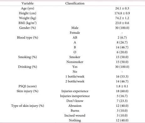
Table 1. The general characteristics of men in their 20s.
Variable Classification
Age (yrs) 24.1 ± 0.3
Height (cm) 174.8 ± 0.9
Weight (kg) 74.2 ± 1.2
BMI (kg/m2) 23.0 ± 0.6
Gender (%) Male 30 (100.0)
Female -
Blood type (%) AB 2 (6.7)
A 8 (26.7)
B 14 (46.7)
O 6 (20.0)
Smoking (%) Smoker 15 (50.0)
Nonsmoker 15 (50.0)
Drinking (%) Yes 30 (100.0)
No -
1 bottle/week 16 (53.3)
2 bottle/week 14 (46.7)
PSQI (score) 1.8 ± 0.1
Skin injury (%) Injuries experience 18 (60.0)
Injuries inexperience 5 (16.7)
Don’t know 7 (23.3)
Type of skin injury (%) Abrasion 12 (40.0)
Burns 3 (10.0)
Incised wound 3 (10.0)
Nothing 12 (40.0)
All data were presented as the mean ± SE. BMI, body mass index; PSQI, Pittsburgh sleep quality index.
was divided into two areas, the shoulder joint, low back, and the knee joint. Repeated measurements were taken for each study subject. Electrical stimulation was conducted at the regions of the shoulder joint, lower back and knee joint using the ICT (ERBE, Elektromedizin Tubingen, Germany), as well as the suc-tion intensity is composed of 1 - 4 intensity, which it used the intensity that the applicasuc-tion feels
comforta-ble with subjects [5]. It was sit used to stimulate line voltage 220 V, Line Frequency 60 Hz, input current
0.35 A and electrical stimulus was conducted for 15 min by setting min 50 Hz - mix 150 Hz frequency [5].
A skin diagnosis meter (SDM, 2016 BOMTECH ver. 3.20) was used to measure the skin’s surface sebum content, moisture, pores, pigmentation, wrinkles, and elasticity of the shoulder, back, and knee. ICT was applied to each region for 15 min, with the distance between the measuring device and the subject’s skin being set at 20 cm. During the measurement of the skin, the normal force level was 0.7 N. The same inves-tigator performed all procedures in a room kept at a constant temperature (22˚C - 24˚C) and humidity (45% - 55%). The skin testing machine is made up of two meters: the E-cam measures sebum,
pigmenta-tion, and pores, and the P-sensor measures moisture and elasticity [16, 17]. The E-cam measures the skin
using an X80 lens and measures the amount of sebum using UV light. The meter measures the dermis
layer of pigmentation, wrinkle length, and pore size using general light [16, 17]. The measurement area is
https://doi.org/10.4236/ns.2018.1011043 439 Natural Science skin reacts to the electricity. It creates a calculation using the values of the skin’s reaction to the electricity.
To measure sebum, the meter uses skin UV to see how much oil is contained in the targeted area [16-18].
2.3. Clinical Assessments
We used the sebum arbitrary units (SAUs); the values are 0 - 650 (a little sebum), 650 - 1200 (nor-mal), 1200 - 5000 (a lot of sebum), and more than 5000 (excessive sebum). To make use of the moisture
arbitrary units (MAUs), set the values to 1% - 100% [16, 17]. These values are 0 - 19 (very dry), 20 - 29
(dry), 30 - 39 (moderate), 40 - 49 (plenty of moisture), and more than 50 (a high amount of moisture) [16,
17]. The P-sensor meter uses a rectangular spring to measure elasticity. When the spring contacts the skin,
it measures how much the sticky part of the spring can be forced into the skin. To make use of the elastici-ty arbitrary units (EAUs), set the values to 1% - 100%. These values are 0 - 24 (very low elasticielastici-ty), 25 - 34
(low), 35 - 44 (moderate), 45 - 54 (high), and more than 55 (very high) [16, 17, 19]. These values were
ex-pressed on a graph. The graph showed the skin surface’s reactivity value according to the distance from the point measured. The data values were measured using Microsoft Office PowerPoint 2007. Based on the x-axis of 0 to 25.5 cm and the y-axis of 0 to 20 cm, each coordinate was set on the basis of each vertex of
the graph. By connecting the set coordinates, we found the area of the graph [16, 17]. Next, each vertex of
the graph was calculated using digital calipers (Industrial Instrument Company, Mitutoyo, Japan), and the
area was calculated using OriginPro 2016 (64 bit). Pigmentation was measured using general light [16, 20,
21]. The pigmentation arbitrary units (PiAUs) are set at values of 1 - 10 grade: 0 - 2 (favorable), 2 - 4 (moderate), 4 - 6 (mild), and 6 - 10 (severe). To make use of the wrinkle arbitrary units (WAUs), set the values to 1 - 10 grade. These are 0 - 2 (favorable), 2 - 3 (early mild wrinkles), 3 - 5 (early severe wrinkles), 5 -
6 (thicker mild wrinkles) [16, 17, 22], and 6 - 10 (thicker severe wrinkles). To make use of the pore
arbi-trary units (PAUs), set the values to 1 - 10 grade. These are 0 - 2 (small size), 2 - 4 (moderate), 4 - 6
(broa-dish), 6 - 8 (large), and 8 - 10 (very large) [16, 17].
3. RESULTS
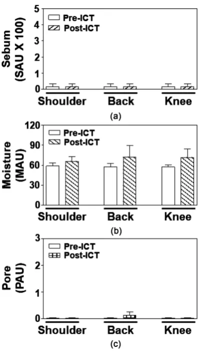
3.1. Changes in the Skin Condition of Sebum and Moisture in Response to the Application of ICT in Relation to Various Body Regions
General characteristics of the thirty healthy subjects are noted in Table 1. No statistical difference in
sebum was found between pre- and post-measured values in the shoulder, back, and shin, which can be
seen in Figure 1(a). There was a statistical difference in moisture levels found between the pre-measured
lower back, which was 66.22 ± 1.07, and the post-measured lower back, which was 68.70 ± 1.45. Also, there was a statistical difference noted in the moisture levels of the knee. The pre-measured moisture level for the knee was 57.38 ± 1.54, and the post-measured level for the knee was 64.18 ± 1.14. In both cases, the
value of p was p < 0.05. The values in the lower back and the knee region are depicted in Figure 1(b).
3.2. Changes in the Skin Condition of Pore and Wrinkle in Response to the Application of ICT in Relation to Various Body Regions
No statistical difference in pore was found between pre- and post-measured values in the shoulder,
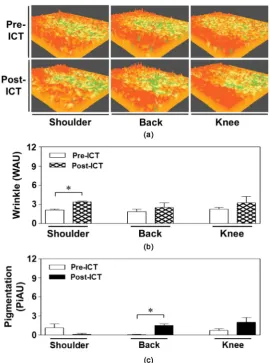
back, and shin, as is demonstrated in Figure 1(c). In contrast, a statistical difference in wrinkle was found
between pre-measured and post-measured values in the shoulder, low back and knee region. The pre-measured values for wrinkles were 3.41 ± 0.27 for the shoulder, 4.19 ± 0.28 for the lower back, and
3.53 ± 0.41 for the knee (Figure 2). The post-measured values were 5.11 ± 0.40 for the shoulder, 5.44 ±
0.27 for the lower back, and 5.12 ± 0.25 for the knee, where p < 0.05 (Figure 2).
3.3. Changes in the Skin Condition of Pigmentation and Elasticity in Response to the Application of ICT in Relation to Various Body Regions
https://doi.org/10.4236/ns.2018.1011043 440 Natural Science
Figure 1. Differences in the sebum,
mois-ture, and pore of the skin’s surface for the shoulder, back, and knee. Each bar represents the mean ± SE. SAU, sebum arbitrary units; MAU, moisture arbitrary units; PAU, pore arbitrary units; ICT, in-terferential current therapy, Pre- and Post-ICT, before and after ICT application.
in the shoulder, lower back and knee region, as can be seen in Figure 2. The pre-measured values of
pig-mentation in the shoulder were 2.48 ± 0.27. In the lower back, the pre-measured value was 2.88 ± 0.33, and the knee was 2.46 ± 0.31. The post-measured value of pigmentation in the shoulder was 5.41 ± 0.69, and in the lower back, it was 4.86 ± 0.46. The post measured value for pigmentation in the knee was 4.78 ± 0.33. The value of p for all three was p < 0.05. A statistical difference in elasticity was found between pre-measured (58.58 ± 2.56) and post-measured (65.11 ± 1.52, where p < 0.05) values in the knee region,
as demonstrated in Figure 3.
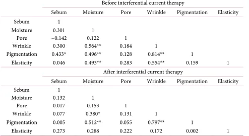
3.4. Correlation of Skin Conditions among Variables before the Application of ICT
The Pearson’s correlation coefficients among components before the application of ICT are shown in
https://doi.org/10.4236/ns.2018.1011043 441 Natural Science
Figure 2. Differences in the wrinkle and pigmentation of
the skin’s surface for the shoulder, back, and knee. Each bar represents the mean ± SE. WAU, wrinkle arbitrary units; PiAU, pigmentation arbitrary units; ICT, interfe-rential current therapy, Pre- and Post-ICT, before and after ICT application. *p < 0.05.
pigmentation (r = 0.433, p < 0.05), moisture levels and wrinkles (r = 0.564, p < 0.01), moisture levels and pigmentation (r = 0.496, p < 0.01), moisture levels and elasticity (r = 0.493, p < 0.01), wrinkles and pig-mentation (r = 0.814, p < 0.01), and wrinkles and elasticity (r = 0.554, p < 0.01) were all positively corre-lated, respectively. The Pearson’s correlation coefficients among each region before the application of ICT
are shown in Table 3.
3.5. Correlation of Skin Conditions among Variables after the Application of ICT
The Pearson’s correlation coefficients among components after the application of ICT are shown in
Table 2. In this study, the variables of sebum, moisture, pore, wrinkle, pigmentation, and elasticity were classified. All variables were input as continuous data. In the correlation analysis results, moisture levels and wrinkles (r = 0.380, p < 0.05), moisture levels and pigmentation (r = 0.512, p < 0.01), and wrinkles and pigmentation (r = 0.797, r < 0.01) were all positively correlated, respectively. The Pearson’s correlation
https://doi.org/10.4236/ns.2018.1011043 442 Natural Science
Figure 3. Differences in the elasticity of the skin’s surface
for the shoulder, back, and knee. Each bar represents the mean ± SE. EAU, elasticity arbitrary units; ICT, interfe-rential current therapy, Pre- and Post-ICT, before and af-ter ICT application.
Table 2. Pearson’s correlation coefficient for skin conditions of before and after ICT.
Before interferential current therapy
Sebum Moisture Pore Wrinkle Pigmentation Elasticity
Sebum 1
Moisture 0.301 1
Pore −0.142 0.122 1
Wrinkle 0.300 0.564** 0.184 1
Pigmentation 0.433* 0.496** 0.128 0.814** 1
Elasticity 0.046 0.493** 0.283 0.554** 0.159 1
After interferential current therapy
Sebum Moisture Pore Wrinkle Pigmentation Elasticity
Sebum 1
Moisture 0.132 1
Pore 0.017 0.153 1
Wrinkle 0.077 0.380* 0.131 1
Pigmentation 0.005 0.512** 0.055 0.797** 1
Elasticity 0.273 0.288 0.222 0.172 0.002 1
[image:7.595.55.550.436.706.2]https://doi.org/10.4236/ns.2018.1011043 445 Natural Science
4. DISCUSSION
This study was a comparative analysis of skin condition differences induced by the application of ICT in healthy male volunteers. We compared the skin condition of Korean men in their 20s before and after the application of ICT. Measurements after the application of ICT showed significantly higher moisture levels as well as significantly higher wrinkle and pigmentation values. These results can be explained by the water retention that occurs within the tissue, which is essential to the maintenance its flexibility and to provide the necessary hydration for the enzymes involved in various aspects of stratum corneum
matura-tion to funcmatura-tion correctly [23]. There is evidence to indicate that ceramide 1 plays a pivotal role in barrier
function [24]. The dry skin condition has been shown to be largely due to the inhibition of the
corneo-desmosomal degradative process, with the resultant retention of these binding complexes in the superficial
layers of the stratum corneum [15, 23]. The underlying cause appears to be a loss of water from the tissue
owing to perturbations of one or both of the systems that are primarily responsible for maintaining
stra-tum corneum water content; namely, the NMF and barrier lipid [15-19, 23, 24]. The cupping therapy is an
ancient form of complementary, alternative medicine. One study reported that dry cupping therapy in-volves stimulation of the skin by suction. In this method, a partial vacuum is created by the production of heat within the cupping glass after it is applied to the skin. With dry or fire cupping, the cups are applied
to intact skin [20, 21]. By creating suction and negative pressure, cupping has been found to affect the
body up to the depth of four inches into the tissues. Thus, it has been used to treat muscle pain and spasms, drain excess fluids and toxins, loosen adhesions, connective tissue and stubborn knots in soft tis-sue, stimulate blood circulation, and bring blood flow to nourish stagnant muscles and skin. In addition, it is said to enhance the flow of energy, stimulate the peripheral nervous system, activate the lymphatic sys-tem, clear colon blockages, help activate and clear the arteries, veins, and capillaries, and improve the
health of varicose veins [9, 20, 21, 25]. Furthermore, it can increase the levels of local blood and lymphatic
circulation to relieve painful muscle tension [9]. Similarly, Noble et al. demonstrated the ability of ICT to
improve blood flow in humans [26]. Such an increase in skin temperature has been previously suggested to
be indicative of vasodilation that is due to decreased sympathetic nerve activity [25]. Ecchymosis and
bleeding were the common immediate skin changes observed in cupping. These result from the breaking of the superficial blood vessels in the papillary dermis because of the negative pressure that creates the dis-tinctive circular cutaneous lesions. The after-effects of cupping often include erythema, edema, and
ecc-hymosis in a characteristic circular arrangement [22]. Most cupping therapies leave marks on the skin
de-pending on the shape of the treatment tool. This is because the negative pressure leaves a mark on the skin
and on the surrounding skin [9, 20, 21, 25]. When we look at the differences in our study before and after
ICT, it is helpful to know that it does change the skin condition based on measurements of healthy Korean men. Our correlation analysis showed that wrinkles and pigmentation were positively related. As the blood
flow and blood vessel velocity changes, the skin is sucked in to the cups due to the negative pressure [21].
Our study is significant because it resulted in reference values and standardized measurements of the skin of various body parts; thus, these values provide knowledge about the skin condition and physiology of healthy Korean men that can be used for comparison in future studies. Further systematic and social scientific studies in the area of integumentary physiotherapy research and therapy are needed.
5. CONCLUSION
In the present study, our data showed that the changes in skin condition are associated with external stimulants. The effect of interferential current therapy on skin may differ depending on the part of the body, and some biophysical properties of skin vary depending on the location on the body.
ACKNOWLEDGEMENTS
https://doi.org/10.4236/ns.2018.1011043 446 Natural Science
CONFLICTS OF INTEREST
The authors declare no conflicts of interest regarding the publication of this paper.
REFERENCES
1. Kim, J. and Kim, B (2010) Identification of Atrophy-Related Proteins Produced in Response to Cast Immobili-zation in Rat Gastrocnemius Muscle. Molecular & Cellular Toxicology, 6, 361-371.
https://doi.org/10.1007/s13273-010-0048-8
2. Beatti, A., Rayner, A., Chipchase, L. and Souvlis, T. (2011) Penetration and Spread of Interferential Current in Cutaneous, Subcutaneous and Muscle Tissues. Physiotherapy, 97, 319-326.
https://doi.org/10.1016/j.physio.2011.01.008
3. Almeida, T.F., Roizenblatt, S., Benedito-Silva, A.A. and Tufik, S. (2003) The Effect of Combined Therapy (Ul-trasound and Interferential Current) on Pain and Sleep in Fibromyalgia. Pain, 104, 665-672.
https://doi.org/10.1016/S0304-3959(03)00139-8
4. Taylor, K., Newton, R.A., Personius, W.J. and Bush, F.M. (1987) Effects of Interferential Current Stimulation for Treatment of Subjects with Recurrent Jaw Pain. Physical Therapy, 67, 346-350.
https://doi.org/10.1093/ptj/67.3.346
5. Lee, L.K., Jeon, H.J., Choi, Y.D., Kim, B. and Kim, J. (2013) Change in the Interferential Current Thera-py-Induced Sensory Threshold on the Bodies of Elderly People. Toxicology and Environmental Health Sciences, 5, 41-47. https://doi.org/10.1007/s13530-013-0154-9
6. Goats, G.C. (1990) Interferential Current therapy. British Journal of Sports Medicine, 24, 87-92. https://doi.org/10.1136/bjsm.24.2.87
7. Fuentes, C.J., Armijo-Olivo, S., Magee, D.J. and Gross, D.P. (2011) A Preliminary Investigation into the Effects of Active Interferential Current Therapy and Placebo on Pressure Pain Sensitivity: A Random Crossover Place-bo Controlled Study. Physiotherapy, 97, 291-301. https://doi.org/10.1016/j.physio.2011.01.001
8. Cheing, G.L., So, E.M. and Chao, C.Y. (2008) Effectiveness of Electroacupuncture and Interferential Eloctrothe-rapy in the Management of Frozen Shoulder. Journal of Rehabilitation Medicine, 40, 166-170.
https://doi.org/10.2340/16501977-0142
9. Rahim, A.A.A. EI., Lkabalawy, M.A., Hassan, A.S.A.D. and Atta, H.K. (2017) Cupping Therapy versus Interfe-rential Cupping Therapy on Mechanical Low Back Pain. International Journal of ChemTech Research, 10, 253-264.
10. Lambert. I., Tebbs, S.E., Hill, D., Moss, H.A., Davies, A.J. and Elliott, T.S. (2000) Interferential Therapy Ma-chines as Possible Vehicles for Cross-Infection. Journal of Hospital Infection, 44, 59-64.
https://doi.org/10.1053/jhin.1999.0647
11. Jeon, H.J., Kim, J.H., Kim, B. and Kim, J. (2012) Analysis of High-Frequency Transcutaneous Electrical Nerve Stimulation-Induced Sensory Threshold from the Elderly People for Healthy Life. Toxicology and
Environmen-tal Health Sciences, 4, 167-172. https://doi.org/10.1007/s13530-012-0132-7
12. Mehta, V. (2015) The Effects of Interferential Stimulation on Pain and Motion after Shoulder Surgery: A Pros-pective Randomized Study. Surgical Science, 6, 464-469. https://doi.org/10.4236/ss.2015.610067
13. Treffene, R.J. (1983) Interferential Fields in a Fluid Medium. Australian Journal of Physiotherapy, 29, 209-216. https://doi.org/10.1016/S0004-9514(14)60672-3
https://doi.org/10.4236/ns.2018.1011043 447 Natural Science 15. Schario, M., Lünnemann, L., Stroux, A., Reisshauer, A., Zuberbier, T., Blume-Peytavi, U. and Garcia Bartels, N.
(2014) Children with Dry Skin and Atopic Predisposition: Daily Use of Emollients in a Participant-Blinded, Randomized, Prospective Trial. Skin Pharmacology and Physiology, 27, 208-216.
https://doi.org/10.1159/000360546
16. Kim, S.J., Yang, S.M., Kim, J.H., Lee, J.U., Kim, M.Y., Lee, L.K., Park, B.S., Lee, W.D., Noh, J.W., Shin, Y.S., Kim, D.H., Kim, I.H. and Kim, J. (2018) Characteristic Dermatologic Changes in the Condition of Skin after Ul-trasonic Stimulation in Healthy Korean Men. Journal of Cosmetics, Dermatological Sciences and Applications, 8 (in Press).
17. Kim, S.J., Kim, J.H., Lee, J.U., Kim, M.Y., Lee, L.K., Park, B.S., Yang, S.M., Lee, W.D., Noh, J.W., Shin, Y.S., Kim, D.H., Kim, I.H. and Kim, J. (2018) Analysis of Skin Parameters of Korean Men According to the Parts of the Body for Integumentary Physiotherapy Research. Health, 10, 467-486.
https://doi.org/10.4236/health.2018.104038
18. Man, M.Q.M., Feingold, K.R., Thornfeldt, C.R. and Elias, P.M. (1996) Optimization of Physiological Lipid Mix-tures for Barrier Repair. Journal of Investigative Dermatology, 106, 1096-1101.
https://doi.org/10.1111/1523-1747.ep12340135
19. Imokawa, G., Abe, A., Jin, K., Higaki, Y., Kawashima, M. and Hidano, A. (1991) Decreased Level of Ceramides in Stratum Corneum of Atopic Dermatitis: An Etiologic Factor in Atopic Dry Skin? Journal of Investigative
Dermatology, 96, 523-526.https://doi.org/10.1111/1523-1747.ep12470233
20. Cao, H., Han, M., Li, X., Dong, S., Shang, Y., Wang, Q., Xu, S. and Liu, J. (2010) Clinical Research Evidence of Cupping Therapy in China: A Systematic Literature Review. BMC Complementary and Alternative Medicine, 10, 70.https://doi.org/10.1186/1472-6882-10-70
21. Yoo, S.S. and Tausk, F. (2004) Cupping: East Meets West. International Journal of Dermatology, 43, 664-665.
https://doi.org/10.1111/j.1365-4632.2004.02224.x
22. Rubaye, K.Q.A.A. (2012) The Clinical and Histological Skin Changes after the Cupping Therapy (AI-Hijamah).
Journal of the Turkish Academy of Dermatology, 6, 1-7.
23. Harding, C.R., Watkinson, A., Rawlings, A.V. and Scott, I.R. (2000) Dry Skin, Moisturization and Corneodes-molysis. International Journal of Cosmetic Science, 22, 21-52.https://doi.org/10.1046/j.1467-2494.2000.00001.x 24. Holleran, W.M., Takagi, Y. and Uchida, Y. (2006) Epidermal Sphingolipids: Metabolism, Function, and Roles in
Skin Disorders. FEBS Letters, 580, 5456-5466.https://doi.org/10.1016/j.febslet.2006.08.039
25. Wong, R.A. and Jette, D.U. (1984) Changes in Sympathetic Tone Associated with Different Forms of Transcu-taneous Electrical Nerve Stimulation in Healthy Subjects. Physical Therapy, 64, 478-482.
https://doi.org/10.1093/ptj/64.4.478
26. Noble, J.G., Henderson, G., Cramp, A.F., Walsh, D.M. and Lowe, A.S. (2000) The Effect of Interferential Ther-apy upon Cutaneous Blood Flow in Humans. Clinical Physiology, 20, 2-7.