warwick.ac.uk/lib-publications
A Thesis Submitted for the Degree of PhD at the University of Warwick
Permanent WRAP URL:
http://wrap.warwick.ac.uk/99591
Copyright and reuse:
This thesis is made available online and is protected by original copyright. Please scroll down to view the document itself.
Please refer to the repository record for this item for information to help you to cite it. Our policy information is available from the repository home page.
The Characterisation of Circulating
Biomarkers and Body Composition
Before and After Cardiac
Resynchronisation Therapy in
Patients with Chronic Heart
Failure and their Role in
Predicting Response
Dr Christopher James McAloon MBChB MRCP PGCME
Doctor of Philosophy,
Translational and Experimental Medicine,
Warwick Medical School,
University of Warwick
Student No: 1365174
2
SUPERVISORS
Dr. Faizel Osman, MD, FRCP, FESC
Consultant Cardiologist /Electrophysiologist Honorary Associate Professor
Department of Cardiology Translational & Experimental Medicine
University Hospital Coventry & Warwick Medical School
Warwickshire NHS Trust University of Warwick
Clifford Bridge Road, Coventry Gibbet Hill Road, Coventry
United Kingdom United Kingdom
Email: [email protected]
Dr. Paul O’Hare, PhD, FRCP
Consultant Physician Reader of Medicine
Warwickshire Institute for the study of Diabetes
Endocrinology & Metabolism (WISDEM) Translational & Experimental Medicine
University Hospital Coventry & Warwick Medical School
Warwickshire NHS Trust University of Warwick
Clifford Bridge Road, Coventry Gibbet Hill Road, Coventry
United Kingdom United Kingdom
3
Prof. Harpal Randeva, PhD, FRCP
Consultant Physician Professor of Medicine
Warwickshire Institute for the study of Diabetes
Endocrinology & Metabolism (WISDEM) Translational & Experimental Medicine
University Hospital Coventry & Warwick Medical School
Warwickshire NHS Trust University of Warwick
Clifford Bridge Road, Coventry Gibbet Hill Road, Coventry
United Kingdom United Kingdom
4
DEDICATION
6
TABLE OF CONTENTS
ACKNOWLEDGEMENTS 19
DECLARATION 27
ABBREVIATIONS 28
ABSTRACT 32
CHAPTER ONE
INTRODUCTION AND BACKGROUND 34
1.1. Heart Failure 35
1.1.1 Reduced Ejection Fraction Heart Failure and Cardiac Dyssynchrony 36
1.2. Cardiac Resynchronisation Therapy 36
1.2.1 The Cost of CRT 37
1.2.2 The Evidence for CRT 38
1.2.3 Left Ventricular Ejection Fraction and CRT 40
1.2.4 New York Heart Association Symptoms Classification and CRT 41
1.2.5 QRS Duration and CRT 41
1.2.6 QRS Morphology and CRT 44
1.2.7 Atrial Fibrillation and CRT 46
1.2.8 Cardiac Dyssynchrony and CRT 47
1.2.9 Cardiac Resynchronisation Therapy Implantation Criteria 49
1.2.10 UK Cardiac Resynchronisation Therapy Implantation 50
1.3 CRT Non-Response 51
1.4 Defining CRT Response 58
1.5 Biomarkers and Cardiac Resynchronisation Therapy 60
1.5.1 Brain Natriuretic Peptide 61
1.5.2 Growth Differentiation Factor-15 63
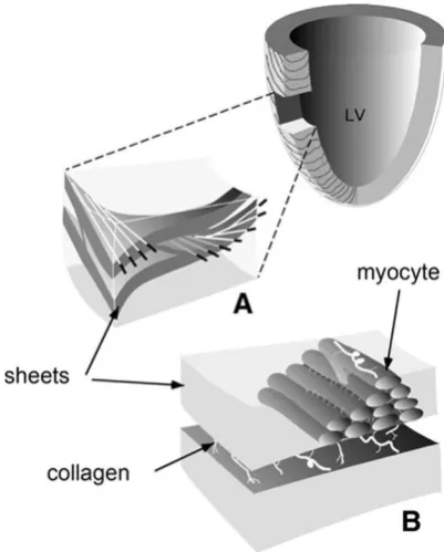
1.6Extracellular Cardiac Matrix 64
1.6.1 Extracellular Cardiac Matrix Structure 64
1.6.2 Extracellular Cardiac Matrix Function 65
1.6.3 Extracellular Cardiac Matrix and Heart Failure 66
1.6.4 Collagen Turnover Biomarkers 67
7
1.6.6. Galnectin-3 69
1.7 MiRNA 70
1.8 Circulating MiRNA 73
1.9 MiRNA and Cardiovascular Disease 77
1.10 MiRNA and Cardiac Remodelling 74
1.10.1 MiR-1 75
1.10.2 MiR-21 76
1.10.3 MiR-29 77
1.10.4 MiR-122 77
1.10.5 MiR-133 78
1.10.6 MiR-210 79
1.11 MiRNA and Heart Failure 79
1.12 MiRNA and Cardiac Resynchronisation Therapy 86
1.13 MiRNA Discussion 95
1.14 Heart Failure Metabolism 96
1.14.1. Adiposity and Heart Failure 97
1.14.2. Body Composition and Heart Failure 99
1.14.3. Conclusion 101
1.15 Publications 101
CHAPTER TWO
GENERAL THESIS HYPOTHESIS AND RESEARCH OBJECTIVES 102
2.1 General Hypothesis 103
2.2 Aim 104
CHAPTER THREE
GENERAL MATERIALS AND METHODOLOGY 107
3.1 Introduction 108
8
3.2.1 Local Service and Patients 108
3.2.2 UHCW CRT Implant Service 110
3.2.3 Implantation Procedure 112
3.2.4 Routine Aftercare 114
3.3 Applied Definitions 115
3.3.1 Outline 115
3.3.2 Electrocardiogram and QRS duration 115
3.3.3 Bundle Branch Block 115
3.3.4 Clinical Assessment 116
3.4 Study I: Retrospective Cohort Study 118
3.4.1 Title 118
3.4.2 Indication for Study 118
3.4.3 Rationale for the Retrospective Study 118
3.4.4 Study Governance 119
3.4.5 Study Overview 120
3.4.6 Clinical Data Acquisition 121
3.4.7 Potential Pre-implant Predictors Model 123
3.4.8 Outcomes 124
3.5 Study II: Explorative Prospective Study 126
3.5.1 Title 126
3.5.2 Research Governance 126
3.5.3 Study Participants 128
3.5.4 Study Outcomes 128
3.5.5 Eligibility Criteria 130
3.5.6 Study Design 132
3.5.7 Assessments 135
3.6 Laboratory Principles and Analysis 150
3.6.1 Enzyme Linked Immunosorbent Assay 151
3.6.2 Electrochemiluminescence Immunoassay 159
3.6.3 MiRNA Profiling 164
9
3.7.1 Study I 174
3.7.2 Study II 175
3.8 Statistical Methods for Analysis of Clinical Studies 176
3.8.1 Database 177
3.8.2 Missing Data 178
3.8.3 Descriptive and Inferential Data Analysis 180
3.8.4 Mixed Between-Within Subjects ANOVA 188
3.8.5 Logistic Regression 189
3.8.6 Survial Analysis 190
3.8.7 Inter-Rater Variability Echocardiography Study 193
CHAPTER FOUR
EXTRACELLULAR CARDIAC MATRIX AND CARDIAC RESYNCHRONISATION THERAPY: A
SYSTEMATIC REVIEW 196
4.1 Introduction 197
4.2 Methodology 197
4.2.1 Eligibility Criteria 197
4.2.2 Database Search Strategies 198
4.2.3 Data Extraction and Management 200
4.2.4 Risk of Bias Assessment 200
4.2.5 Data Synthesis and Analysis 200
4.3 Results 201
4.3.1 Study Design 201
4.3.2 Baseline Characteristics 206
4.3.3. Responder vs Non-Responders 206
4.3.4.Extracellular Matrix Biomarkers in the Heart 207
4.4 Discussion 218
4.5 Contribution of Authors 223
10
CHAPTER FIVE
PRE-IMPLANTATION PREDICTORS AT A UK TERTIARY CENTRE 224
5.1 Introduction 225
5.2 Aims 226
5.3 Concise Methods 226
5.3.1 Study design 226
5.3.2 Data Collection 227
5.3.3 Implant Procedure 228
5.3.4 Potential Pre-implant Predictors Model 228
5.3.5 Outcomes: Clinical Response and Cardiovascular Outcomes 228
5.3.6. Statistical Analysis 229
5.3.7 Missing Data 230
5.4. Results 232
5.4.1 Patient Characteristics 231
5.4.2 Implant details 234
5.4.3 Clinical response 235
5.4.4 Predictors of Clinical Response 236
5.4.5 Predictors of Cardiovascular Outcomes 240
5.4.6 Chronic Kidney Disease 246
5.5 Discussion 246
5.5.1 Study limitations 252
5.5.2 Informing Prospective Cohort Study 253
5.6 Conclusion 254
5.7 Publications 254
CHAPTER SIX
THE CHARACTERISATION OF CIRCULATING BIOMARKERS BEFORE AND AFTER CRT IN
PATIENTS WITH CHF AND THEIR ROLE IN PREDICTING RESPONSE:
THE COVERT-HF STUDY 255
11
6.2 Aim and Objectives 257
6.3 Methodology 258
6.3.1 Study Outcomes 259
6.3.2 Device Implantation 259
6.3.3 Transthoracic Echocardiography 259
6.3.4 Blood Sampling and Laboratory 260
6.3.5 Statistical Analysis 261
6.4 Results 262
6.4.1 Baseline Clinical Characteristics 263
6.4.2 Baseline Biomarker Levels 266
6.4.3 Effects of CRT on Cardiac Function and Biomarker Expression 268 6.4.4 Correlation between change in biomarker expression and cardiovascular
variables following CRT 271
6.4.5 Predicting Functional Response 273
6.4.6 Echocardiographic response and baseline biomarker expression 275
6.4.7 MACE and Baseline Biomarker Expression 276
6.4.8 Coronary Sinus Biomarker Profile 277
6.5 Discussion 279
6.5.1 Study Limitations 283
6.6 Conclusion 284
6.7 Publications 284
CHAPTER SEVEN
BODY COMPOSITION IN HEART FAILURE AND IMPACT OF CARDIAC RESYNCHRONISATION
THERAPY 285
7.1 Introduction 286
7.2 Aim and Objectives 287
7.3 Methodology 287
7.3.1 Device Implantation 288
12
7.3.3 Air-Displacement Plethysmography 289
7.3.4 Blood Sampling and Laboratory Analysis 289
7.3.5 Study Outcomes 289
7.3.6 Statistical Analysis 290
7.4. Results 290
7.4.1 Baseline Sub-Group Characteristics 291
7.4.2 Effect of CRT on Cardiac Function and Body Composition 293
7.4.3 Effects of CRT on Body Composition by Response Status 295
7.4.4 Correlations Analysis between Relative change in Body Composition
and Cardiac Function 297
7.5 Discussion 299
7.5 Study Limiataions 303
7.6 Conclusion 305
7.7 Publications 305
CHAPTER EIGHT
DISCUSSION 306
8.1 Discussion 307
8.2 Limitations 317
8.3 Future Work 321
8.4 Conclusions 323
CHAPTER NINE
13
LIST OF TABLES
Table 1.1 Randomised control trials evaluating CRT in sinus rhythm 39
Table 1.2 NICE 2014 (TA314) Indications for Implantable Cardiac Defibrillator and CRT
in Patients with LVEF<35% 50
Table 1.3 Summary of observational studies examining pre-implant ‘predictors’ of CRT
response 54
Table 1.4 Different CRT response definitions identified by Fornwalt et al1 59
Table 3.1 Elective Complex Cardiac Devices Complications 112
Table 3.2 Criteria for Complete LBBB and RBBB 116
Table 3.3 NYHA symptom classification 117
Table 3.4 Prospective Clinical and Echocardiographic Response Criteria 130
Table 3.5 Serial Dilutions Optical Densities for GDF-15 Microplate 2 156
Table 4.1 Extracellular Cardiac Matrix Candidate Biomarkers in Heart Failure 198
Table 4.2 Summary of included articles study designs and response definitions 204
Table 4.3 Risk of Bias Tables 205
Table 4.4 Baseline characteristics of included articles in systematic review 209
Table 4.5 Extracellular cardiac matrix biomarker analysis summary 210
Table 4.6 Baseline extracellular cardiac matrix associations with CRT response 211
Table 5.1 Selected ‘Pre-Implant’ Predictors Missing Data 231
Table 5.2 Baseline characteristics of overall clinical responders vs non-responders 233
Table 5.3 Baseline characteristics acute response (<12 weeks) response vs
non-responders 237
14
Table 5.5 Univariate and Multivariate Logistic Regression Analysis of Potential
Predictors of Overall Clinical Response 239
Table 5.6 Log-rank (P-value) of Kaplan-Meier Curves 242
Table 5.7 Proportional Hazards Cox Regression for MACE 243
Table 5.8 Proportional Hazards Cox Regression for all-cause mortality 244
Table 5.9 Proportional Hazards Cox Regression for first HF Hospitalisation 245
Table 6.1 Baseline Characteristics 265
Table 6.2 Baseline Biomarker Levels for Functional Responders vs Non-Responders 267
Table 6.3 Behaviour of Functional LV Geometry and Circulating biomarkers Following
CRT Implantation 269
Table 6.4 Comparison of Baseline Biomarker Expression between with and without >15% Reduction in LVESV at 6 Months Following CRT Implantation 275
Table 6.5 Comparison between baseline biomarker expression for participants with and
without MACE at 12 months following CRT implantation 276
Table 7.1 Baseline Characteristics 292
Table 7.2 Changes in Sub-Group Characteristics Over 6 Months 294
Table 7.3 Behaviour of Body Composition Following CRT Implantation 296
Table C1 Chronic Kidney Disease Stage 400
Table L1 The Borg Symptoms Scale 455
15
LIST OF FIGURES
Figure 1.1 Cardiac Resynchronisation Therapy 37
Figure 1.2 Relationship of CRT on all-cause mortality or HF hospitalisation and QRS
duration 43
Figure 1.3 United Kingdom total CRT implant rates 2003-2014 compared with release in
national and international implantation criteria 51
Figure 1.4 Number of publications on ‘CRT and biomarkers’ over 14 years 62
Figure 1.5 The Extracellular Cardiac Matrix 65
Figure 1.6 Normal 3-dimensional representation of cardiac microstructure 66
Figure 1.7 Biogenesis of miRNA 71
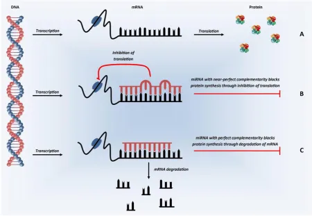
Figure 1.8 MiRNA Mechanisms of Action 72
Figure 1.9 Circulating miRNA associated with a diagnosis HF 80
Figure 1.10 MiRNA Expression Profiles for Responders vs. Non-Responders after one year
of CRT 89
Figure 1.11 Relationship between circulating plasma miR-30d levels and change in LVEF
six months after CRT 93
Figure 3.1 Arden Cardiac Network 2016 109
Figure 3.2 CRT implantation Rates in Arden Cardiac Network compared to National Rate
2010-2012 110
Figure 3.3 Cross Sectional View of Heart and Endocardial CRT Lead Placement 113
Figure 3.4 Patient Screening, Selection and Study Process 121
Figure 3.5 Study Scheme for COVERT-HF 134
Figure 3.6 A Replicated Example of a 6MWT 139
Figure 3.7 A Transthoracic Echocardiogram on a Participant 140
16
Figure 3.9 The BOD POD® in the HRMU at UHCW 146
Figure 3.10 The Process of a Sandwich ELISA 154
Figure 3.11 The 4PL Best-Fit Standard Curve for GDF-15 Plate 2 158
Figure 3.12 The ECL Reaction 161
Figure 3.13 The Process of Sandwich ECLIA 164
Figure 3.14 MiRNA Detection and Quantification 169
Figure 3.15 The Polymerase Chain Reaction Stages 173
Figure 3.16 Examples of Histograms 182
Figure 3.17 Examples of Quantile-Quantile Plots 182
Figure 3.18 A Boxplot of Baseline NT-pro-BNP 184
Figure 3.19 Scatterplot of Change in LVESVi and Fat Mass Over 6 Weeks 188
Figure 3.20 Kaplan-Meier Survival Curves for the CRT Retrospective Registry 193
Figure 4.1 Screening and selection of articles for systematic review 202
Figure 5.1 Screening and selection of all CRT Implantations Eligibile for Study 232
Figure 5.2 CKD status at Implant and Time to Cardiovascular Outcome 241
Figure 5.3 Upgrade Device Status at Implant and Time to All-Cause Mortality 242
Figure 6.1 Patient Recruitment, Flow and Outcomes 263
Figure 6.2 Trends in Functional Variables, LV Geometry and Biomarker Expression Following CRT Implantation in Responders and Non-Responders 270
Figure 6.3 Correlation Analysis of Short and Long-Term Changes Following CRT between Biomarkers vs Functional and Echocardiographic Variables 272
Figure 6.4 Univariate and Multivariate Regression Model of Pre-CRT Implant Variables
17
Figure 6.5 Variation Between Biomarker Expression in Peripheral and Coronary Sinus
Blood 278
Figure 7.1 Recruitment and Flow of Participants through Body Composition
Sub-Study 291
Figure 7.2 Change in Relative Fat Mass in Functional Responders and Non-Responders
over 6 Months 296
18
TABLE OF APPENDICES
APPENDIX A Systematic Review Protocol 389
APPENDIX B Literature Search for Systematic Review 398
APPENDIX C Applied Medical Definitions 399
APPENDIX D Retrospective Cohort Study I Data Collected 401
APPENDIX E Ethical Approval 403
APPENDIX F Clinical Trials Registration Form 408
APPENDIX G Patient Information Sheet 409
APPENDIX H Consent Form 422
APPENDIX I Research Protocol 424
APPENDIX J Clinical and Procedure Data Collected 441
APPENDIX K Data Collection Forms 446
APPENDIX L Six Minute Walk Test Methods 454
APPENDIX M Left Ventricular Dimensions and Function Quantification 456
APPENDIX N Participant Questionnaire 457
APPENDIX O Minnesota Living with Heart Failure® Questionnaire Description 458
APPENDIX P Minnesota Living with Heart Failure® Questionnaire 460
APPENDIX Q Air-Displacement Plethysmography (BOD-POD®) 462
APPENDIX R Selection of Published Abstracts 463
19
ACKNOWLEDGEMENTS
Over the last five years, from the decision to undertake this period of study at University
Hospital Coventry and the University Warwick to the handing in of this thesis, many people
of provided their time and support. It is with no exaggeration that without each and
everyone’s support this research would simply not have been conducted successfully. I have
tried to mention everyone individually who have contributed. I wish to thank everyone who
has had any involvement in my PhD for their help and support.
I owe specific thanks to my principle supervisor Dr Faizal Osman, Consultant Cardiologist
and Electrophysiologist. I met Dr Osman when we shared a taxi in Geneva on the way from
the airport to a basic pacing course I was attending. I spoke then about my intention to
undertake a PhD. Dr Osman showed particular enthusiasm and interest in my planned
period of research. Over the next 12 months Dr Osman created a research program and
developed the initial project that formed the basis of the this body of research. From the
outset he has been accessible, supportive and focused on making my research successful. I
am immensely grateful for the opportunity he has afforded me in taking the next step in my
research career.
I also wish to thank my two other supervisors Dr Paul O’Hare, Reader of Medicine and
Professor Harpal Randeva, Professor of Medicine. Dr O’Hare has been incredibly supportive
as my principle academic supervisor, nurturing my academic development and ensuring
20 knowledge of research practice. I have appreciated his advice navigating several issues that
have arisen during my four years as a postgraduate student. Professor Randeva has been a
very supportive third supervisor; he has funded my tuition fees for which I would not been
able to afford on my own. The expertise in biomarkers has been essential to the
understanding and performance of research in this field. I have been tutored and supported
in undertaking research in the laboratory by Professor Randeva. I have become involved in
his research team in the clinical sciences research laboratory. Professor Randeva has been
supportive of funding my consumable budget for quantification of the extracellular
biomarkers. I am incredibly grateful to for this continued support and advice through the
entire body of research.
I have had the immense pleasure of working alongside a dedicated group of cardiology
research nurses, who not only gave me a home for three years but made me feel part of the
team. Valerie Ansell, Julie Jones and Anntionnette Musa welcomed me in September 2013
and helped give me the training in research conduct that I did not have when I started,
guided me through logistical planning and aided me in every participant visit from
supporting 6-minute walk tests, to making cups of tea or spinning bloods. During my time
there Samantha Hyndman joined the team and was a great source of help and support. They
have all made conduction of the data collection fun and enjoyable. I also had the pleasure of
meeting and working with Josie Goodby who joined the team as a clinical trial practitioner
and took every opportunity to help me with the COVERT-HF project. Josie not only helped
with participant visits, she also over a year undertook the large task of reviewing all my data
21 A late addition to the team was Dr Danish Ali, cardiology research fellow working on
research in heart failure with preserved ejection fraction. Dr Ali assisted me with conducting
my systematic review and performed the role of second reviewer, apraising a large number
of abstracts and articles, including data extraction. I am grateful for his help and assistance
in the conduct of this review. I will forever be grateful to the entire team’s help and for their
friendship.
I worked within the Department of Cardiology, University Hospital Coventry and
Warwickshire NHS trust. I was incredibly supported by the entire department. There are too
many to mention, but everyone made me feel welcome and supported. Miss Asma Qadas,
medical secretary to Dr Osman was an important contact for organsing appointments,
chasing paperwork, performing photocopying, taking messages etc. Without her help my
research simply would not have progressed. I also worked closely with the arrhythmia nurse
team; Sister Helen Eftekhari and Sister Geeta Paul. Both would identify potential patients for
my prospective research study. They would also provide excellent clinical support for our
patients, which would be frequently required. Having their close support meant I could
focus on the research. I am also incredibly humbled working with such a friendly group of
consultants who have all gone out of their way to support my research endeavours. It is a
testament to this support that I chose to continue working in this department after my
research period was completed. I must acknowledge Professor Banerjee, consultant
cardiologist and department research lead for his personal support and enthusiasm for my
efforts. Professor Banerjee has supported me academically and fiscally. I have also found
22 Abdul Maher, consultant cardiologist (George Eliot Hospital) who implanted the majority of
cardiac resynchronisation devices during the prospective research study. Firstly Dr Maher
highlighted patients for potential recruitment and secondly assisted with data collection
from the procedure for the research about the implantations. Finally, when coronary sinus
samples were collected he kindly facilitated this. I want to also thank the entire cardiac
catheter laboratory team and cardiology day unit team for helping me in my data collection
and being as flexible as possible to facilitate my research. The entire cardiology department
has my eternal gratitude for their help during these last four years.
The Department of Cardiac Investigation, University Hospital Coventry and Warwickshire is
full of a group of incredible individuals who always went out of their way to support my
research. The echocardiography team (Kam Rai, Luke Mahoney, Samantha Booth and
Manual De Villa) have provided me with resources to undertake the participant’s
echocardiographic assessments and were always available for advice on image
interpretation. The cardiac technicians were always able to perform an electrocardiograph
when required. The front desk team (Debbie and Lisa) were always so helpful when
organising appointments for participants. The pacing team (Shirley Murray, Chris King, Andy
Read, Ian Patchett and Sam Harvey) were always flexible when arranging pacemaker review
appointments, so they matched research visits. They also provided me with data and
information for the research study. The entire department were essential to the conduct of
each and every research visit and without this group of individuals the research could not
23 I am grateful to the Research, Development and Innovation department at the University
Hospital Coventry and Warwickshire. The entire department led by Professor Chris Imray
and Mrs Ceri Jones has shown faith and confidence in my ability. They have helped me
navigated the world of clinical research. Isabella Petrie and Sonia Kandola have helped my
navigate the governance aspects of the project, helping me apply for ethics and making
amendments when needed. Deborah Griggs and Giovanni Bucci have provided invaluable
advice and help when making grant applications to different funding bodies. Sarah O’Toole
has provided me with constant support regarding my research administration and advice
and where and where to find things. Becky Chadwick has also been a very supportive and
able to help with my many queries over the years. The entire department has always gone
out of their way to help me and support my activities.
Tissue bank has also been a group of individuals that have gone out of their way to help me
over the last four years. The department led my Sean James has gone out of its way to store
all my samples in a safe and secure fashion. Each member of the team; Adrian Fisk, Andrew
White and Parmjit Dahaley have always been happy to help and deal with my frequent
queries quickly and professionally. I am immensely grateful for their help.
I was tutored and supported through the conduct of my advanced research statistics by Dr
Thomas Hamborg, Dr Peter Kamani and Dr Nicholas Parsons, University of Warwick. They
provided support on analysis planning and conduct. Answering questions related to my
24 for his help at the end of the analysis, when immediate assistance was required but not
immediately available from other sources.
During my research I had the opportunity to mentor and work alongside a group of junior
doctors and medical students who had an interest in research and cardiology. Dr Benjamin
Anderson, Core Trainee Year One, Mr Dominic Heining and Mr Gavin Atherton assisted me
in collecting data for my retrospective research study. They assisted in searching anonymous
patient records and extracting data. I also had the opportunity to work with Mr Mark
Theodorsen, Mr Bhaveek Chohan and Mr Luke Boylan on related research projects. All were
extremely dedicated and hard-working. Their contribution to my research work was
invaluable. They will all go on to bigger and brighter things I suspect.
I wish to acknowledge the library team at University Hospital Coventry and Warwickshire
and in particular Mrs Petra Meeson. Mrs Petra Meeson over the last three years has
provided teaching on literature searches and assisted in conducting my systematic review
searches. Petra has also become a good friend and one I could go to for general advice over
a cup of tea. I am very grateful for her help.
I undertook my cardiac extracellular biomarker analysis in the Clinical Sciences Research
Laboratory, University of Warwick within Professor Randeva’s team. I am particularly
25 laboratory techniques required to quantify expression of these biomarkers. Jimiao spent a
lot of time performing the analyses with myself and helped with my interpretation of the
results. I am very grateful for his dedication and help with this analysis.
I was very fortunate to collaborate with Professor Manual Mayr, Professor of Cardiovascular
Proteomics, King's British Heart Foundation Centre, King's College London on miRNA
quantification and analysis. I owe specific thanks to Dr Temo Bawari who was able to guide
me through the miRNA laboratory analysis and tutor me in the techniques involved. Dr
Temo Bawari was critical to the undertaking of this highly complex analysis and certainly
without him I could not have achieved the results we did. Temo’s dedication to this project
was essential to its success, which included working long hours in the laboratory. Personally
I am very grateful for his help, humour and friendship. He remains a constant source of
support and advice one year on. I also wish to thank Professor Mayr for his expert advice
and critiquing of my project. I feel my body of work is better for it. Furthermore, I wish to
thank the entire department who made me feel part of the team and welcomed me in to
the team.
I wish to thank the Research, Development and Innovation Department at the University
Hospital Coventry and Warwickshire for their funding of my post. I would like to Medtronic
Ltd and St Jude Medical for partial funding of my wages and comsumables. Finally, I would
like to thank the Medical and Life Sciences Research Fund for their support on consumable
26 Finally, I wish to thank my friends and family for all their support while I have undertaken
my PhD. This has been a large undertaking over the last 4 years and one that has seen me
work long hours. I wish to thank my wife Dr Anna Gregory for being continually supportive
of my work and giving me the time to complete it. Without her support I simply could not
have performed this body of research. I wish to thank my parents for being continually
supportive in all my endeavours and being there for advice. I wish to thank all my London
based friends for providing me for a place to sleep for the many courses I have undertaken
over the last four years and provided me somewhere to stay. Firstly, I wish to thank Rachel
and Ben Freeman for giving me a place to stay during my seven week secondment to Kings
College London. Secondly I would like to thank Mr Andrew Powell, for providing me with a
place to stay for a course and support while I was away from home. Thirdly, I would like to
thank my sister Laura Shorters and brother-in-law Alex Shorters for providing me a bed and
being ever supportive of my PhD. Finally, I would like to thank Celia and Chris for providing
me with a bed on many occasions over the last few years rent free and for advice on my
27
DECLARATION
This thesis is submitted to the University of Warwick in support of my application for the
degree of Doctor of Philosophy. It has been composed by myself and has not been
submitted in any previous application for any degree .
I am aware of the University regulations governing plagiarism and I declare that this thesis represents my own work except where I have stated otherwise.
The work presented (including data generated and data analysis) was carried out by the author.
All sources are credited by means of referencing.
28
ABBREVIATIONS
4PL Four-Parametic Logistic Regression
6MWD 6-Minute Walk Distance
6MWT Six Minute Walk Test
ACC American College of Cardiology
ACEi Angiotensin Cardioverting Enzyme Inhibitor
AF Atrial Fibrillation
AHA American Heart Association
ANOVA Analysis of variance
AUC Area Under Curve
BBB Bundle Branch Block
BIA Bioelectrical Impedence Analysis
BMI Body Mass Index
BNP Brain Naturetic Peptide
BSA Body Surface Area
BSA Bovine Saline Albumin
CC Cardiac Cachexia
CCS Clinical Composite Score
cDNA complementary DNA
CHF Chronic Heart Failure
CI Confidence Interval
CITP Carboxy-Terminal Telopeptide of Type I Collagen
CKD Chronic Kidney Disease
CMR Cardiac Magnetic Resonace
CRT Cardiac Resynchronisation Therapy
CRT-d CRT-defibrillator
CRT-p CRT-pacemaker
CS Coronary Sinus
CTx Collagen I C-terminal telopeptides
CV Cardiovascular
CV Coefficent of Variability
DNA Deoxyribonucleic Acid
dNTP Deoxynucleoside Triphosphate
dTTP Deoxythymidine triphosphate
ECG Electrocardiogram
29
ELISA Enzyme-Linked Immunosorbent Assays
ESC European Society of Cardiology
GDF-15 Growth Differentiation Factor 15
GFR Glomerular Filtration Rate
HF Heart Failure
HMRU Human Metabolic Research Unit
HRS Heart Rhythm Society
hs-CRP high sensitive-C reactive protein
hs-TnT High Sensitvity Troponin T
ICD Implantable Cardioverter Defirillatior
ICTP Cardoxyterminal Telopeptide of Type I Collagen
IL-18 Interlukin-18
IL-6 Interlukin-6
IQR Interquartile Range
IVCD Interventricular Conduction Disturbance
IVMD Intraventricular Mechanical Delay
LBBB Left Bundle Branch Block
LOCF Last observation carried forward
LV Left Ventricle
LVAD Left Ventricular Assist Device
LVEDD LV End-Diastolic Diameter
LVEDV Left Ventricular End Diastolic Volume
LVEF Left Ventricular Ejection Fraction
LVESV Left Ventricular End Systolic Volume
LVESVI LVESV indexed by body surface area
MACE Major Adverse Cardiovascular Events
MAR Missing at Random
MCAR Missing Completely at Random
MI Myocardial Infarction
miRNA Micro Ribonucleic Acids
MLHFQ Minnesota Living with Heart Failure
Questionnaire
MMP Matrix Metalloproteinases
MNAR Missing Not at Random
MR Mitral Regurgitation
30
MR-proADM pro-adrenomedullin
MR-proANP midregional proANP
NHS National Health Service
NICE National Institute of Health and Care Excellence NIVCD Non-Specific intraventricular Conduction Delay
NYHA New York Heart Association
OMT Optimal Medical Therapy
OR Odds Ratio
PBS Phosphate Buffered Saline
PCI Percutanous Coronary Intervention
PCR Polymerase Chain Reaction
PICP Carboxy-Terminal Propeptide of Procollagen
Type I
PNIIIP Aminoterminal Propeptides of Type III Collagen
PNIP Aminoterminal Propeptides of Type I Collagen
PPP Platelet Poor Plasma
PV Peripheral Venous
QALY Quality Adjusted Life Year
QoL Quality of Life
qPCR quantatative Polymerase Chain Reaction
Q-Q Plots Quartile-Quartile Plots
RA Right Atrium
RAA Right Atrial Appendage
RBBB Right Bundle Branch Block
REC Regional Ethics Committee
RNA Ribonucleiconic Acid
ROC Receiver Operating Characteristics
RQ Relative Quantification
RV Right Ventricle
RVA Right Ventricular Apex
RVS Right Ventricular Septum
SD Standard deviation
SE Standard Error
sST2 Soluble concentration of ST2
TGF-ß1 Transforming Growth Factor-ß1
31
TNC Tenascin-C
TPA Tripropylamine
UHCW University Hospital Coventry and Warwickshire
VE/VCO2 Minute Ventilation/Minute Volume CO2
production
VF Ventricular Fibrillation
VO2 Volume of Oxygen
V̇O2max Oxygen consumption at peak exercise
32
ABSTRACT
Heart failure is a common condition which carries a high mortality and morbidity. Despite
improved medical therapy the outcomes for heart failure with a reduced ejection fraction
remain poor. Cardiac resynchronisation therapy has revolutioned the treatment of patients
with heart failure with a reduced ejection fraction and dyssynchrony, refactory to medical
therapy, improving morbidity and mortality. Unfortunately a significant minority fail to
respond to this expensive therapy, which is challenging for both the patient and society.
Over the last 15 years research has focused on attempting to predict non-response.
Evidence suggests wider QRS duration and bundle branch morphology on the resting
electrocardiograph are the most important predictors of response and outcome following
implantation of a cardiac resynchronisation device. However, the non-response rate
remains unchanged despite extensive research.
Molecular systems have been shown to alter with the development and progression of
heart failure. Many of these systems are now utilised in the diagnosis and prognostication of
heart failure. Cardiac resynctonisation therapy device implantation has been shown to alter
these dysregulated molecular systems. Specific circulating biomarkers reflect these
respective systems. Cardiac extracellular matrix is a dynamic support structure that has
altered turnover in heart failure and is affected when cardiac resynchonisation devices are
implantted. Micro ribonucleic acids have been observed recently to be important in
33 Futhermore altered expression following cardiac resynchronisation therapy device
implantation has been reported. The evidence suggests circulating biomarkers for these
systems have the potential to predict response. Our prospective study examined specific
biomarkers that the literature suggests has the potential to predict response, but the
evidence is currently inconclusive. Moreover we utilised other important patient variables
known to be predictors from the wider literature and our own retrospective cohort analysis
of response to test alongside specific circulating biomarkers. We offer an informed pilot
study to test important circulating biomarkers for their clinical ultility to predict heart failure
34
Chapter One
35 1.1 HEART FAILURE
Chronic Heart Failure (CHF) is a common condition defined as an abnormality in cardiac
structure and function that leads to the inability of the heart to deliver adequate levels of
oxygen to match metabolic demand of the tissues.2 Patients suffer from a plethora of
symptoms including breathlessness, ankle oedema and fatigue.2 Heart Failure (HF) affects
800,000 people in the UK alone.2-4 The estimated lifetime risk of developing HF in the
general population is approximately 1 in 5 for a person aged 40 years.5 Adjusting for age,
the incidence of HF has remained stable over the last 20 years, but the prevalence continue
to increase.5 One of the largest drivers on the increasing burden of HF in the developed
world is ischaemic heart disease (IHD).6 HF is associated with a high mortality with an
estimated 30-40% mortality rate within the first year of diagnosis.7 However, there is an
improving trend in mortality demonstrated by the six month survival rate decreasing from
26% in 1995 to 14% in 2005.8 The HF burden has implications for national health systems as
it accounts for 5% of all acute medical and geriatric hospital admissions and is the
commonest hospital admission cause in the over 65 year old population. It is estimated that
hospital admissions due to HF will rise by 50% over the next 25 years.4,9 The burden has
eased slightly with an age–adjusted hospitalisation rate having decreased by 1–1.5% per
annum since 1992/1993.10 The improvements in mortality and hospitalisation is due to
more effective treatments,2,11-14 however the burden of the aging population and improved
36
1.1.1 Reduced Ejection Fraction Heart Failure and Cardiac Dyssynchrony
Many patients with HF with reduced ejection fractions (HFrEF) develop dyssynchronous
contraction of the heart due to damage of the underlying conduction tissue leading to
inefficient cardiac contraction that leads to symptoms. Cardiac dyssynchrony is a complex
and multifactorial process which impacts function.15 Prolongation of the atrioventricular
interval, can encroach on the starting of systole and filling within early diastole.15 Ventricular
contraction being delayed, the left ventricle (LV) diastolic pressures will exceed the left atrial
pressure during passive filling, leading to the development of functional mitral regurgitation
(MR).15 The impact of reducing ventricular pre-load, leads to a reduction in LV contractility,
by the Starling mechanism.15 Moreover, the occurrence of intra- and inter-ventricular
conduction delays leads to asynchronous contraction on the regional wall segments
(ventricular/mechanical dyssynchrony), which leads to reduced stroke volume, LV ejection
fraction (LVEF) and systolic blood pressure.15 Ventricular dyssynchrony leads to
incoordination of papillary wall contraction and further contributes to the development and
progression of functional mitral regurgitation.15 The development and progression of this
process leads to or contributes to LV adverse remodelling.15
1.2 CARDIAC RESYNCHRONISATION THERAPY
Cardiac Resynchronisation Therapy (CRT) or ‘biventricular pacing’ involves implanting pacing
leads into the heart to pace the left and right heart. The pacemaker leads are implanted via
the transvenous route into the right ventricle (RV) and a branch of the coronary sinus
37 is implanted into the right atrium (RA) to achieve atrioventricular synchrony, this lead is not
necessarily implanted in patients where pacing is not possible (e.g. Chronic Atrial
Fibrillation). Figure 1.1 demonstrates implantation of the CRT pacing leads. CRT can
‘resynchronise’ cardiac contraction through restorations of inter- and intra- ventricular and
atrioventricular dyssynchrony.15 Resynchronisation induces reverse LV remodelling by
[image:38.595.182.414.263.523.2]improving LVEF, contractility and LV filling time.15
Figure 1.1 Cardiac Resynchronisation Therapy. Figure taken from Hare J et al.NEJM, 200216
1.2.1 The Cost of CRT
CRT implantation is a costly intervention with a large up-front cost of an estimated £3,411
for a CRT Pacemaker (CRT-p) and £12,293 for a CRT Defibrillator (CRT-d).17 Additionally there
up-38 front cost is larger than for many other medical devices. Randomised Control Trials (RCT)
have been used to model the quality adjusted life year (QALY) costs of a CRT. It is widely
accepted that this falls below $50,000 per QALY which is the accepted cost of an
intervention in the USA, and equates to approximately £39,000.18 Efforts have focused on
minimising this cost, by better identifying the CHF population who will benefit, streamlining
implantation and distance monitoring to reduce patient visits to hospital 18. However, the
burden of cost to the healthcare system will continue to rise with the growing population of
CHF patients who might benefit from CRT.
1.2.2 The Evidence for CRT
Over the last 25 years CRT has become one of the most effective treatments for CHF and is
applicable to an estimated 25-30% of CHF patients.19 In 1994 Cazeau et al,20 demonstrated a
four lead pacemaker in a 54 year old advancing HF patient improved New York Heart
Association (NYHA) symptoms classification. Since then multiple RCTS have demonstrated
the benefit of CRT for HFrEF patients with mechanical Dyssynchrony; reduced mortality and
hospitalisation,21-23 alongside improved quality of life (QoL),23-27 symptoms,21,28,29 functional
performance28,30 and LV volumes.25,29,31 Table 1.1 summarises the main RCTs demonstrating
39
Table 1.1 Randomised control trials evaluating CRT in sinus rhythm (Adapted15).
Trial (ref) Pts Study Design Inclusion Outcome Main Findings
MUSTIC-SR
2001 21 58
Single-blinded, crossover, randomised - CRT-p vs. OMT,
6months
NYHA III, LVEF<35%,
QRS >150msec
1o: 6MWD
2o: NYHA, QoL, peak VO2, LV volumes,
hospitalisations, mortality
CRT-p ↑6MWD,
↓NYHA, ↑QoL, ↑peak
VO2 & reducedLV
volumes, MR, hospitalisations MIRACLE 2002 28 453 Double-blinded, randomised CRT
vs. OMT, 6 months
NYHA III-IV, LVEF<35%,
QRS >130msec
1o: NYHA, 6MWD, QoL
2o: Peak VO2, LVEDD, LVEF, MR, Clinical Composite response
CRT-pimproved
6MWD, NYHA, QoL, LVEF & reduced LVEDD,
MR
MIRACLE-ICD 2003 30 369
Double-blinded, randomised CRT-d vs. ICD, 6
months
NYHA III-IV, LVEF<35%,
QRS >130msec
1o: NYHA, 6MWD, QoL
2o: Peak VO2, LVEDD, LVEF, MR, Clinical Composite response
CRT-dimproved NYHA,
QoL, peak VO2
CONTAK-CD
2003 25 490
Double-blinded, randomised CRT-d vs. ICD, 6
months
NYHA II-IV, LVEF<35%,
QRS >120msec
1o: Clinical Composite
2o: NYHA, 6MWD, QoL, peak VO2, LV volume,
LVEF
CRT-d improved peak
VO2, 6MWD & reduced
LVEF
MIRACLE-ICD II 2004 29 186
Double-blinded, randomised CRT-d vs. ICD, 6
months
NYHA II, LVEF<35%,
QRS >130msec
1o: Peak VO2
2o: VE/VCO2, NYHA, QoL,
6MWD, LVEF, LV Volumes, Clinical Composite
CRT-d improved NYHA,
VE/VCO2, LVEF &
reduced LV volumes
COMPANION
2004 22 1520
Double-blinded, randomised - OMT vs. CRT-d
/or CRT-p , 15 months
NYHA III-IV, LVEF<35%,
QRS >120msec
1o: All-cause mortality or hospitalisations
2o: All-cause mortality, cardiac mortality
CRT-d & CRT-p reduced
all-cause mortality & hospitalisation
CARE-HF
2005 23 813
Double-blinded randomised - OMT vs. CRT-p
29.4 months
NYHA III-IV, LVEF<35%,
QRS >120msec
1o: All-cause mortality or hospitalisations
2o:All-cause mortality, NYHA, QoL
CRT-p reduced all-cause mortality, hospitalisations &
improved NYHA, QoL
REVERSE
2008 31 610
Double-blinded, randomised - ON vs. CRT-OFF, 12 months
NYHA I-II, LVEF<40%,
QRS >120msec
1o: % worsened HF clinical composite
2o: LVESVi, HF hospitalisations,
All-cause mortality
CRT-p/CRT-d did not
change the primary
endpoint, reduced
LVESVi, HF hospitalisations
MADIT-CRT
2009 27 1820
Single-blinded, randomised - CRT-d vs. ICD, 12 months
NYHA I-II, LVEF<30%,
QRS >130msec
1o: All-cause mortality or HF hospitalisations
2o: All-cause mortality, LVESV
CRT-d reduced the
primary endpoint & LVESV, CRT-d did not
reduce All-cause mortality
RAFT
2010 26 1798
Double-blinded, randomised - CRT-d vs. ICD, 40 months
NYHA II-III, LVEF<30%,
QRS >120msec
1o: All-cause mortality or HF hospitalisations
2o: All-cause mortality & CV death
CRT-d reduced primary
endpoint CRT-d (NYHA III) reduced All-cause
mortality
40 The RCTs for CRT represent benefit from CRT for patients with significantly reduced LVEF
(<35%), symptoms despite optimal medical therapy and a prolonged QRS duration
(>120msec) on resting electrocardiogram in sinus rhythm (Table 1.1). The detailed analysis
of all the RCTs, related systematic review, sub-group analyses and the observational studies
refines the groups of HFrEF patients that derive the most benefit and influence response to
CRT. This evidence is the basis of the international CRT implantation guidelines and the
weighting given to the evidence.
1.2.3 Left Ventricular Ejection Fraction and CRT
Studies have only examined patients with severe LV systolic dysfunction (LVEF <30-40%),
with the specific exception for patients who have a bradycardia pacing indication and
whether a CRT should be implanted over an simple RV pacing only device. Consistent trends
suggest that sustained RV pacing induces deterioration in LV systolic function, therefore
anticipating the deterioration with a CRT might prevent this.15,32-34 The biventricular versus
right ventricular pacing in patients with AV block (BLOCK HF) trial32 was the largest (n=691)
RCT examining RV pacing versus CRT on composite outcomes (all-cause mortality, HF
hospitalisation ↓<15% LV end systolic volume indexed to body surface area (m2)(LVESVi))
for patients with LVEF <50% (mean = 40%) with atrioventricular node block. BLOCK-HF
demonstrated following a 37 month observation period that patients undergoing a CRT
implantation had a 26% greater reduction in outcome occurrence, although it should be
noted some of the echocardiogram data was missing, so censoring of these patients
occurred, excluding end-points from this study.32 The evidence demonstrated that in
41 be implanted in stead of a pacemaker when weighed against the small risks of the
procedure. 32 Comparisons between RV pacing and CRT implantation for preserved systolic
function demonstrated no statistical difference.35,36
1.2.4 New York Heart Association Symptom Classification and CRT
The strength of evidence around CRT implantation for all NYHA symptom classification is
highly variable. The trials tended to recruit a higher proportion of NYHA II/III patients
dependent on the specific inclusion criteria (Table 1.1). In the mild / no symptom trials
(NYHA I-II) with severe LV systolic dysfunction (LVEF<30-40%), QRS duration >120-130msec
improvement in cardiovascular outcomes and reverse LV remodelling was
demonstrated.27,29,31 NYHA I patients represented a small proportion (<20%) of the
participants in all trials and showed a trend towards a benefit to improving cardiovascular
outcomes.27,29,31 Moreover, NYHA IV patients were under represented in the RCTs,
representing between 7% and 15%.15 The evidence for specific outcomes for NYHA IV
patients with LV systolic dysfunction (LVEF<30-40%) and QRS duration >120-130msec is
limited. One retrospective cohort study observed that 5 years survival was 40.4% following
CRT, however this was based upon only 5 patients (n=723).37
1.2.5 QRS Duration and CRT
QRS duration is the most powerful predictor of benefit and response when a patient has a
CRT implanted. Sub-group analyses of the MADIT-CRT27,38 REVERSE31 and RAFT26 RCTs
42 outcomes are those with a QRS duration >150msec. These trials represent patients in NYHA
class I-III and LVEF<30-40%. Cleland et al39 performed a large meta-analysis of individual
patients (n=3782) from 5 Medtronic Ltd (Minneapolis, USA) sponsored RCTs (MIRACLE,28
MIRACLE-ICD,30 CARE-HF,23 REVERSE31 and RAFT26) comparing either CRT with OMT23,28,31 or
CRT-d with implantable cardiac defibrillators (ICD).26 Pre-defined variables were examined
for their ability to predict CRT cardiovascular outcomes (composite all-cause mortality/HF
admissions or all-cause mortality alone).39 For uniformity of the review, patients with atrial
fibrillation (AF) and NYHA I symptoms had individual records removed from the analysis as
they were only present in a small proportion of patients within one RCT cohort. Cleland et
al39 accounted for the influence of having an actual CRT implanted and treated it as a fixed
affect variable in the prediction models. Increasing QRS duration was shown to be beneficial
for patients undergoing CRT and predicted a reduction in cardiovascular outcomes.39Figure
1.2 shows the hazard ratio (HR) and 95% confidence interval (CI) for CRT vs. control for the
composite of all-cause mortality or HF hospitalisation plotted against QRS duration. Cleland
et al39 demonstrate a greater magnitude of benefit from incremental increases in QRS
duration following CRT implantation improving outcomes. The definitive benefit was
observed at >140msec (CI lines cross the HR 1.0).39 The benefit reached a plateau beyond
180msec for composite outcome alone.39 Interestingly left bundle branch block (LBBB)
morphology was associated with broader QRS durations, generating the question of
whether the observed predictive power of QRS duration was due to this cofounding factor.
Cleland et al39 demonstrated non-LBBB had an increased trend towards higher mortality,
however when QRS duration was removed from the multivariable prediction model, little
43 Cleland et al39 meta-analysis is powerful due to the access to individual participant records
across 5 RCTs. Other reviews quoted in this body of work are aggregate reviews and do not
account for individual study confounders. However, only Medtronic Ltd sponsored RCTs
were included, which the authors do acknowledge as a major limitation.
Figure 1.2 Relationship of CRT on all-cause mortality or HF hospitalisation and QRS
duration. Models demonstrating Hazard Ratio’s and 95% confidence interval for CRT vs. controls (OMT/ICD/Back up pacing) against the QRS duration. (Adapted39)
There is definitive evidence demonstrating that patients with a QRS duration 120-129msec
do not benefit from CRT implantation. In 2007, the ‘CRT IN Patients with Heart Failure and
Narrow QRS’ (RethinQ) trial40 recruited patients (n=172) with standard implantation criteria
with QRS complexes <130msec and cardiac dyssynchrony, randomising participants to
therapy CRT-ON or CRT-OFF (after implantation). Over six months, the CRT-ON group
[image:44.595.89.501.222.443.2]44 pre-defined subgroup analysis those with a QRS<120msec demonstrated no difference
(p=0.45).40 The RethinQ40 RCT observation period was too short to be able to examine the
impact of biventricular pacing on a narrow QRS upon morbidity and mortality. More
recently, ‘the Cardiac Resynchronization Therapy in Heart Failure with a Narrow QRS’
Complex (Echo-CRT) trial41 enrolled patients (n=855) across 115 centres who met standard
implantation criteria and had a QRS complex <130msec with evidence of cardiac
dyssynchrony. Following CRT implantation they were randomised to either ON or
CRT-OFF to examine impact on all-cause mortality and HF hospitalisation (treated as a composite
outcome). The Echo-CRT41 RCT was stopped early on ‘the basis of futility with a potential for
harm’. The trial demonstrated CRT-ON had a higher rate of composite primary end-points
occurring compared to CRT-OFF group (28.7% vs. 25.2%), which was not statistically
significant (p=0.15).41 However, when all-cause mortality was examined on its own CRT-ON,
had a statistically higher occurrence than CRT-OFF (11.1% vs. 6.4%, p=0.02).41 The definitive
benefit of CRT is in those with a widened QRS, despite the presence of mechanical cardiac
dyssynchrony.
1.2.6 QRS Morphology and CRT
QRS morphology has been demonstrated to be important in determining response to CRT
implantation. Subsequent sub-group analyses of the MADIT-CRT42, RAFT26 and REVERSE43
trials all identified complete LBBB demonstrated a better outcome on the composite of
all-cause mortality and hospitalisation compared with right bundle branch block (RBBB) and
non-specific intraventricular conduction delay (NIVCD). In 2012 Sipahi et al44 performed a
45 mortality/HF hospitalisations) corresponding with Bundle Branch Block (BBB) morphology.
COMPANION22, CARE-HF23, MADIT-CRT42 and RAFT26 were the only trials that met the
eligibility criteria.44 Sipahi et al44 demonstrated a highly significant reduction in the
composite outcome for patients undergoing CRT implantation with complete LBBB (Risk
Ratio (RR) 0.64, CI (95%) 0.52-0.77, p<0.0001). Within the included RCTs of the
meta-analysis, patients with LBBB tended to have wider QRS’s, which may have confound the
results.44
Cunnington et al45 in a meta-analysis of six landmark CRT RCTs22,23,26-28,31 analysed 6914
participants and compared those with and without LBBB (Non-LBBB was a classification for
QRS morphology in four included trials22,26,27,31 and RBBB was used in the other two RCTs
23,28
). The two trials which classified non-LBBB as RBBB were not involved in the sensitivity
analysis.45 NIVCD was accounted for, in the definition of BBB in four trials.23,26,27,31 The
review summarises that the six trials represented participants with NYHA I-IV, LVEF<30-40%
and QRS>120msec.45 Table 1.1 summarises the characteristics of all the included studies.
Cunnington et al45 demonstrated no benefit from CRT for patients with non-LBBB QRS
morphology for a pooled outcome of all-cause mortality and HF hospitalisation (HR 1.09, CI
(95%) 0.85 – 1.39). It should be noted that Cunnington et al45 only studied cardiovascular
end-points and did not examine symptom, functional or echocardiographic outcomes. It is
also acknowledged that NYHA classes I and IV are underrepresented in the RCTs and the
observations are driven by those with class II and III symptoms. The MADIT-CRT trial27
enrolled 536 non-LBBB participants with NYHA I-II. This RCT demonstrated no clinical benefit
46 1.57, CI (95%) 1.03-2.39). A meta-analysis by Sipahi et al also demonstrated there was
no benefit to implanting CRTs in patients with non-LBBB. The challenge remains how to
treat those with non-LBBB with a widen QRS, which remains an active indication for
implanting a CRT.15,17,47 Cleland et al39 in a large meta-analysis observed that non-LBBB did
not predict cardiovascular outcomes when QRS duration was removed from the analysis.
Different BBB patterns have been demonstrated on recent electrocardiographic activation
mapping studies to have heterogeneous patterns and should be considered as different
entities,24,48 whereas QRS durations can be considered on a continuous spectrum with
incremental benefit the wider the duration when a CRT is implanted.39 Currently QRS
duration represents the most powerful predictor and BBB morphology should be considered
separately with LBBB being more favourable for a successful outcome.
1.2.7 Atrial Fibrillation and CRT
AF commonly co-exists in patients with HF and its presence can reduce the success of CRT.49
Understanding the true influence of AF on the success of CRT is difficult as patients with AF
tend to be older, have more co-morbidities and are more unwell. Comparison between
sinus rhythm and AF is influenced by these confounding factors, which often infer worse
prognosis.15 AF is underrepresented in CRT RCTs and reliance is needed upon meta-analyses.
It has been observed that AF patients receiving CRT have a similar improvement in LVEF
compared with those in sinus rhythm, but have worse symptom and functional response.49
In a large (n=7495) meta-analysis of 33 observational studies, Wilton et al49 compared those
with AF (22.5%) to sinus rhythm receiving CRT and observed a significantly higher all-cause
47 recommendations for CRT in patients with AF remains weak and is based on limited
evidence and expert opinion. However, implantation is favoured if >99% biventricular
pacing percentage can be achieved.15
1.2.8 Cardiac Dyssynchrony and CRT
One of the recent significant changes to the international guidelines was the removal for the
need to demonstrate cardiac dyssynchrony on echocardiography if the patient’s QRS
duration is 120-149msec on resting ECG. The CARE-HF trial23 eligibility criteria required
patients with a QRS duration of 120-149 msec to have cardiac dyssynchrony demonstrated
on echocardiography. Cardiac dyssynchrony was defined as achieving two of three criteria:
aortic pre-ejection delay >140msec, interventricular mechanical delay (IVMD) of 40msec, or
delayed activation of the posterolateral LV wall. The IVMD was calculated as the time
difference between the onset of forward flow in the Aortic pre-ejection time (APET) and
Pulmonary pre-ejection time (PPET) outflow tracts (IVMD = APET – PPET).23 A sub-group
analysis of CARE-HF demonstrated those patients with an IVMD >49.2msec implanted with a
CRT had a reduced composite outcome (all-cause mortality or hospitalisation for a major
cardiovascular event) (HR 0.50, CI (95%) 0.36-0.70).23 A pre-defined sub-study50 of
CARE-HF23 observed that patients with IVMD (>49.2msec) benefitted more from CRT from a
greater reduction in the composite outcome (HR 0.99, 95% CI 0.98–1.00). The
demonstration of the benefit of CRT on patients who demonstrated cardiac dyssynchrony
(as per CARE-HF) formed a part of the implantation guidelines.51
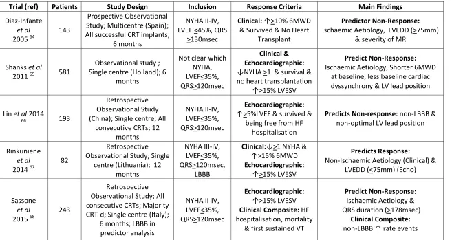
Cardiac dyssynchrony as an indicator of CRT success was seen sceptically, as the data was
48 results of the prospective, multicentre Predictors of Response to Cardiac Resynchronization
Therapy (PROSPECT) study.52 PROSPECT recruited 426 participants who successfully had
CRTs implanted across 53 international centres with standard CRT implantation indications
to measure the ability of 12 pre-defined echocardiographic measures of cardiac
dyssynchrony (including IVMD) abilities to predict response, alongside their validity and
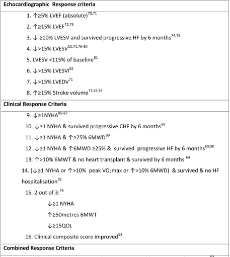
reliability as measurments.52 Two definitions of response were utilised at six months
follow-up; a clinical (HF clinical composite score) and echocardiographic (>↓15% LVESV).52 The 12
predefined cardiac dyssynchrony measures demonstrated wide variability in their ability to
predict clinical and echocardiographic response. For echocardiographic response prediction,
the sensitivity ranged from 9% to 77% and specificity from 31% to 93%.52 None of the
cardiac dyssynchrony variables achieved an area under the receiver operating curve (ROC)
for either response definition <0.62; representing a poor ability to discriminate response.52
PROSPECT also identified a high variability between operators to accurately reproduce
cardiac dyssynchrony measurements.52 Notably only 286 participants had paired baseline
and six month follow-up echocardiogram completed successfully, due to a combination of
poor quality images, presence of AF and mortality events.52 The results of PROSEPCT
demonstrated weak predictive power and high inter-operator variability of cardiac
dyssynchrony echocardiographic markers when used in multiple centres.52 Currently other
echocardiographic measurements of cardiac dyssynchrony continue to be researched,
echocardiographic strain analysis offers some future potential,53-55 however for now the
best discriminator is QRS duration on resting ECGs and this is now used in the current
49
1.2.9 Cardiac Resynchronisation Therapy Implantation Criteria
The current evidence has been significantly modified over the last 15 years of implantation
as more evidence has been produced. The previous section demonstrated the evolution and
refinement of the current evidence, which reflects the current International guidelines
(European Society of Cardiology (ESC)15 and American Heart Association(AHA)47). These
guidelines have recently changed to reflect more recent evidence including now implanting
patients with AF and bradycardia pacemaker indications.15,47,56,57 In June 2014, in the UK,
NICE revised guidance on CRT implantation that reflect the updated international
guidelines.17 Current indications are for CHF (LVEF <35%) with NYHA II/III/IV symptoms on
optimal medical therapy (Angiotensin Converting Enzyme, Beta-Blocker +/-
Mineralocorticoid Antagonist) with a QRS duration on resting ECG with either: 120-149msec
with LBBB or >150msec duration. Patients in atrial fibrillation who can be rate controlled
(medication or AV node ablation) and fulfil the CRT criteria should be considered for a CRT.47
Patients (LVEF <35%) who are anticipated to require ventricular pacing >40% of the time
should be considered for CRT.47 Table 1.2 summarises the NICE 2014 CRT implantation
50
Table 1.2 NICE 2014 (TA314) Indications for Implantable Cardiac Defibrillator and CRT in Patients with LVEF<35%. (Adapted17)
NYHA Class
QRS Interval I II III IV
<120 msec
ICD if there is a high risk of sudden cardiac death
ICD and CRT not clinically indicated
120-149msec without
LBBB ICD ICD ICD CRT-p
120-149msec with LBBB ICD CRT-d CRT-p or CRT-d CRT-P
>150msec CRT-d CRT-d CRT-p or CRT-d CRT-P
The ESC introduced new recommendations for CRT implantation in August 2016 at their
annual conference.58 Referring to the issues raised by the strength of evidence about
implanting CRT into patients with a low QRS 120-130msec raised by Cleland et al39and the
ECHO-CRT41 RCT the new guidelines recommend CRT should only now be implanted with a
QRS>130msec. The NICE 201417 guidelines (Table 1.2) remain applicable currently, however
this is likely to change in the near future.58
1.2.10 UK Cardiac Resynchronisation Therapy Implantation
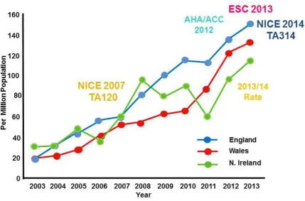
Over the last decade several hundred thousand CRTs have been implanted worldwide.19 In
2013 the UK was the fourth highest total CRT implanter in Western Europe.59 Figure 1.3
demonstrates the increasing CRT implantation year-on-year in the UK, over the last 10
years. Scotland’s national implantation figures are not presented as there was an
incomplete dataset provided to the national audit of cardiac rhythm management.59 These
figures represent a broadening of the international/national implantation guidelines as the
evidence has become more extensive and refined. Figure 1.3 compares the changes in the