http://wrap.warwick.ac.uk/
Original citation:
Valliant, Esther M., Romer, Frederik, Wang, Daming, McPhail, David S., Smith, Mark E.,
Hanna, John V. and Jones, Julian R.. (2013) Bioactivity in silica/poly(γ-glutamic acid)
sol–gel hybrids through calcium chelation. Acta Biomaterialia, Volume 9 (Number 8). pp.
7662-7671.
Permanent WRAP url:
http://wrap.warwick.ac.uk/56415
Copyright and reuse:
The Warwick Research Archive Portal (WRAP) makes this work of researchers of the
University of Warwick available open access under the following conditions. Copyright ©
and all moral rights to the version of the paper presented here belong to the individual
author(s) and/or other copyright owners. To the extent reasonable and practicable the
material made available in WRAP has been checked for eligibility before being made
available.
Copies of full items can be used for personal research or study, educational, or
not-for-profit purposes without prior permission or charge. Provided that the authors, title and
full bibliographic details are credited, a hyperlink and/or URL is given for the original
metadata page and the content is not changed in any way.
Publisher’s statement:
© 2013 Acta Materialia Inc. Published by Elsevier Ltd. All rights reserved.
http://dx.doi.org/10.1016/j.actbio.2013.04.037
A note on versions:
The version presented in WRAP is the published version or, version of record, and may
be cited as it appears here.
Bioactivity in silica/poly(c-glutamic acid) sol–gel hybrids through
calcium chelation
Esther M. Valliant
a, Frederik Romer
b, Daming Wang
a, David S. McPhail
a, Mark E. Smith
b,c, John V. Hanna
b,
Julian R. Jones
a,⇑a
Department of Materials, Imperial College London, South Kensington Campus, London SW7 2AZ, UK
bDepartment of Physics, University of Warwick, Coventry CV4 7AL, UK c
Vice-Chancellor’s Office, University House, Lancaster University, Lancaster LA1 4YW, UK
a r t i c l e
i n f o
Article history:
Received 11 January 2013
Received in revised form 22 March 2013 Accepted 22 April 2013
Available online 28 April 2013
Keywords:
Bioactive Hybrid Sol–gel
Poly(c-glutamic acid) Composite
a b s t r a c t
Bioactive glasses and inorganic/organic hybrids have great potential as biomedical implant materials. Sol–gel hybrids with interpenetrating networks of silica and biodegradable polymers can combine the bioactive properties of a glass with the toughness of a polymer. However, traditional calcium sources such as calcium nitrate and calcium chloride are unsuitable for hybrids. In this study calcium was incor-porated by chelation to the polymer component. The calcium salt form of poly(c-glutamic acid) (cCaPGA) was synthesized for use as both a calcium source and as the biodegradable toughening component of the hybrids. Hybrids of 40 wt.%cCaPGA were successfully formed and had fine scale integration of Ca and Si ions, according to secondary ion mass spectrometry imaging, indicating a homogeneous distribution of organic and inorganic components.29Si magic angle spinning nuclear magnetic resonance data demon-strated that the network connectivity was unaltered with changing polymer molecular weight, as there was no perturbation to the overall Si speciation and silica network formation. Upon immersion in simu-lated body fluid a hydroxycarbonate apatite surface layer formed on the hybrids within 1 week. The poly-mer molecular weight (Mw30–120 kDa) affected the mechanical properties of the resulting hybrids, but all hybrids had large strains to failure, >26%, and compressive strengths, in excess of 300 MPa. The large strain to failure values showed thatcCaPGA hybrids exhibited non-brittle behaviour whilst also incorpo-rating calcium. Thus calcium incorporation by chelation to the polymer component is justified as a novel approach in hybrids for biomedical materials.
Ó2013 Acta Materialia Inc. Published by Elsevier Ltd. All rights reserved.
1. Introduction
Bone regeneration therapies are needed that reduce the reliance on autograft procedures, in which harvesting of the bone can cause severe pain and complications at the donor site[1]. Acellular de-vices, such as synthetic scaffolds that can act as temporary tem-plates for bone regeneration, have potential, but development of a material that can fulfil all the criteria for a regenerative scaffold is a challenge[2–4]. Synthetic bioactive ceramic bone graft substi-tutes that can bond with bone and stimulate osteogenesis are cur-rently used in orthopaedic surgery. Examples are synthetic calcium phosphate-based ceramics, including tricalcium phosphate, e.g. Vi-toss (Orthovita, Malvern, PA), synthetic hydroxyapatite and bioac-tive glass powders, e.g. NovaBoneÒ (NovaBone Products, Jacksonville, FL) and BonAliveÒ (BonAlive, Turku, Finland)[5–7]. However, due to their brittle nature they are limited to non-load-bearing applications[8]. One of the greatest challenges is
develop-ing a bioactive material that has the required mechanical proper-ties and also degrades in a controlled and congruent manner.
One strategy for creating a tough, bioactive material is to com-bine bioactive particles into a polymer matrix to form a composite [9–11]. However, osteoblasts should attach preferentially to the bioactive phase[12], which may be masked by the polymer matrix as it is difficult to control how many bioactive particles are ex-posed at the surface. Also, it is difficult to match the degradation rate of the polymer matrix with that of the bioactive component, which may lead to non-congruent degradation of the composite.
Integrating organic and inorganic components at a finer scale may allow an improved cellular response, mechanical properties and closely matched degradation rates[13,14]. One technique has been to use organo-silica polymers such as polydimethylsiloxane (PDMS)[15], or another option is to form a hybrid from separate or-ganic and inoror-ganic starting materials. Hybrids have the potential to achieve fine scale interactions and improved properties through interpenetrating networks (IPNs) that allow the inorganic and or-ganic species to interact at the molecular level[16]. Through the sol–gel process hybrids can be formed by incorporating a polymer into the sol, allowing a silica network to form around the polymer
1742-7061/$ - see front matterÓ2013 Acta Materialia Inc. Published by Elsevier Ltd. All rights reserved.
http://dx.doi.org/10.1016/j.actbio.2013.04.037
⇑Corresponding author. Tel.: +44 2075946749.
E-mail address:[email protected](J.R. Jones).
Contents lists available atSciVerse ScienceDirect
Acta Biomaterialia
chains. Additional bonding between the components can be pro-vided by covalent coupling, creating a Class II hybrid material[13].
[image:3.595.344.532.581.731.2]Class II hybrids have been formed with organic polymers and sil-ica. Chitosan and gelatin have been successfully reacted with 3-glycidoxypropyltrimethoxysilane (GPTMS) to form hybrid materials [17,18]and porous structures[19,20]. Poly(c-glutamic acid) has also been functionalized with 3-aminopropyltriethoxysilane (APTES) [21]. The concentration of APTES or GPTMS dictated both the degree of covalent coupling and the silica content. To control the degree of covalent coupling between the inorganic and organic components independently of the organic/inorganic ratio tetraethyl orthosilicate (TEOS) was added as the silica precursor. This allowed GPTMS to act solely as a coupling agent in gelatin systems[16].
While poly(c-glutamic acid) has been shown to nucleate apatite in simulated body fluid (SBF)[22], the release of calcium ions from a hybrid should enhance the rate of bone bonding. Calcium ions and soluble silica should be released from hybrids to stimulate new bone growth[8]. However, it is not trivial to effectively incorporate calcium into a hybrid[23]. The traditional method for incorporation of calcium into the sol–gel process is to use calcium nitrate (Ca(NO3)2) as the calcium precursor[24]. The nitrate by-products are toxic to the body so sol–gel glasses are heated to a minimum stabilization temperature of 600°C to remove the nitrates[23,25]. This temperature will destroy polymer-containing materials, thus calcium nitrate is not a viable option for hybrid materials.
Calcium chloride has been used as a calcium source in several hybrids, including chitosan/GPTMS/silica[26], polyvinyl alcohol/ GPTMS/silica[27],
cPGA/GPTMS/silica
[28]andcPGA/APTES
[21]. However, the calcium was not incorporated into the silica network and recrystallized on the surface during drying of the hybrids [23,28]. The calcium was therefore rapidly released from the hy-brid materials upon immersion[28]. In other hybrids rapid calcium release led to reduced cell attachment[26], probably due to high dose exposure of calcium causing a pH burst. Calcium alkoxides are another calcium source for the sol–gel process[23]. This has been introduced into a poly(L-lactic acid)/silica system, but leadsto very short gelling times[29].
cPGA is a polypeptide which has a secondary amide in the
poly-mer backbone and is degradable by enzyme action[30]. A pendant carboxylic acid group in the repeating unit is available for function-alization.cPGA is a naturally occurring biopolymer that is best
known as the health food Natto in Japan[31]. It is also produced by a fermentation reaction[32–34]and the polymer has been clas-sified as ‘‘generally regarded as safe’’ by the US Food and Drug Administration [35]. Previous work by Poologasundarampillai et al. used the free acid form of poly(c-glutamic acid), which is only soluble in the organic solvent dimethylsulfoxide (DMSO) [28], however, the salt forms (e.g. Ca, Na and K salt forms) of the poly-mer are soluble in water[28].This work examines the calcium salt form of poly(c-glutamic acid) (cCaPGA) as the organic component of the hybrid and as a source for calcium incorporation into the hybrid at a relatively low temperature whilst keeping it available for bioactivity. At low temperatures, rather than trying to incorporate the calcium di-rectly in the silica network itself the calcium is coordinated to the polymer. This method of calcium incorporation may also provide further strengthening of the material by forming ionic crosslinking between the polymer chains.
2. Materials and methods
2.1. Materials
Poly(c-glutamic acid) in the free acid form (cHPGA) was pur-chased from Natto Biosciences (cosmetic grade). All other
chemi-cals were purchased from Sigma–Aldrich Co. Ltd. (Gillingham, Dorset, UK).
2.2. Preparation of
c
CaPGAThe first step was to synthesize the calcium salt form of
cPGA
from the free acid (cHPGA,Mw120 kDa).cHPGA was mixed with
deionized water to make a 20 wt.% slurry. Ca(OH)2was added to 45 mol.% and mixed for 1 h.cCaPGA was then ready to be
incorpo-rated into the sol–gel process and hybrid synthesis.2.3. Polymer chain scission
cHPGA was purchased from Natto Biosciences, but it was not
available in a variety ofMw. A chain scission reaction was carried out to obtaincCaPGA with a range of molecular weights that were
used as the starting organic component of the hybrid materials. An alkaline pH and elevated temperatures were used to reduce the molecular weight ofcCaPGA. A slurry was made of 50 g of
cHPGA
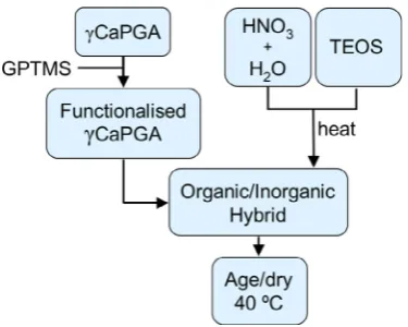
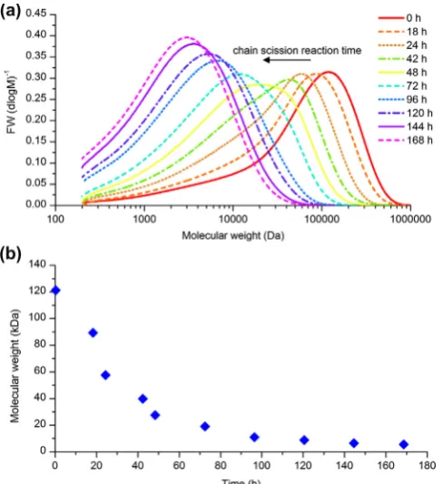
in 140 ml deionized water at 90°C, to which calcium hydroxide was added to 45 mol.%. After mixing for 1 h at 90°C, 1.86 g of so-dium hydroxide was dissolved in 10 ml of deionized water con-taining 0.05 wt.% sodium azide (bacteriocide). At 16, 20, 24, 42 and 48 h a portion of the solution was removed and the pH was immediately reduced to pH 4.8 with 50 vol.% HNO3. The unreacted polymer and chain scission products were submitted forMw anal-ysis by conventional gel permeation chromatography (GPC) by Smithers Rapra Technology Ltd (Shawbury, UK). Molecular weight distributions are shown in Fig. 2a. TheMwof the polymers de-creased with reaction time (Fig. 2b) and were found to be 120, 100, 80, 60, 40 and 30 kDa. ThesecCaPGA solutions were used
di-rectly in the hybrid synthesis process (Fig. 1), to which GPTMS was added.2.4. Synthesis of
c
CaPGA/silica hybridsClass II hybrids using the calcium salt form of
cPGA and silica
(40 wt.%cPGA, 60 wt.% SiO
2) were synthesized with varyingMw ofcCaPGA (Fig. 1). The final calcium content of the hybrids was
5 wt.% for a final composition of 44 wt.%cCaPGA and 56 wt.%
SiO2. GPTMS was added so that every tenth repeating unit ofcPGA
was functionalized and the solution mixed for 4 h. This degree of covalent coupling was chosen to give the best properties from ini-tial testing (see Appendix B), as the amount of GPTMS was found to affect both the dissolution and mechanical properties of the hy-brids. Meanwhile, the silica sol was prepared. TEOS was hydrolysed with anRratio (TEOS:water) of 26 in 0.016 M nitric acid for 30 minuntil clear. The small quantities of nitric acid were used to catalyse the hydrolysis reaction.
cCaPGA was not soluble in hydrolysed
TEOS solution ascPGA is very insoluble in ethanol. Since ethanol
was produced during the hydrolysis reaction of TEOS this ethanol had to be removed in order to combine the polymer with the inor-ganic sol. As ethanol was the component with the lowest boiling point it was removed from the hydrolysed TEOS by evaporation. The sol was heated at 70°C (boiling point of ethanol) for approxi-mately 1 h until 40% of the original aqueous volume had evapo-rated. The lost volume was replaced with deionized water to prevent premature gelation and to maintain theRratio. The silica solution was cooled to room temperature and added to the func-tionalized polymer solution. The hybrid sol was placed in sealed polystyrene moulds and aged for 3 days at 40°C, followed by dry-ing open to the atmosphere at 40°C.2.5. Characterization methodology
2.5.1. Immersion in SBF solution
Apatite formation was tested by immersing the hybrids in SBF and subsequently examining the dissolution profile, along with the formation of hydroxycarbonate apatite (HCA) on the surface of the material.
SBF was formed following the method described by Kokubo and Takadama[36]. Each sample was measured in triplicate, where a constant ratio of sample mass to SBF volume was used with agita-tion, as defined by Jones et al.[37]: 0.15 g of hybrid was immersed in 100 ml of SBF and mixed at 120 r.p.m. in an orbital shaker held at 37°C. Samples of 1 ml were removed at 1, 2, 4, 24, 72, 168 and 336 h and replaced by 1 ml of fresh SBF. The final solids were col-lected using filter paper (particle retention 5–13
lm), washed with
acetone and dried overnight at 40°C.The solids were then examined by X-ray diffraction (XRD) and the solutions analysed by inductively coupled plasma spectroscopy (ICP).
2.5.2. Immersion in Tris buffer solution
Tris(hydroxymethyl)aminomethane (Tris) buffer solution is a physiological pH solution which was used to measure dissolution in a solution that is free of salts (including Ca2+) [38]. The pH of 0.062 M Tris buffer was adjusted to pH 7.3 at 37°C with 2 N HCl [39]. 75 mg of hybrid was immersed in 50 ml of TRIS buffer solu-tion and mixed at 120 r.p.m. in an orbital shaker held at 37°C for 8, 24, 72, 168, 336 and 672 h. The solutions were passed through a filter paper (particle retention 5–13
lm) and the solids washed
with acetone and dried overnight at 40°C. Ca and Si concentrations in solution were determined by ICP with optical emission spec-trometry (OES) and thecPGA concentration was measured using
the bicinchoninic acid (BCA) assay.2.5.3. BCA assay
A modified protocol for the PierceÒBCA Protein Assay Kit (Ther-mo Scientific) was used to determine the
cPGA release profile.
Cal-ibration solutions were made usingcCaPGA in Tris buffer solution
at concentrations of 0, 50, 100, 200, 300, 500 and 750lg ml
1cCaPGA. 150
ll of the filtered supernatant solutions from the TRIS
immersion test were placed into each well of a 96-well microplate. This was followed by 150ll of working reagent (Reagent
A:Re-agent B 1:20) and then mixed. The well plate was sealed and placed in a water bath at 60°C for 30 min. It was removed, cooled to room temperature and the absorbance was immediately mea-sured using a Molecular Devices Spectra Max M5 plate reader at 562 nm. The calibration standards were measured in triplicate and repeated for each analysis.2.5.4. Composition analysis
Lithium metaborate fusion was carried out on the hybrid sam-ples, with 0.1 g of finely ground sample for analysis. The hybrid sample and 0.5 g of lithium metaborate were placed in the cruci-ble, and heated to 1400°C for 30 min. The melt was dissolved in 100 ml of 10% analytical grade HNO3. The solution was analysed by ICP.
2.5.5. ICP
ICP measurements were taken with a Thermo Scientific iCAP 6300 Duo ICP-OES analyser with an auto sampler. Samples eachwere diluted by a factor of 10 with analytical grade 2 M HNO3. Mixed standards of silicon, phosphorus and calcium were prepared at 0, 2, 5 and 20
lg ml
1for the calibration curve. Silicon and phosphorus were measured in the axial direction of the plasma flame, whereas calcium was measured in the radial direction.2.5.6. Scanning electron microscopy (SEM)
SEM was performed in a Leo 1525 with Gemini column with a gun voltage of 5 keV upon hybrid fracture surfaces coated with chromium.
2.5.7.29Si magic angle spinning nuclear magnetic resonance (MAS NMR) spectroscopy
29Si MAS NMR data were measured at ambient temperatures on
a Varian InfinityPlus 300 MHz (7.05 T) spectrometer operating at a Larmor frequency of 59.62 MHz. All29Si MAS NMR experiments were enabled using a Bruker 7 mm MAS probe spinning at 5 kHz. A 5.0
ls (45
°tip angle) pulse and 4 min recycle delay were used to facilitate quantitative study of the Si speciation by ensuring the29Si nuclear spins were fully relaxed. All29Si MAS NMR chem-ical shifts were referenced to kaolinite as a secondary solid refer-ence which was located at d 92 ppm relative to the primary IUPAC reference TMS atd0 ppm. The proportion of Q and T species in each sample was determined by fitting each resonance in the 29 [image:4.595.38.282.66.333.2]Si MAS NMR data using Gaussian peaks in the DMFIT simulation program[40,41].
Fig. 2.(a) Molecular weight distributions of chain scission reaction products as determined by conventional GPC and (b) averageMwas a function of reaction time,
2.5.8. Time of flight secondary ion mass spectrometry (TOF-SIMS)
SIMS analysis was carried out with a TOF-SIMS 5 time of flight instrument (ION-TOF GMbH, Münster, Germany). Hybrid samples were prepared by grinding and polishing to 1
lm. The sample
was sputtered for 1 s per scan with a C60+ beam at 10 keV (1.9 nA) to form a 250lm crater, after which an image was taken
with a 25 keV Bi+analysis beam (1.24 pA) at a resolution of 5lm. A
100100lm area in the crater centre was analysed to provide an
image of 256256 pixels for a total of 410 scans. A low energy 20 eV pulsed electron flood gun was used for charge compensation.2.5.9. XRD
XRD was performed in a PANalytical X’Pert Pro MPD with an X’Celerator detector. The radiation used was Ni-filtered CuKa at 40 kV, 40 mA. The diffraction was measured between 2h= 5°and 70°, with a 0.03°step size and a counting time of 25 ms for each step with a secondary graphite crystal monochromater. The sam-ple was ground and placed on an amorphous silicon disk.
2.5.10. Compression testing
Cylindrical monoliths (diameter 7 mm, height 2 mm) were compressed using a Zwick 1474 fitted with a 100 kN load cell at 0.5 mm min 1. The Young’s modulus, maximum strength and strain to failure were determined. This test was repeated eight times and the mean and standard deviation were reported.
3. Results and discussion
Molecular weight affects many of the properties of polymers, including strength and dissolution[42]. Smaller polymers are pre-ferred for use in implanted medical devices because molecules lar-ger than 50 kDa should not be released into the body as they have a limited ability to pass into and out of the vascular system[43]. Non-degradable polymers with molecular weights greater than 30 kDa may restrict filtration through the kidneys [43]. Thus a range of molecular weights of
cCaPGA from 30 to 120 kDa were
used in hybrid synthesis, as this represents molecular weights above and within this target range.3.1. Uniformity analysis
Hybrids of
cCaPGA and silica were successfully formed through
the sol–gel process usingcCaPGA with
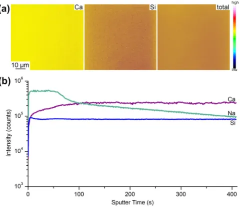
Mwof 30 to 120 kDa. All hy-brids were transparent (Fig. 3), amorphous and shrank in volume during drying by 85–90%. The composition was confirmed by lith-ium metaborate fusion, which found the calclith-ium content to be 4.4 ± 0.8 wt.%, compared to the theoretical value of 5 wt.%.The fine scale integration of the organic and inorganic compo-nents was confirmed by TOF-SIMS for hybrids made with 40 wt.% 120 kDa
cCaPGA (Fig. 4). Both the silicon and calcium
concentra-tion maps showed fairly uniform ion distribuconcentra-tions laterally for all the hybrids made with polymers of 30–120 kDa (data not shown). As calcium was coordinated to the polymer in the hybrids the cal-cium distribution also indicated the distribution of the polymer in the hybrid material. Since there are no silica- or polymer-rich do-mains in the hybrids these were true hybrid materials with the components present as interpenetrating networks.
From an examination of the ion concentration with depth there was a clear, defined surface layer, indicated by the initial high so-dium content (Fig. 4b). Once this surface layer was removed (130 s) the Si:Ca ratio was constant, and a strong sodium signal was also detected throughout the depth of the hybrids. This so-dium contamination could be due to handling of the samples, pol-ishing the samples to a smooth surface or preparation in the open atmosphere. Signal intensity does not convert directly to ion con-centration as the detector registers a stronger response to certain ions. Sodium was an expected background contaminant in SIMS analysis due to the high sensitivity of SIMS to the sodium ion. The constant Si:Ca ratio indicates a uniform distribution and fine scale integration of polymer and silica throughout the bulk of the hybrid.
3.2. NMR of the silicate network
[image:5.595.318.559.515.723.2]cCaPGA hybrids were also examined using solid state
29 Si MAS NMR, which indicated thatMwhad no effect on the structure of the silica network and the formation of bridging oxygen bonds (Fig. 5 andTable 1). AQnspecies is defined as a Si–O tetrahedral (4 coor-dinate) environment withnbridging oxygen bonds to other Si tet-rahedra in the glass network[44]. In contrast, Tn species have a silicon atom bonded to a carbon chain and n bridging oxygen bonds. The proportion ofQandTspecies in each sample was deter-mined by fitting using a series of Gaussian peaks in the DMFIT sim-ulation program [41]. From the intensities extracted for each species the distribution is shown to be very similar within the error (Table 1). The hybrids had a range ofQnspecies (Q4,Q3,Q2), which formed from condensation of the hydrolysed TEOS. The network modifier is likely to be H+, as the other cations were coordinated to the polymer. As the network connectivity was unchanged with different polymerMw, changes inMwdid not disrupt silica network formation. It is interesting to compare these observations withFig. 3.Hybrid of 40 wt.% 120 kDacCaPGA, where every tenth repeating unit of cPGA was functionalized with GPTMS.
[image:5.595.98.240.613.731.2]those of the network connectivity previously observed by29Si MAS NMR of sol–gel produced bioactive calcium silicates and, in partic-ular, Class II
cPGA/silica hybrids in which the organic/inorganic
content[28]was the same as used here. In their work Poologasun-darampillai et al. reported that theTnspecies detected were, as ex-pected, directly correlated with the GPTMS content. In terms of the GPTMS content used here, one GPTMS molecule to every 10 repeat-ing units ofcCaPGA, this was expected to have a total
Tnintensity of <4%, which was exactly what was observed here. TheQn distri-bution was also broadly in agreement with that observed in previ-ous studies, indicating that the silica network, although intimately mixed with the polymer here, is largely similar in its atomic scale connectivity with previously produced hybrids.As all hybrids were found to be uniform and homogeneous over the entire range of molecular weights, they were further tested by immersion in Tris and SBF buffer solutions.
3.3. Immersion in Tris buffer solution
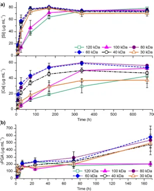
Hybrids were subjected to a dissolution study in Tris buffer solution to determine the effect of molecular weight on hybrid dis-solution. Tris was used as it was calcium free, so the release rate of calcium ions into solution could be observed. Upon immersion in Tris buffer solution all of the hybrids (cCaPGA Mw 30–120 kDa) had similar positive slope dissolution curves, with the majority of the ion release occurring in the first 3 days (Fig. 6a). Encourag-ingly, calcium was released from the hybrids, indicating that the chelation was reversible. Ion release was not proportional to molecular weight, as the hybrids synthesized with mid-range molecular weight
cCaPGA of 60 and 80 kDa had the highest total
calcium release, at 54 and 56
lg ml
1after 4 weeks. The polymer release profiles were similar for all of the hybrids up to 3 days, with approximately 35% (190lg ml
1) of the
cPGA dissolving in 8 h
(Fig. 6b). After 1 week the concentration ofcPGA released into
solution remained at 204lg ml
1 for hybrids made fromcPGA
withMwof 100 and 120 kDa. For all other hybrids the amount of polymer dissolved increased by300lg ml
1. Hybrids made with 100 and 120 kDa polymer also released soluble silica more slowly than the other hybrids (Fig. 6a). All of the other hybrids had reached their maximum silicon release of75lg ml
1by 3 days, whereas hybrids made with 100 and 120 kDa polymer had only re-leased 68% and 61% of this value by this time point. Thus hybrids synthesized from 120 and 100 kDa polymers had the slowest dis-solution in Tris, while hybrids made with 80 and 60 kDa polymer had the fastest.Dissolution of the hybrid upon immersion in Tris buffer solution was also monitored by SEM (Fig. 7). After 2 weeks the appearance of the hybrid fracture surface changed, becoming rougher, so that it looked like small closely packed spheres (Fig. 7c and e). The texture resembled a pure inorganic silica/calcium sol–gel glass, which had been synthesized and dried under similar conditions to the hybrid (Fig. 7f)[45]. By 3 days this change in appearance had occurred in all hybrids, except that made with 120 kDa
cCaPGA, in which not
all regions had changed (Fig. 7b). This suggests that the polymer was dissolving preferentially and the exposed silica network was observed by SEM. For use in a scaffold for bone tissue engineering the components must degrade slowly and at a similar rate. The rate of dissolution of thesecCaPGA hybrids must be slowed before they
can be used clinically.The hybrid made with the largest molecular weight (120 kDa) was the most stable in Tris buffer solution; it had the lowest total
cPGA release, the slowest soluble silica release and one of the
slowest Ca release rates. This was in contrast to the behaviour of the hybrids produced with mid-range Mw polymer of 80 and 60 kDa, which had the mostcPGA release and one of the highest
total Si and Ca ion releases. Thus decreasing the molecular weight did not cause a corresponding linear increase in mass loss in Tris.3.4. Bioactivity testing in SBF solution
SBF has an ionic content similar to blood plasma as it contains salts in similar concentrations. Tris buffer solution does not contain metal ions. Immersion studies in Tris buffer allow the examination of dissolution and ion release from the material (e.g. Ca2+) into a buffer solution without interference by ions in the solution. SBF is used to approximate how a solution containing physiological ion concentrations will interact with the material.
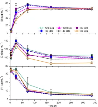
The effect ofMwon apatite deposition on the hybrids in SBF solution was examined (Fig. 8). Most of the hybrids followed sim-ilar concentration profiles with soluble silica release, phosphorus concentration decrease and an initial calcium release followed by a decrease in calcium concentration. Of particular note, the hybrid synthesized with polymer of the lowestMw(30 kDa) had the
slow-Fig. 5.29
Si MAS NMR showing the silica speciation for hybrids with 40 wt.% cCaPGA of varyingMw.
Table 1
Silicon speciation obtained by29
Si MAS NMR (relative intensityI) for hybrids made with 40 wt.%cCaPGA of varyingMwwhere average errors are ±2 ppm for the chemical shift (d)
and ±1% forI.
Mw(kDa) T2 T3 Q2 Q3 Q4
d(ppm) I (%) d(ppm) I (%) d(ppm) I (%) d(ppm) I (%) d(ppm) I (%)
120 –52.2 2 62.0 1 92.2 9 101.5 33 111.3 55
100 55.1 2 63.5 1 92.4 8 101.6 35 111.0 54
80 53.1 2 62.6 1 91.8 7 101.4 35 111.2 55
60 53.3 3 62.0 1 92.6 11 101.5 31 111.1 54
40 56.2 2 64.1 2 92.5 9 101.6 33 111.0 54
est kinetics of silicon release, whereas the hybrid formed from the polymer with the highestMw(120 kDa) had the fastest kinetics. In the calcium profile, the hybrid containing the polymer with the lowestMw(30 kDa) had the lowest final calcium concentration of
[image:7.595.146.455.64.448.2]88
lg ml
1 at 336 h and the hybrid made with the highest M w polymer (120 kDa) has the highest calcium concentration of 107lg ml
1 at 336 h. This could be due to finer integration be-tween the inorganic and organic components in the interpenetrat-Fig. 6.(a) Ca and Si profile in Tris buffer solution for up to 4 weeks and (b) polymer release profile for up to 1 week for hybrids of 40 wt.%cCaPGA of varyingMw. [image:7.595.143.459.486.666.2]ing network asMwdecreased. As the degree of coupling remained constant (every tenth repeating unit of the polymer), the total number of coupling units on each individual polymer chain was greater on the larger molecular weight polymers. This could lead to a more rigid structure that was more likely to fracture due to the stress of water absorption, and thus led to faster silicon release. However, this trend differed from the dissolution behaviour in Tris buffer solution, where the hybrids made from
cPGA with
molecu-lar weights of 60 and 80 kDa had the fastest silicon release.All hybrids produced a reduction in phosphorus concentration from 33
lg ml
1 in the initial SBF to 8lg ml
1 after 1 week immersion in SBF. When combined with a corresponding decrease in the calcium concentration, the decrease in phosphorus concen-tration indicated precipitation of calcium phosphate. HCA forma-tion was confirmed by XRD and SEM (Fig. 9). This indicates that the hybrids might be bioactive, even though they only contained 5 wt.% calcium, which was much lower than conventional bioac-tive glasses (17.5 wt.% Ca in the original BioglassÒ).3.5. Compression testing
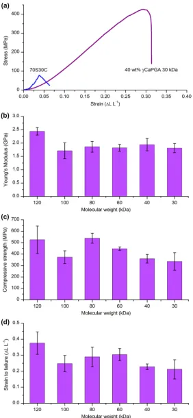
The fine scale integration of interpenetrating networks of silica and the polymer at the molecular level was expected to result in improved mechanical properties over bioactive glasses. Composite materials tend to fail at the interface between components. Thus removing this interface and having covalent coupling between the components should lead to improved mechanical properties. All stress–strain curves for the hybrids had a linear elastic region followed by slight plastic deformation before failure (Fig. 10a),
[image:8.595.135.450.69.414.2]reaching compressive strengths greater than 300 MPa and a strain to failure of >26% (Fig. 10c and d). This is a significant improvement over sol–gel glasses, which are quite brittle with a strain to failure of 4% and compressive strength of 66 MPa (70S30C sintered to 800°C). When comparing the stress–strain curves of a hybrid and sol–gel glass (Fig. 10a) there was a difference in the shapes of the curves as well as the ultimate stress and strain values. The hybrid had a gradually increasing slope at low strain values and a gradual decrease in slope just before failure, whereas the sol– Fig. 8.Si, Ca and P concentration profiles in SBF solution for up to 2 weeks for hybrids of 40 wt.%cCaPGA of varyingMw.
[image:8.595.307.548.566.722.2]gel glass only underwent linear elastic behaviour before failure. This deformation of the hybrids showed that incorporation of
cPGA
[image:9.595.161.444.64.676.2]into the sol–gel process had eliminated the brittle nature of the sol–gel glass.
Fig. 10.Compression testing for hybrids of 40 wt.%cCaPGA of varyingMw. (a) Sample stress–strain curve for 30 kDacPGA hybrid and a sample stress–strain curve for a
The maximum compressive strength of the
cCaPGA hybrids was
540 MPa for a hybrid synthesized withcCaPGA of 80 kDa. This was
a greater compressive strength than that of cortical bone at 190– 209 MPa, bearing in mind that cortical bone does contain some pores[46]. This same hybrid had a Young’s modulus of 1.9 GPa, which was an order of magnitude lower than that of cortical bone at 17.4 GPa[47]. The compression testing inFig. 10was carried out on hybrid monoliths, not on porous scaffolds. However, in bone tis-sue engineering applications a scaffold should be porous with interconnects large enough for vascularisation. The compressive strength will decrease once the hybrid materials have been formed into a porous scaffold.From examining the mechanical properties of hybrids made from polymers with varying molecular weights it could be seen that the strength of the hybrids generally increased as molecular weight increased (Fig. 10c). This left the hybrids made from 100 kDa
cPGA as an erroneous data point. To confirm this, the
hy-brid of 40 wt.%cCaPGA made with the 100 kDa polymer was
syn-thesized again and subjected to compression testing. The repeated test yielded the same mechanical properties, as they were equiva-lent within error (maximum compressive strength 377 ± 57 vs. 371 ± 59 MPa), which confirmed this as a true value and not simply synthesis error. The hybrid was synthesized with the 120 kDa polymer, as received from Natto Biosciences, but all of the lower molecular weight polymers (30–100 kDa) were the products of a chain scission reaction. This suggests that there was something inherent in the chain scission process that has adversely affected the mechanical properties of the resulting hybrids. Upon comparing the hybrids made with polymer from the chain scission reaction, those made with the 80 kDa polymer had the best (statis-tically significant atp< 0.05) mechanical properties. A lowerMw may encourage finer scale integration of the components in the hy-brid, but this was counterbalanced by the lower strength of the polymer as the Mwdecreased. However, the hybrids made with the 80 and 60 kDa polymers had the fastest dissolution in Tris and SBF. It is important to balance the polymer strength and integration.For all hybrids the strain to failure was >26%, which is a signif-icant improvement over sol–gel glasses and desirable for a cal-cium-containing material suitable for bone tissue engineering. However, the stability in solution must be increased in order for the
cCaPGA hybrids to be used in bone tissue engineering. This
work shows that calcium chelating polymers can be successfully incorporated into the sol–gel process as a calcium source to encourage bioactive behaviour.4. Conclusions
Calcium salt
cPGA was used as a calcium source and a glass
toughening agent. Hybrids of 40 wt.%cCaPGA were successfully
formed and found to be transparent and homogeneous. There was a uniform distribution of Ca and Si ions throughout the hy-brids, as measured by SIMS, which indicated a uniform distribution of the organic and inorganic components. The successful incorpo-ration of calcium aided the formation of HCA within 1 week in SBF. In Tris buffer solutioncPGA was found to dissolve
preferen-tially compared with silica for hybrids made with allMwpolymer. In compression the hybrids ofcCaPGA all had large strains to
fail-ure (>26%) (cPGAMw30–120 kDa), with a maximum compressive strength of 540 MPa and a strain to failure at 40% for the hybrids synthesized with 80 kDacPGA. This showed that
cCaPGA sol-gel
hybrids are of particular interest for bone tissue engineering as have non-brittle behaviour and encapsulate calcium.Acknowledgements
This research has been funded in part by the Natural Sciences and Engineering Research Council of Canada, Canadian Centennial Scholarship Fund UK, the Department of Materials, Imperial Col-lege London, the EPSRC (EP/E057098/1, EP/E051669/1, Challenging Engineering Grant EP/I020861/1) and the Philip Leverhulme Prize for Engineering. J.V.H. and M.E.S. acknowledge support for the so-lid-state NMR facilities at Warwick used in this research, which were funded by EPSRC and the University of Warwick. N.M.R. was also partially funded through the Birmingham Science City projects which were supported by Advantage West Midlands and the European Regional Development Fund. J.V.H. and M.E.S. acknowledge EPSRC support for FR via project EP/I004688/1.
Appendix A. Figures with essential colour discrimination
Certain figures in this article, particularly Figs. 1–4, 6 and 8–10 are difficult to interpret in black and white. The full colour images can be found in the on-line version, athttp://dx.doi.org/10.1016/ j.actbio.2013.04.037.
Appendix B. Supplementary data
The amount of covalent coupling was found to affect both the dissolution and mechanical properties of the silica/cCaPGA hybrids (Supplementary figures 1-3). As the amount of covalent coupling increased, from no coupling (no GPTMS) to coupling at every tenth repeating unit of the polymer (10 EC), the rate of dissolution of sil-ica, calcium and
cPGA decreased. As covalent coupling was
in-creased further, to bond at every other repeating unit of the polymer (2 EC), the rate of ion and polymer release increased. Thus covalent coupling at every tenth repeating unit of polymer was chosen to produce hybrids with the optimum mechanical and dis-solution properties. Supplementary data associated with this arti-cle can be found, in the online version, at http://dx.doi.org/ 10.1016/j.actbio.2013.04.037.References
[1]Sasso RC, LeHuec JC, Shaffrey C, The Spine Interbody Research Group. Iliac crest bone graft donor site pain after anterior lumbar interbody fusion: a prospective patient satisfaction outcome assessment. J Spinal Disord Tech 2005;18:S77–81.
[2]Jones JR. New trends in bioactive scaffolds: the importance of nanostructure. J Eur Ceram Soc 2009;29:1275–81.
[3]Jones JR. Review of bioactive glass: from Hench to hybrids. Acta Biomater 2013;9:4457–86.
[4]Hench LL, Polak JM. Third-generation biomedical materials. Science 2002;295:1014–7.
[5]Seebach C, Schultheiss J, Wilhelm K, Frank J, Henrich D. Comparison of six bone-graft substitutes regarding to cell seeding efficiency, metabolism and growth behaviour of human mesenchymal stem cells (MSC) in vitro. Injury 2010;41:731–8.
[6]Detsch R, Mayr H, Ziegler G. Formation of osteoclast-like cells on HA and TCP ceramics. Acta Biomater 2008;4:139–48.
[7]Ducheyne P, Qiu Q. Bioactive ceramics: the effect of surface reactivity on bone formation and bone cell function. Biomaterials 1999;20:2287–303. [8]Hench LL. The story of BioglassÒ
. J Mater Sci Mater Med 2006;17:967–78. [9]Li H, Chang J. PH-compensation effect of bioactive inorganic fillers on the
degradation of PLGA. Compos Sci Technol 2005;65:2226–32.
[10]Niemelä T, Niiranen H, Kellomäki M, Törmälä P. Self-reinforced composites of bioabsorbable polymer and bioactive glass with different bioactive glass contents. Part I: initial mechanical properties and bioactivity. Acta Biomater 2005;1:235–42.
[11]Rezwan K, Chen QZ, Blaker JJ, Boccaccini AR. Biodegradable and bioactive porous polymer/inorganic composite scaffolds for bone tissue engineering. Biomaterials 2006;27:3413–31.
[13]Novak BM. Hybrid nanocomposite materials – between inorganic glasses and organic polymers. Adv Mater 1993;5:422–33.
[14]Valliant EM, Jones JR. Softening bioactive glass for bone regeneration: sol–gel hybrid materials. Soft Matter 2011;7:5083–95.
[15]Tsuru K, Ohtsuki C, Osaka A, Iwamoto T, Mackenzie JD. Bioactivity of sol–gel derived organically modified silicates. J Mater Sci Mater Med 1997;8:157–61. [16]Mahony O, Tsigkou O, Ionescu C, Minelli C, Ling L, Hanly R, et al. Silica–gelatin hybrids with tailorable degradation and mechanical properties for tissue regeneration. Adv Funct Mater 2010;20:3835–45.
[17]Shirosaki Y, Tsuru K, Hayakawa S, Osaka A, Lopes MA, Santos JD, et al. Physical, chemical and in vitro biological profile of chitosan hybrid membrane as a function of organosiloxane concentration. Acta Biomater 2009;5:346–55. [18]Ren L, Tsuru K, Hayakawa S, Osaka A. Sol–gel preparation and in vitro
deposition of apatite on porous gelatin–siloxane hybrids. J Non-Cryst Solids 2001;285:116–22.
[19]Shirosaki Y, Okayama T, Tsuru K, Hayakawa S, Osaka A. Synthesis and cytocompatibility of porous chitosan–silicate hybrids for tissue engineering scaffold application. Chem Eng J 2008;137:122–8.
[20]Ren L, Tsuru K, Hayakawa S, Osaka A. Novel approach to fabricate porous gelatin–siloxane hybrids for bone tissue engineering. Biomaterials 2002;23:4765–73.
[21]Koh MY, Ohtsuki C, Miyazaki T. Modification of polyglutamic acid with silanol groups and calcium salts to induce calcification in a simulated body fluid. J Biomater Appl 2011;25:581–94.
[22]Sugino A, Miyazaki T, Ohtsuki C. Apatite-forming ability of polyglutamic acid hydrogels in a body-simulating environment. J Mater Sci Mater Med 2008;19:2269–74.
[23]Yu B, Turdean-Ionescu CA, Martin RA, Newport RJ, Hanna JV, Smith ME, et al. Effect of calcium source on structure and properties of sol–gel derived bioactive glasses. Langmuir 2012;28:17465–76.
[24]Jones JR, Ehrenfried LM, Hench LL. Optimising bioactive glass scaffolds for bone tissue engineering. Biomaterials 2006;27:964–73.
[25]Lin S, Ionescu C, Pike KJ, Smith ME, Jones JR. Nanostructure evolution and calcium distribution in sol–gel derived bioactive glass. J Mater Chem 2009;19:1276–82.
[26]Shirosaki Y, Tsuru K, Hayakawa S, Osaka A, Lopes MA, Santos JD, et al. In vitro cytocompatibility of MG63 cells on chitosan–organosiloxane hybrid membranes. Biomaterials 2005;26:485–93.
[27]Pereira MM, Jones JR, Orefice RL, Hench LL. Preparation of bioactive glass– polyvinyl alcohol hybrid foams by the sol–gel method. J Mater Sci Mater Med 2005;16:1045–50.
[28]Poologasundarampillai G, Ionescu C, Tsigkou O, Murugesan M, Hill RG, Stevens MM, et al. Synthesis of bioactive class II poly([gamma]-glutamic acid)/silica hybrids for bone regeneration. J Mater Chem 2010;20:8952–61.
[29]Poologasundarampillai G, Yu B, Jones JR, Kasuga T. Electrospun silica/PLLA hybrid materials for skeletal regeneration. Soft Matter 2011;7:10241–51. [30]King EC, Blacker AJ, Bugg TDH. Enzymatic breakdown of poly-c-D-glutamic
acid in Bacillus licheniformis: identification of a polyglutamyl c-hydrolase enzyme. Biomacromolecules 2000;1:75–83.
[31]Ho G, Ho T, Hsieh K, Su Y, Lin P, Yang J, et al. Gamma-Polyglutamic acid produced by Bacillus subtilis (natto): structural characteristics, chemical properties and biological functionalities. J Chin Chem Soc 2006;53:1363. [32]Schallmey M, Singh A, Ward OP. Developments in the use of Bacillus species
for industrial production. Can J Microbiol 2004;50:1–17.
[33]Inbaraj BS, Chien JT, Ho GH, Yang J, Chen BH. Equilibrium and kinetic studies on sorption of basic dyes by a natural biopolymer poly([gamma]-glutamic acid). Biochem Eng J 2006;31:204–15.
[34]Inbaraj BS, Chiu CP, Ho GH, Yang J, Chen BH. Effects of temperature and pH on adsorption of basic brown 1 by the bacterial biopolymer poly([gamma]-glutamic acid). Bioresour Technol 2008;99:1026–35.
[35] Lake LR. Carbohydrase and protease enzyme preparations derived from
Bacillus subtilisorBacillus amyloliquefaciens: affirmation of GRAS status as direct food ingredients. Department of Health and Human Services; 1999. p. 19887–19895.
[36]Kokubo T, Takadama H. How useful is SBF in predicting in vivo bone bioactivity? Biomaterials 2006;27:2907–15.
[37]Jones JR, Ehrenfried L, Saravanapavan P, Hench LL. Controlling ion release from bioactive glass foam scaffolds with antibacterial properties. J Mater Sci Mater Med 2006;17:989–96.
[38]Gomori G. Buffers in the range of pH 6.5–9.6. Proc Soc Exp Biol Med 1946;62:33–4.
[39]Brauer DS, Mneimne M, Hill RG. Fluoride-containing bioactive glasses: flouride loss during melting and ion release in tris buffer solution. J Non-Cryst Solids 2011;357:3328–33.
[40] De Angelis AA, Capitani D, Crescenzi V. Synthesis and13C CP-MAS NMR
characterization of a new chitosan-based polymeric network. Macromolecules 1998;31:1595–601.
[41]Massiot D, Fayon F, Capron M, King I, Le Calvé S, Alonso B, et al. Modelling one-and two-dimensional solid-state NMR spectra. Magn Reson Chem 2002;40:70–6.
[42]Bahadur P, Sastry NV. Principles of polymer science. New York: CRC Press; 2002. p. 135–40.
[43]Yamaoka T, Tabata Y, Ikada Y. Body distribution of intravenously administered gelatin with different molecular weights. J Control Release 1994;31:1–8.
[44]MacKenzie KJD, Smith ME. Multinuclear solid state NMR of inorganic materials. Oxford: Pergamon; 2002.
[45]Valliant EM, Turdean-Ionescu CA, Hanna JV, Smith ME, Jones JR. Role of pH and temperature on silica network formation and calcium incorporation into sol– gel derived bioactive glasses. J Mater Chem 2012;22:1613–9.
[46]Currey JD. Ontogenetic changes in compact bone material properties. In: Cowin SC, editor. Bone mechanics handbook. Boca Raton, FL: Informa Healthcare CRC Press; 2001. p. 19.5–9.