organic papers
o1486
Clegg and Jamieson C16H16N2S doi:10.1107/S160053680501264X Acta Cryst.(2005). E61, o1486–o1488 Acta Crystallographica Section E
Structure Reports
Online
ISSN 1600-5368
Pifithrin-
b
William Clegg* and Clare Jamieson
School of Natural Sciences (Chemistry), University of Newcastle upon Tyne, Newcastle upon Tyne NE1 7RU, England
Correspondence e-mail: [email protected]
Key indicators
Single-crystal X-ray study
T= 160 K
Mean(C–C) = 0.002 A˚ Disorder in main residue
Rfactor = 0.038
wRfactor = 0.103
Data-to-parameter ratio = 16.8
For details of how these key indicators were automatically derived from the article, see http://journals.iucr.org/e.
#2005 International Union of Crystallography
Printed in Great Britain – all rights reserved
The title compound, 2-p-tolyl-5,6,7,8-tetrahydrobenzo[d ]-imidazo[2,1-b]thiazole, C16H16N2S, is a condensation product
of pifithrin-, which has been previously reported as an inhibitor of the tumour suppressor protein p53. The molecule contains a planar fused pair of heterocyclic five-membered rings and the attached p-tolyl substituent is also essentially coplanar.
Comment
The compound pifithrin- [2-(2-imino-4,5,6,7-tetrahydro-benzothiazol-3-yl)-1-(4-methylphenyl)ethanone, (1)] was previously reported to inhibit,in vitro, a number of processes involving the tumour suppressor protein p53; it was thus of interest in the development of cancer therapies (Komarovet al., 1999). During a further evaluation of the effectiveness of pifithrin-, a crystalline sample was obtained and its structure investigated by X-ray diffraction. However, the material was found to be a condensation product of pifithrin-, from which water had been eliminated in a ring closure. The product, referred to as pifithrin-, (2), is more stable than pifithrin-in tissue culture medium. The revelation of this transformation through crystallographic identification of the condensation product has led to an expansion of the original evaluation of pifithrin-to include also pifithrin-, and the recognition that some of the inhibitory effects previously ascribed to pifithrin-are probably due instead to pifithrin-or a combination of the two compounds (Waltonet al., 2005).
The molecule of pifithrin- (Fig. 1) contains three fused rings with a p-tolyl substituent. The cyclohexene ring (or tetrahydrobenzo group) is disordered over two conformations, in which the two saturated C atoms furthest from the double bond lie one on each side of the mean plane of the other atoms of the ring [by 0.377 (4) and 0.375 (4) A˚ for the major component]; the two disorder components have opposite senses of twist for this CH2CH2 segment (see the torsion
angles in Table 1). The fused thiazole and imidazole rings are individually planar (r.m.s. deviations < 0.003 A˚ ) and form a
single planar unit [r.m.s. deviation 0.009 A˚ ; dihedral angle between the two five-membered rings = 1.32 (5)]. Such
imidazo[2,1-b]thiazole fused ring systems have been found in ten other crystal structures in the Cambridge Structural Database (Version 5.26 plus one update, February 2005; Allen, 2002), and they are all planar.
The benzene ring of thep-tolyl substituent makes a dihedral angle of 2.32 (5) with the imidazole ring to which it is attached. The whole molecule, excluding H atoms and the disordered CH2CH2linkage, is thus essentially planar, with an
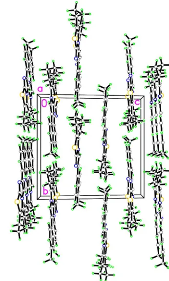
r.m.s. deviation of 0.039 A˚ . There are no notable inter-molecular interactions in the crystal structure. The molecules lie in almost planar sheets parallel to (001) (Fig. 2).
Experimental
The compound was prepared by a condensation reaction of , initially unintentionally during a study of anti-tumour agents, and subsequently by refluxing in aqueous methanol (Waltonet al., 2005). It was recrystallized from methanol.
Crystal data
C16H16N2S
Mr= 268.37
Monoclinic,P21=c a= 7.1729 (4) A˚ b= 13.5386 (8) A˚ c= 14.3530 (8) A˚
= 103.917 (2)
V= 1352.92 (13) A˚3 Z= 4
Dx= 1.318 Mg m 3
MoKradiation Cell parameters from 9390
reflections
= 2.1–28.5
= 0.23 mm1 T= 160 (2) K Block, colourless 0.440.300.26 mm
Data collection
Bruker SMART 1K CCD diffractometer Thin-slice!scans
Absorption correction: multi-scan (SADABS; Sheldrick, 2003) Tmin= 0.91,Tmax= 0.94
11454 measured reflections
3222 independent reflections 2871 reflections withI> 2(I) Rint= 0.022
max= 28.5
h=9!9 k=17!17 l=18!18
Refinement
Refinement onF2
R[F2> 2(F2)] = 0.038 wR(F2) = 0.103
S= 1.05 3222 reflections 192 parameters
H-atom parameters constrained
w= 1/[2(F
o2) + (0.0542P)2
+ 0.5147P]
whereP= (Fo2+ 2Fc2)/3
(/)max< 0.001 max= 0.32 e A˚
3 min=0.25 e A˚
3
Table 1
Selected geometric parameters (A˚ ,).
N1—C1 1.3949 (17) N1—C2 1.3147 (17) N2—C2 1.3666 (16) N2—C8 1.3991 (16) N2—C9 1.3770 (17)
S1—C2 1.7391 (13) S1—C3 1.7650 (14) C1—C9 1.3760 (18) C3—C8 1.3447 (19)
C1—N1—C2 103.49 (11) C2—N2—C8 115.28 (11) C2—N2—C9 106.49 (11) C8—N2—C9 138.21 (11) C2—S1—C3 90.36 (6) N1—C1—C9 111.16 (11) N1—C1—C10 121.52 (11) C9—C1—C10 127.32 (12) N1—C2—N2 113.56 (11)
N1—C2—S1 136.19 (10) N2—C2—S1 110.23 (10) S1—C3—C4 123.59 (10) S1—C3—C8 112.18 (10) C4—C3—C8 124.22 (13) N2—C8—C3 111.96 (11) N2—C8—C7 122.51 (11) C3—C8—C7 125.52 (12) N2—C9—C1 105.31 (11)
C3—C4—C5—C6 45.8 (3) C4—C5—C6—C7 64.3 (3) C3—C4—C5X—C6X 47.9 (8) C4—C5X—C6X—C7 61.1 (10)
C5X—C6X—C7—C8 41.1 (9) C5—C6—C7—C8 45.5 (3) N1—C1—C10—C15 1.16 (19) C9—C1—C10—C11 1.5 (2)
H atoms were positioned geometrically (C—H = 0.95–0.99 A˚ ) and refined with a riding model (including free rotation about the C—C bond for the methyl group), and withUisovalues constrained to be 1.2
organic papers
Acta Cryst.(2005). E61, o1486–o1488 Clegg and Jamieson C
[image:2.610.347.520.73.361.2]16H16N2S
o1487
Figure 1
The molecular structure of (2) with atom labels and 50% probability ellipsoids for non-H atoms. The minor disorder component has been omitted.
Figure 2
[image:2.610.46.298.73.268.2](1.5 for methyl groups) times Ueq of the carrier atom. Twofold
disorder was resolved and refined for the central CH2CH2linkage of
the cyclohexene ring, with occupancy factors 0.766 (6):0.234 (6). Data collection:SMART(Bruker, 2001); cell refinement:SAINT
(Bruker, 2001); data reduction: SAINT; program(s) used to solve structure: SHELXTL (Sheldrick, 2001); program(s) used to refine structure:SHELXTL; molecular graphics:SHELXTL; software used to prepare material for publication:SHELXTLand local programs.
We thank the EPSRC for financial support and Dr Ian Hardcastle for supplying the sample.
References
Allen, F. H. (2002).Acta Cryst.B58, 380–388.
Bruker (2001).SMARTandSAINT.Bruker AXS Inc., Madison, Wisconsin, USA.
Komarov, P. G., Komarova, E. A., Kondratov, R. V, Christov-Tselkov, K., Coon, J. S., Chernov, M. V. & Gudkov, A. V. (1999).Science,285, 1733– 1737.
Sheldrick, G. M. (2001).SHELXTL.Version 6. Bruker AXS Inc., Madison, Wisconsin, USA.
Sheldrick, G. M. (2003).SADABS.University of Go¨ttingen, Germany. Walton, M. I., Wilson, S. C., Hardcastle, I. R., Mirza, A. R. & Workman, P.
(2005).Mol. Cancer Ther. In the press.
organic papers
o1488
Clegg and Jamieson Csupporting information
sup-1
Acta Cryst. (2005). E61, o1486–o1488
supporting information
Acta Cryst. (2005). E61, o1486–o1488 [https://doi.org/10.1107/S160053680501264X]
Pifithrin-
β
William Clegg and Clare Jamieson
2-p-tolyl-5,6,7,8-tetrahydrobenzo[d]imidazo[2,1-b]thiazole
Crystal data
C16H16N2S
Mr = 268.37 Monoclinic, P21/c
a = 7.1729 (4) Å
b = 13.5386 (8) Å
c = 14.3530 (8) Å
β = 103.917 (2)°
V = 1352.92 (13) Å3
Z = 4
F(000) = 568
Dx = 1.318 Mg m−3
Mo Kα radiation, λ = 0.71073 Å Cell parameters from 9390 reflections
θ = 2.1–28.5°
µ = 0.23 mm−1
T = 160 K Block, colourless 0.44 × 0.30 × 0.26 mm
Data collection
Bruker SMART 1K CCD diffractometer
Radiation source: sealed tube Graphite monochromator
Detector resolution: 8.192 pixels mm-1
thin–slice ω scans
Absorption correction: multi-scan (SADABS; Sheldrick, 2003)
Tmin = 0.91, Tmax = 0.94
11454 measured reflections 3222 independent reflections 2871 reflections with I > 2σ(I)
Rint = 0.022
θmax = 28.5°, θmin = 2.1°
h = −9→9
k = −17→17
l = −18→18
Refinement
Refinement on F2
Least-squares matrix: full
R[F2 > 2σ(F2)] = 0.038
wR(F2) = 0.103
S = 1.05 3222 reflections 192 parameters 36 restraints
Primary atom site location: structure-invariant direct methods
Secondary atom site location: difference Fourier map
Hydrogen site location: inferred from neighbouring sites
H-atom parameters constrained
w = 1/[σ2(F
o2) + (0.0542P)2 + 0.5147P]
where P = (Fo2 + 2Fc2)/3
(Δ/σ)max < 0.001
Δρmax = 0.32 e Å−3
Δρmin = −0.25 e Å−3
Fractional atomic coordinates and isotropic or equivalent isotropic displacement parameters (Å2)
x y z Uiso*/Ueq Occ. (<1)
N1 0.58299 (16) 0.17620 (8) 0.14058 (8) 0.0257 (2)
N2 0.69620 (16) 0.02495 (8) 0.12171 (8) 0.0242 (2)
supporting information
sup-2
Acta Cryst. (2005). E61, o1486–o1488
C1 0.76869 (18) 0.18220 (10) 0.12686 (9) 0.0237 (3)
C2 0.54752 (18) 0.08082 (10) 0.13678 (9) 0.0242 (3)
C3 0.4914 (2) −0.10047 (10) 0.13244 (9) 0.0268 (3)
C4 0.4184 (2) −0.20417 (11) 0.13456 (11) 0.0326 (3)
H4A 0.3239 −0.2189 0.0736 0.039* 0.766 (6)
H4B 0.3535 −0.2114 0.1877 0.039* 0.766 (6)
H4X 0.2834 −0.2088 0.0971 0.039* 0.234 (6)
H4Y 0.4246 −0.2247 0.2014 0.039* 0.234 (6)
C5 0.5846 (3) −0.27551 (14) 0.1484 (2) 0.0333 (6) 0.766 (6)
H5A 0.5345 −0.3435 0.1347 0.040* 0.766 (6)
H5B 0.6590 −0.2732 0.2161 0.040* 0.766 (6)
C6 0.7167 (4) −0.25056 (15) 0.0826 (2) 0.0316 (6) 0.766 (6)
H6A 0.6409 −0.2503 0.0152 0.038* 0.766 (6)
H6B 0.8164 −0.3023 0.0890 0.038* 0.766 (6)
C5X 0.5584 (10) −0.2755 (5) 0.0866 (7) 0.035 (2) 0.234 (6)
H5X1 0.5311 −0.3456 0.0980 0.042* 0.234 (6)
H5X2 0.5273 −0.2647 0.0163 0.042* 0.234 (6)
C6X 0.7701 (13) −0.2555 (6) 0.1274 (8) 0.0367 (19) 0.234 (6)
H6X1 0.8478 −0.3011 0.0981 0.044* 0.234 (6)
H6X2 0.8033 −0.2670 0.1976 0.044* 0.234 (6)
C7 0.8147 (2) −0.14939 (10) 0.10651 (11) 0.0304 (3)
H7A 0.9181 −0.1539 0.1661 0.036* 0.766 (6)
H7B 0.8721 −0.1278 0.0537 0.036* 0.766 (6)
H7X 0.9395 −0.1306 0.1494 0.036* 0.234 (6)
H7Y 0.8282 −0.1451 0.0396 0.036* 0.234 (6)
C8 0.66567 (19) −0.07719 (9) 0.11945 (9) 0.0249 (3)
C9 0.84012 (19) 0.08993 (10) 0.11508 (10) 0.0258 (3)
H9 0.9626 0.0744 0.1046 0.031*
C10 0.86652 (18) 0.27755 (10) 0.12710 (9) 0.0241 (3)
C11 1.05548 (19) 0.28174 (10) 0.11660 (10) 0.0291 (3)
H11 1.1197 0.2224 0.1077 0.035*
C12 1.1500 (2) 0.37143 (11) 0.11911 (10) 0.0315 (3)
H12 1.2785 0.3722 0.1122 0.038*
C13 1.0616 (2) 0.46007 (10) 0.13146 (9) 0.0288 (3)
C14 0.8730 (2) 0.45609 (11) 0.14183 (10) 0.0317 (3)
H14 0.8092 0.5156 0.1507 0.038*
C15 0.7773 (2) 0.36671 (10) 0.13934 (10) 0.0294 (3)
H15 0.6487 0.3661 0.1461 0.035*
C16 1.1659 (2) 0.55676 (11) 0.13264 (11) 0.0360 (3)
H16A 1.3047 0.5455 0.1534 0.054*
H16B 1.1261 0.6024 0.1772 0.054*
H16C 1.1348 0.5854 0.0680 0.054*
Atomic displacement parameters (Å2)
U11 U22 U33 U12 U13 U23
N1 0.0233 (5) 0.0267 (5) 0.0276 (5) 0.0019 (4) 0.0071 (4) 0.0006 (4)
supporting information
sup-3
Acta Cryst. (2005). E61, o1486–o1488
S1 0.02331 (18) 0.02947 (19) 0.0411 (2) −0.00072 (12) 0.01156 (14) 0.00096 (14)
C1 0.0233 (6) 0.0262 (6) 0.0220 (6) 0.0019 (5) 0.0060 (5) 0.0012 (5)
C2 0.0214 (6) 0.0265 (6) 0.0253 (6) 0.0021 (5) 0.0069 (5) 0.0010 (5)
C3 0.0279 (6) 0.0256 (6) 0.0266 (6) 0.0007 (5) 0.0059 (5) 0.0017 (5)
C4 0.0337 (7) 0.0292 (7) 0.0348 (7) −0.0068 (6) 0.0081 (6) 0.0019 (6)
C5 0.0387 (11) 0.0231 (9) 0.0372 (16) −0.0024 (7) 0.0074 (9) 0.0046 (8)
C6 0.0373 (13) 0.0232 (9) 0.0350 (14) 0.0043 (8) 0.0098 (10) −0.0003 (10)
C5X 0.039 (4) 0.026 (3) 0.037 (5) 0.000 (2) 0.005 (3) 0.003 (3)
C6X 0.042 (4) 0.031 (3) 0.032 (4) 0.000 (3) 0.000 (3) −0.001 (3)
C7 0.0314 (7) 0.0255 (6) 0.0358 (7) 0.0029 (5) 0.0114 (6) 0.0013 (5)
C8 0.0285 (6) 0.0219 (6) 0.0244 (6) 0.0011 (5) 0.0064 (5) 0.0018 (5)
C9 0.0236 (6) 0.0249 (6) 0.0305 (6) 0.0013 (5) 0.0094 (5) 0.0019 (5)
C10 0.0250 (6) 0.0254 (6) 0.0215 (6) 0.0003 (5) 0.0048 (5) 0.0007 (5)
C11 0.0268 (7) 0.0293 (7) 0.0320 (7) 0.0014 (5) 0.0083 (5) −0.0032 (5)
C12 0.0249 (6) 0.0383 (8) 0.0319 (7) −0.0043 (5) 0.0079 (5) −0.0028 (6)
C13 0.0323 (7) 0.0305 (7) 0.0219 (6) −0.0070 (5) 0.0033 (5) −0.0005 (5)
C14 0.0337 (7) 0.0254 (7) 0.0368 (7) 0.0010 (5) 0.0101 (6) 0.0012 (5)
C15 0.0261 (7) 0.0270 (6) 0.0366 (7) 0.0014 (5) 0.0101 (5) 0.0017 (5)
C16 0.0391 (8) 0.0340 (7) 0.0332 (7) −0.0118 (6) 0.0053 (6) −0.0003 (6)
Geometric parameters (Å, º)
N1—C1 1.3949 (17) C5X—C6X 1.515 (12)
N1—C2 1.3147 (17) C6X—H6X1 0.990
N2—C2 1.3666 (16) C6X—H6X2 0.990
N2—C8 1.3991 (16) C6X—C7 1.518 (8)
N2—C9 1.3770 (17) C7—H7A 0.990
S1—C2 1.7391 (13) C7—H7B 0.990
S1—C3 1.7650 (14) C7—H7X 0.990
C1—C9 1.3760 (18) C7—H7Y 0.990
C1—C10 1.4689 (18) C7—C8 1.4926 (18)
C3—C4 1.5014 (19) C9—H9 0.950
C3—C8 1.3447 (19) C10—C11 1.4005 (18)
C4—H4A 0.990 C10—C15 1.3971 (18)
C4—H4B 0.990 C11—H11 0.950
C4—H4X 0.990 C11—C12 1.387 (2)
C4—H4Y 0.990 C12—H12 0.950
C4—C5 1.510 (2) C12—C13 1.389 (2)
C4—C5X 1.658 (7) C13—C14 1.397 (2)
C5—H5A 0.990 C13—C16 1.5061 (19)
C5—H5B 0.990 C14—H14 0.950
C5—C6 1.528 (3) C14—C15 1.388 (2)
C6—H6A 0.990 C15—H15 0.950
C6—H6B 0.990 C16—H16A 0.980
C6—C7 1.540 (3) C16—H16B 0.980
C5X—H5X1 0.990 C16—H16C 0.980
supporting information
sup-4
Acta Cryst. (2005). E61, o1486–o1488
C1—N1—C2 103.49 (11) C5X—C6X—C7 109.4 (7)
C2—N2—C8 115.28 (11) H6X1—C6X—H6X2 108.2
C2—N2—C9 106.49 (11) H6X1—C6X—C7 109.8
C8—N2—C9 138.21 (11) H6X2—C6X—C7 109.8
C2—S1—C3 90.36 (6) C6—C7—H7A 110.1
N1—C1—C9 111.16 (11) C6—C7—H7B 110.1
N1—C1—C10 121.52 (11) C6—C7—C8 107.94 (13)
C9—C1—C10 127.32 (12) C6X—C7—H7X 108.8
N1—C2—N2 113.56 (11) C6X—C7—H7Y 108.8
N1—C2—S1 136.19 (10) C6X—C7—C8 113.9 (3)
N2—C2—S1 110.23 (10) H7A—C7—H7B 108.4
S1—C3—C4 123.59 (10) H7A—C7—C8 110.1
S1—C3—C8 112.18 (10) H7B—C7—C8 110.1
C4—C3—C8 124.22 (13) H7X—C7—H7Y 107.7
C3—C4—H4A 109.8 H7X—C7—C8 108.8
C3—C4—H4B 109.8 H7Y—C7—C8 108.8
C3—C4—H4X 110.4 N2—C8—C3 111.96 (11)
C3—C4—H4Y 110.4 N2—C8—C7 122.51 (11)
C3—C4—C5 109.43 (13) C3—C8—C7 125.52 (12)
C3—C4—C5X 106.7 (2) N2—C9—C1 105.31 (11)
H4A—C4—H4B 108.2 N2—C9—H9 127.3
H4A—C4—C5 109.8 C1—C9—H9 127.3
H4B—C4—C5 109.8 C1—C10—C11 120.64 (12)
H4X—C4—H4Y 108.6 C1—C10—C15 121.77 (12)
H4X—C4—C5X 110.4 C11—C10—C15 117.59 (12)
H4Y—C4—C5X 110.4 C10—C11—H11 119.6
C4—C5—H5A 109.4 C10—C11—C12 120.84 (13)
C4—C5—H5B 109.4 H11—C11—C12 119.6
C4—C5—C6 111.4 (2) C11—C12—H12 119.2
H5A—C5—H5B 108.0 C11—C12—C13 121.61 (13)
H5A—C5—C6 109.4 H12—C12—C13 119.2
H5B—C5—C6 109.4 C12—C13—C14 117.66 (13)
C5—C6—H6A 109.2 C12—C13—C16 120.75 (14)
C5—C6—H6B 109.2 C14—C13—C16 121.58 (14)
C5—C6—C7 112.1 (2) C13—C14—H14 119.4
H6A—C6—H6B 107.9 C13—C14—C15 121.12 (13)
H6A—C6—C7 109.2 H14—C14—C15 119.4
H6B—C6—C7 109.2 C10—C15—C14 121.18 (13)
C4—C5X—H5X1 109.0 C10—C15—H15 119.4
C4—C5X—H5X2 109.0 C14—C15—H15 119.4
C4—C5X—C6X 112.7 (6) C13—C16—H16A 109.5
H5X1—C5X—H5X2 107.8 C13—C16—H16B 109.5
H5X1—C5X—C6X 109.0 C13—C16—H16C 109.5
H5X2—C5X—C6X 109.0 H16A—C16—H16B 109.5
C5X—C6X—H6X1 109.8 H16A—C16—H16C 109.5
C5X—C6X—H6X2 109.8 H16B—C16—H16C 109.5
supporting information
sup-5
Acta Cryst. (2005). E61, o1486–o1488
C2—N1—C1—C10 −179.28 (11) C2—N2—C8—C7 −178.23 (12)
C1—N1—C2—N2 0.03 (14) C9—N2—C8—C3 178.36 (15)
C1—N1—C2—S1 177.90 (12) C9—N2—C8—C7 −0.2 (2)
C8—N2—C2—N1 178.58 (11) C6—C7—C8—N2 −166.19 (16)
C8—N2—C2—S1 0.15 (14) C6—C7—C8—C3 15.5 (2)
C9—N2—C2—N1 −0.08 (15) C6X—C7—C8—N2 167.5 (5)
C9—N2—C2—S1 −178.51 (9) C6X—C7—C8—C3 −10.8 (5)
C3—S1—C2—N1 −178.32 (15) N1—C1—C9—N2 −0.08 (15)
C3—S1—C2—N2 −0.39 (10) C10—C1—C9—N2 179.18 (12)
C2—S1—C3—C4 179.36 (12) C2—N2—C9—C1 0.10 (14)
C2—S1—C3—C8 0.57 (11) C8—N2—C9—C1 −178.09 (14)
S1—C3—C4—C5 −162.66 (15) N1—C1—C10—C11 177.71 (12)
S1—C3—C4—C5X 164.7 (3) N1—C1—C10—C15 −1.16 (19)
C8—C3—C4—C5 16.0 (2) C9—C1—C10—C11 −1.5 (2)
C8—C3—C4—C5X −16.7 (4) C9—C1—C10—C15 179.65 (13)
C3—C4—C5—C6 −45.8 (3) C1—C10—C11—C12 −178.43 (12)
C4—C5—C6—C7 64.3 (3) C15—C10—C11—C12 0.5 (2)
C3—C4—C5X—C6X 47.9 (8) C10—C11—C12—C13 −0.4 (2)
C4—C5X—C6X—C7 −61.1 (10) C11—C12—C13—C14 0.3 (2)
C5X—C6X—C7—C8 41.1 (9) C11—C12—C13—C16 −179.17 (13)
C5—C6—C7—C8 −45.5 (3) C12—C13—C14—C15 −0.3 (2)
S1—C3—C8—N2 −0.59 (14) C16—C13—C14—C15 179.12 (13)
S1—C3—C8—C7 177.88 (11) C13—C14—C15—C10 0.5 (2)
C4—C3—C8—N2 −179.37 (12) C1—C10—C15—C14 178.36 (13)