Original Article
Suppression of the CXCL4-JNK pathways contributes to
the attenuation of DSS-induced acute colitis in mice
Jie Song, Yajie Hu, Yunguang Hu, Lei Guo, Jingjing Wang, Jiaqi Li, Yancui Wang, Ruotong Ning, Huiwen Zheng, Haijing Shi, Donghong Tang, Junjie Mei, Longding Liu
Institute of Medical Biology, Chinese Academy of Medical Science and Peking Union Medical College, Kunming, China
Received February 22, 2016; Accepted May 21, 2016; Epub July 1, 2016; Published July 15, 2016
Abstract: Inflammatory bowel diseases (IBD) are chronic disorders of the gastrointestinal tract with a complex ae
-tiology. Recently, the incidence of the disease has been on the rise and has received additional attention through -out the world. The chemokine CXCL4 was reported to induce directed cellular migration and was involved in the
development and progression of inflammatory diseases. Although it has been reported that CXCL4 expression is significantly increased in patients with IBD, the precise role and mechanism of CXCL4 in IBD remain obscure. This study aims to investigate the role of CXCL4 in dextran sulfate sodium (DSS)-induced acute colitis mouse model. The results showed that the serum level of CXCL4 was markedly increased in the acute colitis mice challenged
with DSS compared to the control group. Additionally, compared to the control group, the weight loss, survival time, disease activity, colorectal length, and pathological injuries were remarkably increased in the acute colitis mice;
moreover, the phosphorylation Jun N-terminal kinase (p-JNK) and the proinflammatory cytokines were significantly
up-regulated in the acute colitis mice compared to the control group. In contrast, the above symptoms were notably
ameliorated in the DSS-challenged CXCL4 knockout mice. Therefore, the results in the present study indicate that CXCL4 may have a crucial impact on the progression of the inflammatory response during acute colitis through JNK-mediated signalling, and it may be a candidate molecule for targeted therapy of IBD.
Keywords: Acute colitis, CXCL4, JNK, inflammatory cytokines
Introduction
Inflammatory bowel disease (IBD), which pri -marily includes Crohn’s disease (CD) and ulcer-ative colitis (UC), is a chronic inflammatory dis -order of the gastrointestinal tract that is char-acterized by alternating phases of the clinical manifestation, including as abdominal pain, vomiting, diarrhoea, rectal bleeding, and weight loss, relapse and remission [1]. Although the precise underlying etiologies and pathogenesis of IBD have not been fully elucidated, several reports have indicated that sustained intestinal infections, mucosal barrier defects, mucosal immune dysregulation, and genetic and envi-ronmental factors are involved in the disease process [2-4]. In recent years, the incidence and prevalence of the disease have persistent -ly increased. Moreover, there is still a lack of reliable and effective methods for diagnosing and monitoring this disease. Thus, it has
gradu-ally been appreciated as an important public health problem [5, 6]. Accumulating evidence has demonstrated that cytokines, surface mol -ecules, and signal transduction molecules co- uld play crucial roles in the initiation and pro-gression of IBD. In addition, numerous research -ers have been searching for a key molecule to identify, diagnose, monitor or even treat IBD [7-10].
modu-lation, the inflammatory response, vascular endothelial cell proliferation, haematopoiesis and angiogenesis [13]. The specific and coop -erative signals that regulate the CXCL4-mediated activation of different cell types will also be diverse. In neutrophils, the CXCL4-induced activation of c-Jun N-terminal kinase (JNK)/c-jun promotesstrong adhesion of neu -trophils to vascular endothelial cells. Additi- onally, in monocytes, CXCL4 mediated reactive oxygen species (ROS) formation by promoting the rapid activation of phosphatidyl inositol 3-kinase (PI3K), Src-tyrosine kinase (Syk), and p38 mitogen-activated protein (MAP) kinase. CXCL4 also promoted cell survival and cytokine release by increasing the activity of extracellu -lar signal-regulated kinase (Erk) and facilitating JNK activation, respectively [12]. Recently, mo- unting evidence has shown that CXCL4 expres-sion is dramatically elevated in colorectal can-cer patients, and it may be a tumour suppres-sor gene that inhibits the development and progression of colorectal cancer by recruiting leukocytes to the inflammation site, attracting pro- or anti-inflammatory cytokines, and res-tricting tumour cell growth, invasion and metas-tasis [14]. Moreover, it has been demonstrated that the plasma concentrations of CXCL4 were markedly elevated in patients with IBD [15]. Furthermore, an analysis of the serum protein spectrum showed that there was a positive cor-relation between the CD activity index and CXCL4 expression. CXCL4 was identified as a biomarker for IBD [15, 16]. Nevertheless, the function of CXCL4 in IBD is still unknown.
The JNK protein kinase, a member of the MAP kinase family, may contribute to multiple physi -ological processes, including the regulation of cell proliferation and apoptosis [17]. Although p38 MAP kinase, another MAP kinase, was thought to regulate an important primary sig-nalling pathway that mediates inflammatory signals in the past, recent studies have shown that the JNK pathway was also implicated in the inflammatory response, particularly in mono -cytes [12]. Moreover, it was revealed that the cytokines produced by activation of the JNK signalling pathway could, in turn, further trigger the JNK signalling pathway and ultimately lead to a cytokine storm that promotes the inflam -matory response [12]. Thus, it was presumed that the JNK signalling pathway may act in con-cert with the p38 MAP kinase pathway to
mod-ulate the inflammatory response in various inflammatory diseases. As described above, activated CXCL4 could also stimulate the JNK signalling pathway [18]. However, it is not known whether the CXCL4 and JNK interaction is involved in the progression of acute colitis.
In this study, acute colitis was induced in mice by administering DSS in the drinking water for five consecutive days. To further verify the role of CXCL4 in the progression of acute colitis, we compared the inflammatory and immune responses in CXCL4-/- and wild type mice. The
results demonstrated that the CXCL4-/- mice
were resistant to the development of acute coli -tis compared to the wild type mice.
Materials and methods
Animal groups and DSS challenge
All experiments were conducted under an ani-mal welfare protocol approved by the Institute of Medical Biology (IMB), Chinese Academy of Medicine Science (CAMS). Wild-type (WT) mice and heterozygous CXCL4 mice on the C57BL/6 background were provided by The Children’s Hospital of Philadelphia and were subsequently housed and bred in specific pathogen-free (SPF) cages. The animals were fed irradiated ch-ow and pure water. The homozygous CXCL4
-/-mice were obtained from the heterozygous CXCL4 mice by interbreeding and were identi-fied by PCR and ELISA (in press). Adult mice of the same sex and genotype were co-housed in groups of no more than 5 mice per SPF cage, with 12-hour light/dark cycles, constantly mon-itored temperature and humidity conditions. Husbandry was performed by the IMB staff. The WT mice and homozygous CXCL4 mice were randomly divided into the following 4 groups: WT+H2O group (n=3), WT+DSS group (n=5), CXCL4-/-+ H
2O group (n=3) and CXCL4-/-+
DSS group (n=5). Acute colitis was induced in the WT+DSS and CXCL4-/-+ DSS groups by
administering 3% (wt/vol) DSS (36-50 kDa, MP Biomedical, California, USA) dissolved in the drinking water for five consecutive days, fol -lowed by regular drinking water for four days, according to the method adopted by He et al [9]. The WT+H2O group and CXCL4-/-+ H
2O group
CXCL4 and cytokine levels. Subsequently, the colons were removed to measure the length (cm) from the rectum to the cecum, and then the colons were cut into two sections. One was used for the Western blotting analysis, and the other was fixed in 10% formalin for the histo -logical examination.
Detection of the serum levels of CXCL4
The serum levels of CXCL4 were measured using the CXCL4 test kit (SINO-American Biotechnology, China) in accordance with the manufacturer’s instructions. We first added 50 μl of the Standards and Samples to the appro -priate wells. The Blank well was empty. Then, we added 100 μl of horseradish peroxidase (HRP)-conjugated reagent to the Standard and Sample wells, but not the Blank well, and incu -bated them for 1 h at 37°C. After 4 rinses with the Wash Solution, chromogens A and B were successively added to each well and incubated for 15 min at 37°C in the dark. Subsequently, the Stop Solution was added to each well and the optical density (O.D.) was read at 450 nm within 15 min using a microtiter plate reader.
Body weights, survival rate and clinical assess-ment
The animals’ body weights were monitored every two days and then averaged. Additionally, the number of dead mice was recorded every two days. Nevertheless, the progression of acute colitis was clinically evaluated daily by measuring the volume of water consumed, measuring the body weights, evaluating the stool consistency, and observing the presence of blood in the faeces, as described previously. In addition, a previously validated clinical dis-ease activity index (DAI) was also calculated using the following parameters: stool consis -tency (0-4), presence or absence of blood in the faeces (0-4), and body weight loss (0-4). The maximum possible score is 12 [14].
Morphological observation and histopathologi-cal scoring
HE staining was applied to observe the patho-logical changes in the colon. The fixed tissues were embedded in paraffin and stained with haematoxylin and eosin (H&E). The mean degree of acute colitis in the colon was calcu -lated from observations of 20 different visual
fields of H&E-stained longitudinal sections of the colon from each animal. A histopathological score was obtained in a blinded fashion using a widely used grading tool to examine the crypts, epithelia, goblet cells, cellular infiltration, and edema. Scores of 0-1 reflect a normal morphol -ogy, and scores of 2-4, 5-7, and 8-10 represent mild, moderate, and severe colitis, respectively, as previously described [8].
Immunohistochemical examination
Induced nitric oxide synthase (iNOS) was used to detect the degree of inflammation in the colon. The longitudinal sections of the colon were deparaffinized with xylene and a gradient of alcohol. The deparaffinized sections were subjected to antigen retrieval by heating in a microwave for 15 min and were then treated with 3% H2O2 in methanol for 15 min at room temperature. Subsequently, the sections were blocked with 5% goat serum in PBS for 1 h at 37°C and incubated with a diluted anti-iNOS antibody (1:200, Boster, China) or PBS over -night at 4°C. Some sections were incubated with PBS as a negative control. After three washes in Tris-buffered saline (TBS), the sec -tions were incubated with an HRP-conjugated secondary antibody (ZSGB-Bio, China) for 1 hour at 37°C, followed by three washes in TBS. Then, the peroxidase activity was visualized using diaminobenzidine (DAB, TIANGEN, China). After two washes with water, the nuclei in the sections were stained with haematoxylin (Solarbio, China). Finally, the sections were washed once in water and sealed with neutral resin (Solarbio, China). An Olympus microscope (CX41, Japan) attached to a digital camera (DP71, Olympus, Japan) was used to photo-graph the slides at × 10 and × 40 magnifica -tions. The images were recorded with a com-puter-assisted video-imaging system (Olympus DP controller, Japan).
Western blot assay
reader. Equal amounts of protein (p-JNK: 40 μg/20μl; β-actin: 20μg/10μl) were separated by SDS-PAGE and transferred to polyvinylidene fluoride membranes (Milipore, USA). After blocking with TBST (Tris-buffered saline, pH 7.6, 0.05% Tween 20) containing 5% non-fat dried milk for 1 h, the membranes were incu-bated with antibodies specific for p-JNK (1:1000 dilution, Beyotime Biotechnology, China) and β-actin (1:2000 dilution, Boster, China) ove-rnight at 4°C. After 3 washes with TBST, the me-mbranes were incubated with an HRP-conju- gated secondary antibody (1:10000 dilution, Abmart, China) for 1 h and developed using an Enhanced Chemiluminescence Kit (Beyotime, China). The p-JNK levels were compared to those of β-actin based on the grey values calculated by Image J.
Detection of the plasma cytokine levels
The cytokine assays were performed using a Bio-Plex Mouse Cytokine 23-Plex Panel (1×96-well) (Bio-Rad, USA), according to the manufac -turer’s instructions and as previously described [19]. The plasma samples were diluted 1:4 with mouse sample diluent. The desired number of wells was pre-wet with 100 μl of Bio-Plex assay buffer. The multiplex bead working solution was vortexed for 15-20 s and 50 μl were pipetted into each well. Then, the buffer was immedi -ately removed using the Bio-Plex Pro Magnetic Wash Station. The wells were washed twice by
adding 100 μl of wash buffer. 50 μl of the dilut -ed standards or prepar-ed samples were add-ed to each well and the plate was incubated for 30 min. Then, the plate was washed three times with 100 μl of Bio-Plex wash buffer. 25 μl of vor -texed Bio-Plex Detection Antibody working solu -tion were added to each well, incubated for 30 min, and then washed three times with Bio-Plex wash buffer. 50 μl of vortexed 1× streptavidin-PE was added to each well, incubated for 30 min, and then washed three times with Bio-Plex wash buffer. The beads were resuspended in 125 μl assay buffer, incubated for 30 s, and then immediately read on the Bio-Plex 200 System. The cytokine concentrations were cal-culated using the standard curve and the Bio-Plex manager software. The samples were run in duplicate.
Statistical analysis
All data are expressed as the means ± S.E.M and were analysed using SPSS software (ver -sion 18.0). The serum CXCL4 levels, weight changes, disease activity scores, colorectal lengths, histopathological scores and Western blot data were analysed using one-way ANOVA, followed by the post hoc least significant differ -ence (LSD) test. Statistical significance was set at P<0.05.
Results
Serum CXCL4 levels
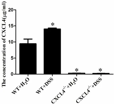
Several studies have reported that the CXCL4 levels were elevated in patients with IBD [15]. To investigate whether the trend in the CXCL4 levels in the DSS-induced acute colitis mice coincided with that in the IBD patients, we detected the serum levels of CXCL4 by ELISA. The concentration of serum CXCL4 in the WT+DSS group of mice was significantly increased compared to the WT+H2O group of mice (14.03±0.58 μg/ml vs. 9.44±2.95μg/ml,
P<0.05) (Figure 1). There was very little serum CXCL4 in the CXCL4-/-+ DSS and CXCL4-/-+ H
2O
groups of mice (Figure 1).
The effects of DSS-induced acute colitis on the animals’ body weights, survival rate and clini-cal symptoms
IBD, a chronic inflammatory disease of the gas -trointestinal tract, is characterized by abdomi-Figure 1. Detecting the concentration of CXCL4 in the
mice. The concentrations of CXCL4 were detected by
ELISA. The data represent the means ± SEM (n=3).
*P<0.05, compared to the WT+H
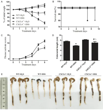
[image:4.612.91.288.71.251.2]nal pain, diarrhoea, weight loss and rectal bleeding [1]. Although the body weights of the WT+DSS and CXCL4-/-+ DSS mice progressively
decreased, the decrease in the body weights of the WT+DSS mice was significantly higher than
the CXCL4-/-+ DSS mice on days 5, 7 and 9
(Figure 2A). Moreover, the survival time of the WT+DSS mice is 2 days less than the CXCL4-/-+
[image:5.612.92.515.74.523.2]DSS mice (Figure 2B). In addition, the clinical symptom assessment in the WT+DSS mice Figure 2. DSS-induced colitis in the WT and CXCL4-/- mice. A. Changes in the body weights of the H
2O- and
DSS-challenged WT and CXCL4-/- mice over nine days of treatment. The data are expressed as the percentage of the
original weight prior to treatment. The data represent the means ± SEM (n=3). *P<0.05, compared to the WT+H 2O group. B. Survival rate of the H2O- and DSS-challenged WT and CXCL4-/- mice over the nine days of treatment. The data are expressed as the percentage of total mice in each group. C. Disease activity index of the WT and CXCL4 -/- mice over the nine days of treatment. The disease activity index was generated by evaluating the various pre
-sentations typically observed in the DSS-induced colitis model, including reduced mobility, vocalization, and group interactions. *P<0.05, compared to the WT+H
2O group. D. The colon lengths were measured from the anus to the
caecum. *P<0.05, compared to the WT+H
2O group; #<0.05, compared to the WT+DSS group; Δ<0.05, compared to
the CXCL4-/-+ H
using the DAI was significantly higher than the CXCL4-/-+ DSS mice on days 5, 7 and 9 (Figure
2C). Furthermore, the lengths of the colons from the WT+DSS mice were significantly short -er than those of the CXCL4-/-+ DSS mice (Fig-
ure 2D and 2E). Thus, these results suggested that the DSS-induced acute colitis in the WT+DSS mice was more intense compared to the CXCL4-/-+ DSS mice.
Morphological changes
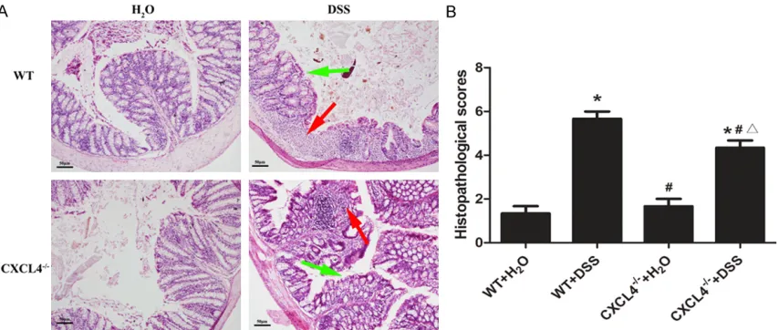
H&E staining revealed no obvious histopatho-logical abnormalities in the WT and CXCL4-/-
mice that were not challenged with DSS. However, histological damage was observed in the DSS-treated mice. H&E staining demon-strated that the epithelial destruction and inflammatory cell infiltration in the CXCL4-/-+
DSS mice was noticeably reduced compared to the WT+DSS mice (Figure 3A). The histopatho-logical scores demonstrated that the CXCL4-/-+
DSS mice develop a less severe inflammatory injury than the WT+DSS mice (Figure 3B). Hence, the results suggested that CXCL4 could aggravate acute colitis in mice.
The expression of iNOS in the colon
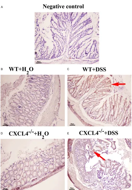
The immunohistological observations indicated that the WT mice produced more iNOS than the CXCL4-/- mice after the DSS treatment (Figure
4). Nevertheless, the level of iNOS expression
between the WT+H2O and CXCL4-/-+ H
2O groups
was not significantly different (Figure 4). iNOS is a marker of inflammation. Therefore, it was concluded that CXCL4 may promote the inflam -matory response during acute colitis in mice.
Level of the p-JNK protein in the colon
To determine the role of CXCL4 in the pathogen -esis of acute colitis, we examined whether the JNK signalling pathway, which is intimately associated with inflammation, was regulated by CXCL4. Western blotting was utilized to detect the levels of the p-JNK protein, the activated form of JNK. The p-JNK protein levels were significantly up-regulated in the WT+DSS mice compared to the WT+ H2O mice. Moreover, the DSS-treated CXCL4-/- mice exhibited a dramatic
increase in the p-JNK protein levels compared to the CXCL4-/-+ H
2O mice (Figure 5A). In
addi-tion, the expression of p-JNK in the WT+DSS mice was significantly higher than that in the CXCL4-/-+ DSS group (P<0.05) (Figure 5B).
Thus, these data illustrated that CXCL4 may exacerbate the development of acute colitis by activating JNK signalling.
Cytokines released in serum
[image:6.612.94.521.72.253.2]As part of a complex biological response, inflammation elicits the release of numerous inflammatory mediators, particularly cytokines [20]. Meanwhile, inflammation plays a major Figure 3. Histopathological changes in the colons of the WT and CXCL4-/- mice challenged with H
2O and DSS. A: Histopathological changes in the body weights of the H2O- and DSS-challenged WT and CXCL4-/- mice over the nine days of treatment. B: Average histopathological scores of the colons from the WT and CXCL4-/- mice challenged with
Figure 4. iNOS expression levels inthe WT and CXCL4-/- mice challenged with H
2O and DSS. The levels of iNOS in the colon tissues from the H2O- and DSS-challenged WT and CXCL4-/- were measured using immunohistochemistry. A: Negative control; B: WT+H2O; C: WT+DSS; D: CXCL4-/-+ H
role in the pathogenesis of colitis [21]. We dem -onstrated that the levels of proinflammatory cytokines were increased in the WT mice fol -lowing DSS challenge, but this response was more modest in the CXCL4-/-+ DSS group
com-pared to the WT group (Figure 6). Thus, these results further demonstrated that the inflam -matory process is reduced in the DSS-chall- enged CXCL4-deficient mice, and this trend
cated that CXCL4 could exacerbate the pro-gression of acute colitis by activating the JNK signalling pathway, which caused the subse-quent activation of downstream cytokines and led to an increased inflammatory response.
[image:8.612.93.522.74.205.2]The experimental DSS-induced murine colitis model is a well-established and consistent method used to investigate the development, Figure 5. p-JNK expression levels in the WT and CXCL4-/- mice challenged with H
2O and DSS. A: Western blot analysis of the expression of p-JNK and β-actin in the WT and CXCL4-/- mice challenged with H
2O and DSS. B: The level of p-JNK relative to β-actin in the four groups was calculated with Image J. *P<0.05, compared to the WT+H
2O group; #<0.05, compared to the WT+DSS group; Δ<0.05, compared to the CXCL4-/-+ H
2O group.
Figure 6. Cytokine expression profile in the serum from the WT and
CXCL4-/- mice challenged with H
2O and DSS. Cytokines, includingMCP-1,
GM-CSF, G-CSF, 9, 3, 13, 10, 6, 5, 4, TNF-a, IFN-g, 17,
IL-12 (p70), IL-IL-12 (p40), IL-2, IL-1², and IL-1±, were detected using a Bio-Plex Mouse Cytokine Panel. The coloured bar represents the fold change in the
cytokine levels relative to the WT+H2O group.
was similar to the p-JNK le- vels.
Discussion
[image:8.612.91.378.283.523.2]indi-progression and changes in acute colitis in the gut [23]. We adopted this colitis model and observed that the serum levels of CXCL4 were remarkably up-regulated. This result exhibited the same tendency as that found in Meuwis’ laboratory [15]. Thus, we further generated a CXCL4 knockout mouse to investigate the func -tion of CXCL4 during the progression of acute colitis. In the present study, we verified that the weight loss, survival time, disease activity and colorectal lengths of the DSS-treated CXCL4 knockout mice were notably decreased com-pared to the normal mice subjected to the same treatment. In the clinic, weight loss, sur-vival time, and disease activity are closely associated with the degree of IBD, and weight loss is one of clinical symptoms of IBD [24]. Therefore, these findings supported the hypoth -esis that CXCL4 could accelerate the develop-ment of acute colitis. In addition, it has been reported that there is a direct correlation between the outward signs of disease and the pathological changes in the gut tissue [23]. In our study, we also observed a disruption of the mucosal structures and a massive infiltration of inflammatory cells in the colons of the WT+DSS group, but these pathological injuries were ameliorated in the CXCL4-/-+ DSS group.
Moreover, iNOS was expressed at higher levels in the WT+DSS group than in the CXCL4-/-+ DSS
group. iNOS, a marker of inflammation, is capa -ble of synthesizing superoxides, particularly nitric oxide (NO), which has been recognized as a gaseous signalling molecule involved in the regulation of diverse physiological and patho -physiological mechanisms in the cardiovascu-lar, nervous and immune systems [25]. NO can act as a cytotoxic agent in inflammatory disor -ders, and the inhibition of iNOS may be a ben -eficial treatment for inflammatory disease [26]. Together, our results further suggested that CXCL4 exacerbated the inflammatory response in the DSS-treated mice.
Based on the above phenomenon, we analysed the pathway downstream of CXCL4 to elaborate the underlying mechanism of CXCL4 in acute colitis. The pathways involved in the CXCL4-mediated signal transduction cascade can result in the induction of a broad spectrum of acute and delayed functions, including phago -cytosis, a respiratory burst, survival, and the secretion of cytokines [12]. Moreover, it was
confirmed that the immediate CXCL4-stimula-ted monocyte functions (oxygen radical forma -tion) were regulated by p38 MAP kinase, Syk, and PI3K, whereas the delayed functions (sur -vival and cytokine expression) were controlled by Erk and JNK [27]. Additionally, the JNK-induced expression of pro-inflammatory cyto -kines could cause a cytokine storm, which dis-turbed homeostasis and provoked an inflam-matory response [28]. Furthermore, accumu-lating literature has revealed that the cytokine responses, including proinflammatory cyto -kines such as tumour necrosis factor-α (TNF-α), interferon-γ (IFN-γ), interleukin (IL)-1, IL-6, and IL-12 and anti-inflammatory cytokines such as IL-4, IL-10, and IL-11, are the key pathophysio-logic elements of IBD [20, 29]. Therefore, we examined the levels of the p-JNK protein and confirmed that the p-JNK levels were signifi -cantly increased in the WT+DSS group com-pared to the WT+H2O group, but the p-JNK lev-els were significantly reduced in the CXCL4-/-+
DSS group compared to the WT+DSS group. Furthermore, cytokine release, which is related to inflammation, was increased in the WT+DSS group compared to the CXCL4-/-+ DSS group.
These results implied that CXCL4 may activate JNK; subsequently, the JNK signal accelerated cytokine release. Following CXCL4 knockout, JNK activation and cytokines release were sup-pressed to a certain extent. Among all of the tested cytokines, the changes in IL-1α expres -sion were the most significant. Nevertheless, the binding of IL-1α to its receptor can rapidly initiate proinflammatory cytokine release by activating JNK [28]. Thus, we presumed that the increased cytokine release could, in turn, trigger additional JNK signalling and exacer-bate the inflammatory response.
Acknowledgements
This work was supported by National Natural Sciences Foundation of China (81373142 and 128001003), Important National Science & Technology Specific Projects (2014ZX09102-042) and Science and Technology Project of Yunnan Province (2012ZA009 and 2015GA- 010). The funders had no role in study design, data collection and analysis, decision to pub-lish, or preparation of the manuscript.
Disclosure of conflict of interest
None.
Address correspondence to: Drs. Longding Liu and
Junjie Mei, Institute of Medical Biology, Chinese Academy of Medical Science and Peking Union
Medical College, 935 Jiaoling Road, Kunming 650118, Yunnan, China. Tel: +86-871-68335905; Fax: +86-871-68334483; E-mail: longdingl@gmail. com (LDL); [email protected] (JJM)
References
[1] Kim H, Im JP, Kim JS, Kang JS and Lee WJ.
Alloferon Alleviates Dextran Sulfate Sodium-in -duced Colitis. Immune Netw 2015; 15: 135-141.
[2] Cosnes J, Gower-Rousseau C, Seksik P and
Cortot A. Epidemiology and natural history of inflammatory bowel diseases. Gastroenterolo -gy 2011; 140: 1785-1794.
[3] Loftus EV Jr. Clinical epidemiology of inflamma -tory bowel disease: Incidence, prevalence, and
environmental influences. Gastroenterology
2004; 126: 1504-1517.
[4] de Souza HS and Fiocchi C.
Immunopathogen-esis of IBD: current state of the art. Nat Rev
Gastroenterol Hepatol 2016; 13: 13-27. [5] Nielsen OH. New strategies for treatment of in
-flammatory bowel disease. Front Med (Laus -anne) 2014; 1: 3.
[6] Molodecky NA, Soon IS, Rabi DM, Ghali WA,
Ferris M, Chernoff G, Benchimol EI, Panac
-cione R, Ghosh S, Barkema HW and Kaplan GG. Increasing incidence and prevalence of the inflammatory bowel diseases with time,
based on systematic review. Gastroenterology 2012; 142: 46-54, e42; quiz e30.
[7] Xu Y, Hunt NH and Bao S. The role of granulo
-cyte macrophage-colony-stimulating factor in acute intestinal inflammation. Cell Res 2008;
18: 1220-1229.
[8] Chami B, Yeung AW, van Vreden C, King NJ and Bao S. The role of CXCR3 in DSS-induced coli -tis. PLoS One 2014; 9: e101622.
[9] Wei J, Wei C, Wang M, Qiu X, Li Y, Yuan Y, Jin C, Leng L, Wang J, Yang X and He F. The GTPase-activating protein GIT2 protects against colitis by negatively regulating Toll-like receptor sig-naling. Proc Natl Acad Sci U S A 2014; 111: 8883-8888.
[10] Radulovic K and Niess JH. CD69 is the crucial
regulator of intestinal inflammation: a new tar
-get molecule for IBD treatment? J Immunol
Res 2015; 2015: 497056.
[11] Deutsch E, Johnson SA and Seegers WH. Dif
-ferentiation of certain platelet factors related
to blood coagulation. Circ Res 1955; 3: 110-115.
[12] Kasper B and Petersen F. Molecular pathways of platelet factor 4/CXCL4 signaling. Eur J Cell Biol 2011; 90: 521-526.
[13] Van Raemdonck K, Van den Steen PE, Liekens
S, Van Damme J and Struyf S. CXCR3 ligands
in disease and therapy. Cytokine Growth Fac-tor Rev 2015; 26: 311-327.
[14] Vandercappellen J, Van Damme J and Struyf S. The role of the CXC chemokines platelet fac -tor-4 (CXCL4/PF-4) and its variant (CXCL4L1/
PF-4var) in inflammation, angiogenesis and
cancer. Cytokine Growth Factor Rev 2011; 22: 1-18.
[15] Meuwis MA, Fillet M, Geurts P, de Seny D,
Lut-teri L, Chapelle JP, Bours V, Wehenkel L, Be -laiche J, Malaise M, Louis E and Merville MP.
Biomarker discovery for inflammatory bowel disease, using proteomic serum profiling. Bio -chem Pharmacol 2007; 73: 1422-1433. [16] Takeyama H, Mizushima T, Iijima H, Shinichiro
S, Uemura M, Nishimura J, Hata T, Takemasa I, Yamamoto H, Doki Y and Mori M. Platelet Acti-vation Markers Are Associated with Crohn’s Disease Activity in Patients with Low C-Reac-tive Protein. Dig Dis Sci 2015; 60: 3418-3423. [17] Weston CR and Davis RJ. The JNK signal
trans-duction pathway. Curr Opin Cell Biol 2007; 19:
142-149.
[18] Kasper B, Brandt E, Ernst M and Petersen F.
Neutrophil adhesion to endothelial cells
in-duced by platelet factor 4 requires sequential activation of Ras, Syk, and JNK MAP kinases. Blood 2006; 107: 1768-1775.
[19] Sawin EA, De Wolfe TJ, Aktas B, Stroup BM,
Murali SG, Steele JL and Ney DM.
Glycomacro-peptide is a prebiotic that reduces Desulfovi
-brio bacteria, increases cecal short-chain fatty acids, and is anti-inflammatory in mice. Am J
Physiol Gastrointest Liver Physiol 2015; 309: G590-601.
[20] Neurath MF. Cytokines in inflammatory bowel
disease. Nat Rev Immunol 2014; 14: 329-342. [21] Baars JE, Nuij VJ, Oldenburg B, Kuipers EJ and
van der Woude CJ. Majority of patients with in
have mucosal inflammation. Inflamm Bowel
Dis 2012; 18: 1634-1640.
[22] Plevy S and Mayer L. Meeting summary: Signal
transduction pathways in immune and inflam -matory cells. November 30-December 3,
2000, Amelia Island, Florida, U.S.A. Inflamm Bowel Dis 2003; 9: 28-33.
[23] Kessler SP, Obery DR and de la Motte C. Hyal-uronan Synthase 3 Null Mice Exhibit
De-creased Intestinal Inflammation and Tissue
Damage in the DSS-Induced Colitis Model. Int
J Cell Biol 2015; 2015: 745237.
[24] Hendrickson BA, Gokhale R and Cho JH. Clini
-cal aspects and pathophysiology of inflamma -tory bowel disease. Clin Microbiol Rev 2002; 15: 79-94.
[25] Lirk P, Hoffmann G and Rieder J. Inducible ni
-tric oxide synthase--time for reappraisal. Curr Drug Targets Inflamm Allergy 2002; 1: 89-108.
[26] Forstermann U and Sessa WC. Nitric oxide
syn-thases: regulation and function. Eur Heart J
2012; 33: 829-837, 837a-837d.
[27] Kasper B, Winoto-Morbach S, Mittelstadt J, Brandt E, Schutze S and Petersen F. CXCL4-in -duced monocyte survival, cytokine expression,
and oxygen radical formation is regulated by
sphingosine kinase 1. Eur J Immunol 2010; 40: 1162-1173.
[28] Akira S, Uematsu S and Takeuchi O. Pathogen recognition and innate immunity. Cell 2006; 124: 783-801.
[29] Strober W and Fuss IJ. Proinflammatory cyto
-kines in the pathogenesis of inflammatory