Original Article
Role of sex hormone on morphological and histological
changes in benign prostatic hypertrophy rats
Renn Lovett, Michael Banta, Nidal Shkarni, Xeuying Chen, Suguru Nakamura
Department of Biological Sciences, Murray State University, Murray, KY, USA
Received December 8, 2016; Accepted September 8, 2017; Epub November 1, 2017; Published November 15, 2017
Abstract: Background: The prostate, the key secondary male reproductive organ, serves an important function of alkalizing seminal fluid and protecting genetic information in the acidity of the vaginal tract. As males age, the most common urologic condition manifests as an enlargement of the prostate known as benign prostatic hypertrophy (BPH). The purpose of this study is to examine the relationship between hormonal regulation and the morphologi-cal changes in BPH. Furthermore, we examine whether the ion-transport pump, H-K-ATPase (HKA), mediates such hormonal regulation. The experiments were designed to test the effects of the primary male androgen, testosterone propionate (TP), as well as the female hormone, estradiol (E2). Methods: The rats were divided into three groups; control group, TP group, and TP+E2 group. Both the TP and the E2 were diluted in vegetable oil and covered to eliminate light exposure. A subcutaneous injection of TP at 3 mg/mL was administered to induce BPH in each rat. After 6 weeks of TP-induced BPH, we divided these rats into two groups. In one group of BPH rats, we injected 60 µg of E2, and in another group of BPH rats, we injected 120 µg of E2 subcutaneously. The rats were sacrificed under anesthesia, and the prostate specimens were dissected. The rat’s body weight and the prostate tissue weight were measured as the organ quotient. Results: The data indicate significant hypertrophy of the luminal cells in rats with 3 mg TP compared to the control (524.542 ± 4.637 vs. 350.583 ± 1.996, P-value < 0.005). Whereas, the group with 60 µg E2 on TP-induced BPH showed significant inhibitory effects compared to TP-induced BPH (385.571 ± 7.265 vs. 524.542 ± 4.637, P-value < 0.005). The experimental group with 120 µg E2 on TP-induced BPH also showed significant inhibitory effects compared to TP-induced BPH (465.857 ± 8.259 vs. 524.542 ± 4.637, P-value < 0.005). The inhibitory effects of the 60 µg E2 group were more significant than the inhibitory effects of the 120 µg E2 group (385.571 ± 7.265 vs. 465.857 ± 8.259, P-value < 0.005), suggesting the importance of maintaining a proper E2:TP ratio. Western blot analysis shows up-regulation of specific bands for HKA alpha subunit at ~97 kDa for TP-induced BPH and down-regulation of HKA in the TP+E2 treatment groups. Conclusions: The results show that TP induces benign prostate hypertrophy. Whereas, E2 is shown to inhibit BPH; the effect of E2 inhibition on BPH requires the optimal ratio between E2 and TP. If such a ratio is not reached, then BPH inhibition will not occur or will be less effective by E2. Both the induction and inhibition of hypertrophic cells suggest that the prostate is under hormonal regulation. The proper E2:TP ratio plays a crucial role in the pathogenesis of BPH. The ratio of E2:TP may lead to new approaches to preventing and treating BPH disease in the future.
Keywords:Benign prostatic hypertrophy (BPH), H-K-ATPase, prostate, ATPase, hormonal regulation
Introduction
Encircling the first part of the urethra, just infe -rior to the bladder, is a walnut-sized organ of the male reproductive system: the prostate. The main function of the prostate is to secrete a milky fluid that is slightly alkaline and con -tains an androgen-regulated protein known as prostate-specific antigen (PSA). Being one of the most abundant prostate-derived proteins in seminal fluid, PSA is in charge of enhancing motility and protecting genetic information [1].
adenocarci-noma that typically manifests from epithelial cells of the peripheral zone; however, BPH and PCa can co-exist in the transition zone [4, 5]. Although there is no clear evidence regarding a molecular and genetic relationship between BPH and PCa, research suggests that the com-mon denominators of increased incidence with increased age, hormone dependence, and inci-dence with prostatic inflammation, make for the need of further research [6].
The etiology of BPH is poorly understood, mak-ing treatment and prevention complex. It is believed that aging and endocrine dysfunction of the hypothalamic-pituitary-gonadal axis con-tribute to the development of BPH. The primary androgen in males is testosterone. The pros-tate converts this testosterone into another androgen, dihydrotestosterone (DHT), by the enzyme 5-α-reductase, which is shown to have a 2-3 times greater androgen receptor affinity than testosterone itself [7] and facilitates cel-lular differentiation and proliferation of the prostate [8]. Furthermore, we see that luminal epithelial cells are the primary cells in which the androgen receptor is expressed [9]. Abn- ormal prostate hypertrophy occurs when there is a disruption of the androgen profile of the prostate, specifically acting on DHT and encour -aging proliferation of the tissue [5, 10].
However, proper estrogen levels may play more of a role in prostate growth and development than previously thought. Estrogens, specifically estradiol, play an important role in males, in- cluding regulation of gonadotropin feedback, bone maturation, and lipid metabolism. Tes- tosterone is one of the main factors influencing estrogen levels in men. Such changes in estro-gen-androgen equilibrium, which occur in mid- life, have been subject to study recently and are referred to as endropause [11]. One study revealed a decrease in serum testosterone with a relative rise in serum estradiol levels with advancing age in patients with BPH [12]. With the prostate being an androgen-depen-dent gland, there is further evidence suggest-ing that estrogen is necessary in growth and functional maintenance of the prostate [13-15]. The role of a proper androgen-estrogen ratio needs to be established to further under-stand the physiology and pathology of the human prostate.
The prostate consists of potassium-dependent
ed in the plasma membrane and function as cation pumps that transfer K+ into the cell in
exchange for Na+ or H+. Nongastric H-K-ATPase
(HKA) is composed of a catalytic α-subunit (αng) that performs ATP hydrolysis and ion translo- cation [16, 17]. Furthermore, ATP12A (former ATP1AL1) genes encode αng and interact str- ongly with Na-K-ATPase (NKA) β-isoform (β1) [7], which is crucial for structural and functional maturation. It is also important for modulation of enzymes’ affinities for cations thus making HKA an αng-β1 complex [16, 18, 19], meaning that HKA can mediate Na+/K+ transport and K+
homeostasis in vivo under K+ or Na+ deprived
conditions [18]. Of note, the function of ATP- 12A protein in the rodent prostate is that of acidification of the semen [7], which is impor -tant for the survival of semen in the highly acidic vaginal fluid. Furthermore, ATP12A null mice display loss of acidification and K+
absorp-tion in the prostate and the kidney [20-22]. Specifically, non-gastric HKA is required for acidification of luminal prostate fluids and is confined to the lumen-exposed surface of the epithelium [21, 23]. Furthermore, we see that such luminal epithelial cells are the primary cells in which the androgen receptor is expre- ssed [9] and that androgens promote down-regulation of NKA β1 [24], which may suggest an androgen-dependence of ATP12A expres-sion and function. It is important to see if androgen and estrogen dependence is a factor involved in regulation of non-gastric HKA ex- pression and derangement in the lateral lobes of the rat prostate. The purpose of the current experiment is to examine the relationship be- tween hormonal regulation and the morphologi-cal changes in BPH. Furthermore, we examine whether the ion-transport pump, HKA, medi-ates such hormonal regulation.
Methods
Animals
Male and female Sprague-Dawley outbred rats were obtained from Harlan® Laboratory, In-
dianapolis, Indiana. Animals were kept two rats per cage (one male, one female) with free access to food and water. The rats then mated at will and the males were separated from the females postpartum. After 3 weeks, the off-spring were separated from the mother and placed two per cage with free access to food and water. The rats were divided into three groups, including one control, one experimental with TP only, and one experimental with TP and E2.
Hormones
Preparation of testosterone propionate (TP): A 500 mL Erlenmeyer flask was filled with 1,500 mg TP (oil soluble: solid: C22H32O3). It was then covered with aluminum foil to avoid light exposure and filled with 500 mL of pure vegeta -ble oil to obtain a 3 mg/mL concentrated TP solution. The solution was then stirred for 48 hours.
Preparation of estradiol (E2): A 250 mL Erl- enmeyer flask was filled with 6 mg E2 (oil solu -ble: solid: C18H24O2). It was then covered with aluminum foil to avoid light exposure and filled with 100 mL of pure vegetable oil to obtain a 60 µg/mL concentrated E2 solution. The solu-tion was stirred for 48 hours.
Injections and time sequencing: Both the TP and E2 solutions were given subcutaneously (SQ) at a 45-degree angle in the adipose tissue of the rear distal flank. The solution was slightly warmed, and a restraint was used to ensure adequate administration. Needles (22 gauge, 1” length) and syringes (5 cm3) were replaced
after each injection. The rats were then marked on their tail to distinguish groupings.
Exactly 7 weeks after initial TP administration, the TP-induced group was sacrificed, and the prostates were dissected and defined as the TP group. The remaining rats were separated into two groups. The first grouping was adminis -tered 60 µg of E2 (TP+E2a), while the second
was administered 120 µg of E2 (TP+E2b). After
3 additional weeks under E2 regulation (60 µg vs. 120 µg), the TP+E2a and the TP+E2b groups
were sacrificed and the prostates were dissect
-ed and defin-ed as the TP+E2a (60 µg) and the
TP+E2b (120 µg) groups.
Euthanasia and prostatectomy
Rats were placed in a large container with a covered cotton ball soaked with isoflurane (inhalational anesthesia). After one minute of exposure to isoflurane, the rat was removed from the container and placed supine on a clean glass plate. The left thoracic cavity was palpated for location of the heart. An injec- tion of 1 mL sodium pentobarbital (euthanasia) was given via cardiac puncture at a 90-degree angle with aspiration to assess for heart punc-ture. Pressure was maintained over the injec-tion site until expiry was confirmed. Separate needles (20 gauge, 1” length) and syringes (5 cm3) were used for each rat. After euthanasia,
the rat’s total body mass was measured. The prostate was then removed, and the urethra and seminal vesicles were detached. The weight of the prostate was measured and divid-ed by the total weight of the rat to determine the organ quotient.
Histopathology
Histopathology methods were used to obtain prostate cell sections, which were placed on microscopic slides for further analysis. The cell images were taken at 100 × and 400 × magni-fication using a Nikon TE 300 and used to show hormonal effects on the prostate at the cellular level. A calibrated ocular micrometer eyepiece, combined with a stage micrometer, was used to take measurements of prostatic cell lengths in the lateral lobes of the prostate. Fixation and embedding
sections, which were then mounted on a glass microscope slide. After mounting, the tissue sections were stained using hematoxylin (base) and eosin (acid) (H&E). The final step was to permanently mount the sections under a cover-slip using Premount mounting resin. The tissue was then labeled and ready for microscopic examination.
SDS-PAGE & western blot
In order to separate the proteins, the sodium dodecyl sulfate polyacrylamide gel electropho-resis (SDS-PAGE) method was used. 100 µL sample with 100 µL of Laemmli’s sample buf-fer were mixed and incubated at room tempera-ture for 1 hr (50 µL 2-mercaptoethanol + 950 µL Laemmli’s sample buffer). The gel cassette was prepared by removing the tape at the bot-tom of the gel and assembling the mini protean tetra cell electrophoresis module. The assem-bly was filled with SDS running buffer up to the edge of the outer plate. The first well was load -ed with 20 µL standard protein sample, and small volumes of sample protein (20 µL) dis-solved in gel loading buffer were added to each individual well. The gel ran at 200 V for 40 min and was removed gently when finished.
While running the gel, a piece of polyvinylidene difluoride (PVDF) membrane was cut to the same size as the filter paper. Two plastic dishes were prepared-one filled with methanol and the other filled with Western blot transfer buffer. The membrane was incubated in the methanol for 15 sec and then equilibrated for 5 min in the transfer buffer. The transfer stack was assembled, and the whole tank was filled with Western blot transfer buffer. Ice and a stirrer were then placed in the tank, as well. The gel ran at 100 V and < 350 mA for 1 hr. To reduce nonspecific binding, the membranes were soaked in 10% skim milk (15 mL PBS + 1.5 g dry milk, pH 7.2) for 1 hr at room temperature and again overnight at 4°C. The membrane was incubated with primary antibody-anti-pro-ton pump/HKAα (anti-HKAα, code no.-D031-3, clone-1H9, subclass-Mouse IgG1, quantity-100 µg, concentration-1 mg/mL; Medical and Biological Laboratory Co., Ltd., Japan)-diluted with PBS (pH 7.2) containing 1% skim milk for 1 hr at room temperature. Aliquot 100 µL of anti-HKAα into 20 tubes, each 5 µL of antibody. The application was to put 5 µL anti-HKAα into 5 mL PBS + 1% skim milk. The membrane was then
washed with PBST (0.05% Tween-20 in PBS) 3 times for 5 min each time. Then the membrane was incubated with 1:10,000 horseradish per-oxidase (HRP)-conjugated anti-mouse IgG dilut-ed with 1% skim milk (in PBS, pH 7.2) for 1 hr at room temperature (20 mL PBS + 2 g milk + 2 µL secondary antibody). The membrane was was- hed with PBST 6 times for 5 min each time and incubated with chemiluminescence reagent for 1 min. The extra reagent was removed from the membrane by dabbing with a paper towel and sealed in a plastic wrap to avoid sensitive light. The membrane was exposed to an X-ray film in a dark room for 5 min and developed as usual (developer solution + water-fixer solution). Re-sultant band densities were analyzed using ImageJ® computer software and converted into
graphs comparing area densities using Mic- rosoft Excel®.
Statistics
The data is expressed as mean ± standard error where appropriate. For the statistical analysis of luminal cell size, the micrometer readings were measured, and the statistical analysis was performed. The Western blot analysis was performed using ImgaeJ soft- ware. Analysis of variance and t-test were used where appropriate to determine statistical sig-nificance. P-values < 0.05 were considered sta-tistically significant.
Results
The experimental groups of rats we selected were TP-induced and TP+E2-induced. All rats were under normal dietary conditions. The rats were then separated into four groups depend-ing on hormonal regulation: control, TP-induced, TP+E2a-induced, and TP+E2b-induced. Follow
the histopathology procedure (mentioned in the methods section). Cell size was measured by ocular and stage micrometer. An average and standard deviation of the total measure-ments per slide were calculated to determine hypertrophy in each tissue section. These results were then compared to the control group under no external hormonal administra- tion.
Organ quotient
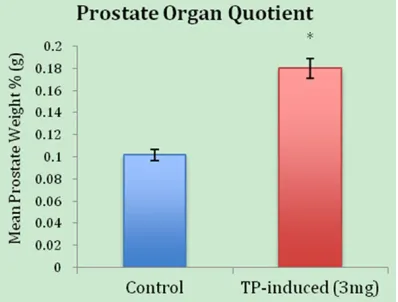
dis-sected prostate (after removal of the urethra and the seminal vesicles) by the total body weight of the rat (before dissection of the pros-tate) and multiplying by 100. In the rats, the OQ of the control group (no hormone treated) vs. the TP-induced (3 mg) BPH showed 0.102 ± 0.013 g (n = 6) vs. 0.180 ± 0.010 g (n = 5), P = 0.0006 (Table 1 and Figure 1). A statistical comparison of the OQ for the control group vs. the TP-induced BPH suggests that TP at a concentration of 3 mg/mL induces significant hypertrophy in overall organ size.
Cell size
An analysis of the micrometer measurements of cell length in the lateral lobe of the prostate examines the association of TP and E2 with BPH. In the rats, the epithelial cell sizes of the control group vs. the TP-induced (3 mg) BPH group were 350.583 ± 1.996 μm (n = 72) vs. 524.542 ± 4.637 μm (n = 83) (Figure 2). The results suggest that TP at a concentration of 3 mg/mL induces significant hypertrophy in cell size. The cell sizes of the rats treated with
conditions by E2a or E2b (Figure 3) shows the
inhibitory effects of the 60 µg E2 group were more significant than the inhibitory effects of the 120 µg E2 group (385.571 ± 7.265 vs. 465.857 ± 8.259, P-value < 0.005), suggesting the importance of maintaining a proper E2:TP ratio.
Western blot
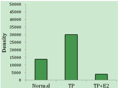
By using anti-HKAα primary antibody, we were able to detect bands at ~97 kDa in epithelial cells of the lateral lobe in the rodent prostate and the stomach (Figure 4). HKAα levels were increased (P < 0.005) by 2-fold at 5 min in the TP-induced treatment group. Furthermore, we observed HKAα levels decreased (P-value < 0.005) in the TP+E2-induced group as com-pared to the TP-induced group (Figures 5, 6).
Discussion
The development of BPH is related to particu- lar histological changes that take place in the prostate cells as a result of changes in the estrogen/androgen ratio. With increasing age, the serum level of estrogen/androgen in elderly men is normally 1/120-1/80 and, in extreme cases, the prostate can reach serum levels of 1/8 [25]. Therefore, a change in the E2:TP ratio could induce BPH. The results of this study col-laborate with the previous research findings. Relative prostate masses show that BPH of the lateral lobe was induced by 3 mg injections of TP after just 3 weeks. Significant hypertrophy was observed in the luminal cells of the lateral lobe of the 3 mg TP groups.
[image:5.612.91.372.84.154.2]The action of estrogens as regulators of the function and development of the prostate is significant. With the prostate being the model organ for androgen-dependent growth and de- velopment, estrogens are also responsible for regulating growth, development, and function through interactions with and maintenance of
Table 1. Effects of TP and E2 on organ and cell size
Hormonal Regulation Organ Quotient (g) Cell Size (μm) Control 0.102 ± 0.013 (n = 6) 350.583 ± 1.996 (n = 72) TP 0.180 ± 0.010 (n = 5) 524.542 ± 4.637 (n = 83)
TP+E2a -- 385.571 ± 7.265 (n = 14)
TP+E2b -- 465.857 ± 8.259 (n = 14)
Abbreviations are: TP, testosterone propionate; E2, estradiol. aTP = 3 mg, E2 = 60
μg; bTP = 3 mg, E2 = 120 μg.
Figure 1. Organ quotient (OQ) of the weight of the prostate compared to the total body weight of the rat (before dissection). The 3 mg/mL TP-induced BPH group yielded a 0.18% OQ compared to the control group at 0.10% (P-value = 0.0006).
TP+E2a (3 mg TP, 60 μg E2)
vs. TP+E2b (3 mg TP, 120 μg
[image:5.612.90.289.197.348.2]particular ratios of androgens [26]. Biologically, estrogens repress the hypothalamic-pituitary-gonadal axis, a process that has direct effects on the testes [26]. As a result, androgen
[image:6.612.92.522.72.335.2]pro-many different theories speculating the spe- cifics regarding hormonal deregulation (DHT hypothesis, embryonic reawakening theory, stem cell theory), further research is needed to Figure 2. Images of the cell size in the lateral lobe of the rodent prostate. Significant hypertrophy of the luminal cells is seen in the TP-induced (3 mg/mL) group (524.542 ± 4.637), while significant inhibition of luminal hypertrophy is seen in the both TP+E2-induced (E2a: 60 µg/mL vs. E2b: 120 µg/mL) groups (385.571 ± 7.265 vs. 465.857 ± 8.259, respectively). *A = 100 × zoom, B = 400 × zoom.
Figure 3. An analysis of the micrometer measurements of the cell length in the lateral lobe of each group shows that 3 mg/mL TP significantly induces cellular hypertrophy (P-value < 0.005), while the TP+E2 groups shows sig-nificant inhibition on TP-induced hypertrophy. Then, the ratio of the TP+E2a (3 mg TP, 60 μg E2) group shows better inhibitory effects on hypertrophy compared to the ratio of the TP+E2b (3 mg TP, 120 μg E2) group. This dem -onstrates the importance of maintaining a proper E2:TP ratio in control over prostate enlargement.
[image:6.612.93.372.412.570.2]determine the particular mechanism(s) of hor-monal imbalance.
In this study, significant findings were observed between the experimental groups and the con-trol group. Specifically, the rat prostate-body weight percentage, or the organ quotient (OQ), of the control was 0.10% vs. 0.18% in the TP-induced BPH group. Furthermore, the epi-thelial cell lengths show that TP-induced BPH (3 mg) increases the hypertrophy of the lateral prostate cells, confirming that androgens, in particular TP, and estrogens, in particular E2, are vital in the development of BPH.
The two groups of rats injected with both TP and E2 (E2a: 60 μg and E2b: 120 μg) 3 weeks
after TP-induced (3 mg) BPH showed a sig- nificant inhibition of BPH. It was observed that the lateral lobes of the TP+E2 groups were much smaller than the TP-induced BPH group, similar to that of the control group. Furthermore, E2 at a concentration of 60 μg (TP+E2a) showed
significantly more inhibition than the group with an E2 concentration of 120 μg (TP+E2b). Higher
E2 concentration achieves less inhibition and may actually induce BPH by potentially stimu-lating androgen receptors (AR). This suggests that not only is it pivotal in maintaining the balance of androgens and estrogens but, more specifically, the proper E2:TP ratio plays an important role in the pathogenesis of BPH. If an optimal ratio is not maintained, meaning a fluc -tuation in TP and/or E2, BPH is likely to occur. Our studies indicate that TP increases hyper- trophy of the luminal prostate cells and may demonstrate an alteration in enzymatic acti- vity of non-gastric HKA, consistent with results from Nakamura et al. [29]. According to Streif et al., cellular apoptosis is characterized in part by intracellular acidification, K+ loss and cell
shrinkage [7, 30]. HKA activation is important in antagonizing intracellular acidification and may attenuate cell apoptosis by countering K+
[image:7.612.91.283.74.245.2]loss and apoptotic cell shrinking [7]. HKA plays Figure 4. Protein expression of H-K-ATPase
[image:7.612.324.521.76.273.2]alpha-subunit in the lateral lobe of the transition zone in normal rodent prostate and stomach detected by (A) Western blot, and (B) band density measured by Im-ageJ software.
Figure 6. Protein expression of non-gastric H-K-ATPase alpha-subunit in the lateral lobe of the tran-sition zone in TP-induced and TP+E2-induced BPH rodent prostate detected by (A) Western blot, and (B) band density measured by ImageJ software.
[image:7.612.89.288.328.476.2]an important role in K+ homeostasis and
medi-ation of Na+/K+ transport as a α
ng-β1 complex
[16, 18]. In this study, we identified up regula -tion of HKAα (~97 kDa) in TP-induced BPH rats (Figure 6), which suggests that HKA mediates hormonal regulation and expression. Further, we identified down regulation of HKAα in the TP+E2-induced groups compared to the TP- induced BPH group. Such deranged expression pattern might be from an alteration in gene transcription.
While the study conducted shows valuable evi-dence of organ growth in TP-induced BPH, it was limited in some manners. Foremost, a larg-er sample size of rats would increase the confi -dence and decrease the uncertainty, especially in the TP+E2 groups. Also, fluctuating more dosages (5-10 each) of hormone variation wo- uld provide more insight on the specific E2:TP ratio. With this being said, future research needs to be done increasing the sample size and increasing the amount of hormonal varia-tion (both TP and E2) the rat receives. From there, further research should be conducted towards establishing an optimal ratio and a potential link between BPH and prostate can-cer at a molecular level.
Conclusion
Both BPH and carcinogenesis of prostate can-cer are accepted to be in the transition zone of the prostate. The cell images and measure-ments thus were taken from the lateral lobes of the transition zone in rodents, and TP-induced BPH was found to hold true through our experi-mental results. Rats who were injected with 3 mg/mL TP show a significant increase in pros -tate growth. Furthermore, epithelial cell lengths show that 3 mg/mL TP increases the hypertro-phy of the lateral prostate cells. Such a finding suggests that androgens serve a vital function in the development of BPH and potentially other prostate dysplasias, like PCa. Our studies indicate that TP-induced BPH demonstrates up regulation while the TP+E2-induced groupings demonstrate down regulation in enzymatic activity of H-K-ATPase, suggesting that HKA mediates hormonal regulation in the prostate. Furthermore, rats injected with 60 μg/mL E2 and 3 mg/mL TP (TP+E2a) show a significant
inhibition of BPH. Both induction and inhibition of hypertrophied cells suggest that the pros-tate is under hormonal regulation. With this
being said, the group injected with 120 μg/mL E2 and 3 mg/mL TP (TP+E2b) shows significant
inhibition of BPH, but significantly less inhibi -tion compared to the TP+E2a group. The proper
E2:TP ratio plays an important role in the patho-genesis of BPH. If the optimal ratio is not main-tained, it can lead to BPH and possibly other pathological conditions like PCa. Such knowl-edge of optimizing E2:TP in humans may help to prevent or cure BPH in the future.
Acknowledgements
These studies were supported by the Office of Research and Creative Activity (ORCA) pro-gram at Murray State University. We would like to thank Dr. Leon Duobinis-Gray (Murray State, Parasitology) for his help in identification of the lobes of the prostate.
Disclosure of conflict of interest
None.
Address correspondence to: Suguru Nakamura, Department of Biological Sciences, Murray State University, Murray, KY, USA. E-mail: snakamura@ murraystate.edu
References
[1] Crawford ED, DeAntoni EP, Ross CA. The role of prostate-specific antigen in the chemopreven -tion of prostate cancer. J Cell Biochem Suppl 1996; 25: 149-55.
[2] Prostate enlargement: benign prostatic hyper-plasia. National Institute of Diabetes and Di-gestive and Kidney Diseases 2014; 14: 1-20. [3] De Nunzio C, Kramer G, Marberger M,
Mon-tironi R, Nelson W, Schröder F, Sciarra A, Tuba-ro A. The contTuba-roversial relationship between benign prostatic hyperplasia and prostate can-cer: the role of inflammation. Eur Urol 2011; 60: 106-17.
[4] McNeal JE. Normal histology of the prostate. Am J Surg Pathol 1988; 12: 619-33.
[5] Miah S, Catto J. BPH and prostate cancer risk. Indian J Urol 2014; 30: 214-8.
[6] Alcaraz A, Hammerer P, Tubaro A, Schröder FH, Castro R. Is there evidence of a relationship between benign prostatic hyperplasia and prostate cancer? Findings of a literature re-view. Eur Urol 2009; 55: 864-73.
[8] Andriole GL, Bostwick DG, Brawley OW, et al. Effect of dutasteride on the risk of prostate cancer. N Engl J Med 2010; 362: 1192-202. [9] Banerjee PP, Banerjee S, Brown TR. Increased
androgen receptor expression correlates with development of age-dependent, lobe-specific spontaneous hyperplasia of the brown Norway rat prostate. Endocrinology 2001; 142: 4066-75.
[10] Dobrek L, Thor PJ. Benign prostatic hyperpla-sia-progress in pathophysiology and manage-ment. Pol Merkur Lekarski 2015; 39: 263-70. [11] Gillenwater JY, Grayhack JT, Howards SS,
Mi-chell ME. Adult and pediatric urology. 4 th edition. Philadelphia: Lippincott Williams & Wilkins; 2002.
[12] Tan MO, Karabiyik I, Uygur MC, Diker Y, Erol D. Serum concentrations of sex hormones in men with severe lower urinary tract symptoms and benign prostatic hyperplasia. Int Urol Nephrol 2003; 35: 357-63.
[13] Xiang-Yun L, Ying-Wen X, Chen-Jing X, Jiu-Jiu W, Qi P, Bo G, Zu-Yue S. Possible mechanism of benign prostatic hyperplasia induced by andro-gen-estrogen ratios in castrated rats. Indian J Pharmacol 2010; 42: 312-7.
[14] Pettersson K, Gustafsson JA. Role of estrogen receptor beta in estrogen action. Annu Rev Physiol 2001; 63: 165-92.
[15] Fujimoto N, Suzuki T, Honda H, Kitamura S. Es-trogen enhancement of androgen-responsive gene expression in hormone-induced hyperpla-sia in the ventral prostate of F344 rats. Cancer Sci 2004; 95: 711-5.
[16] Pestov NB, Korneenko TV, Radkov R, Zhao H, Shakhparonov MI, Modyanov NN. Identifica -tion of the beta-subunit for nongastric H-K-ATPase in rat anterior prostate. Am J Physiol Cell Physiol 2004; 286: C1229-37.
[17] Gumz ML, Lynch IJ, Greenlee MM, Cain BD, Wingo CS. The renal H+-K+-ATPases: physiolo-gy, regulation, and structure. Am J Physiol Re-nal Physiol 2010; 298: F12-21.
[18] Crambert G, Horisberger JD, Modyanov NN, Geering K. Human nongastric H+-K+-ATPase: transport properties of ATP1al1 assembled with different beta-subunits. Am J Physiol Cell Physiol 2002; 283: C305-14.
[19] Codina J, Delmas-Mata JT, DuBose TD Jr. The alpha-subunit of the colonic H+,K+-ATPase assembles with beta1-Na+,K+-ATPase in kid-ney and distal colon. J Biol Chem 1998; 273: 7894-9.
[20] Kone BC, Higham SC. A novel N-terminal splice variant of the rat H+-K+-ATPase alpha2 sub-unit. Cloning, functional expression, and renal adaptive response to chronic hypokalemia. J Biol Chem 1998; 273: 2543-52.
[21] Pestov NB, Korneenko TV, Shakhparonov MI, Shull GE, Modyanov NN. Loss of acidification of anterior prostate fluids in Atp12a-null mu -tant mice indicates that nongastric H-K-ATPase functions as proton pump in vivo. Am J Physiol Cell Physiol 2006; 291: C366-74.
[22] Greenlee MM, Lynch IJ, Gumz ML, Cain BD, Wingo CS. The renal H,K-ATPases. Curr Opin Nephrol Hypertens 2010; 19: 478-82.
[23] Pestov NB, Korneenko TV, Adams G, Tilleker-atne M, Shakhparonov MI, Modyanov NN. Non-gastric H-K-ATPase in rodent prostate: lobe-specific expression and apical localization. Am J Physiol Cell Physiol 2002; 282: C907-16. [24] Blok LJ, Chang GT, Steenbeek-Slotboom M,
van Weerden WM, Swarts HG, De Pont JJ, van Steenbrugge GJ, Brinkmann AO. Regulation of expression of Na+,K+-ATPase in androgen-de-pendent and androgen-indeandrogen-de-pendent prostate cancer. Br J Cancer 1999; 81: 28-36.
[25] Castro JE. Treatment of prostatic hypertrophy and neoplasia. New York: Springer Science and Business Media; 2013.
[26] Harkonen PL, Makela SI. Role of estrogens in development of prostate cancer. J Steroid Bio-chem Mol Biol 2004; 92: 297-305.
[27] Vermeulen A, Kaufman JM, Goemaere S, van Pottelberg I. Estradiol in elderly men. Aging Male 2002; 5: 98-102.
[28] Isaacs JT, Coffey DS. Etiology and disease pro-cess of benign prostatic hyperplasia. Prostate Suppl 1989; 2: 33-50.
[29] Nakamura S. H+-ATPase activity in selective disruption of H+-K+-ATPase alpha 1 gene of mice under normal and K-depleted conditions. J Lab Clin Med 2006; 147: 45-51.