Original Article
Comparison of tissue changes caused by microwave
and radio frequency energy in an experimental liver
ablation
Nguyen Quoc Vinh1,2, Tohru Tani3, Shigeyuki Naka1,3, Soichiro Tani1,3
1Department of Surgery, Shiga University of Medical Science, Seta-Tsukinowa, Otsu City, Shiga 520-2192, Japan; 2Department of General Surgery, University of Medicine and Pharmacy, Ho Chi Minh City, 217 Hong Bang District
5, Ho Chi Minh City, Vietnam; 3Biomedical Innovation Center, Shiga University of Medical Science, Seta-Tsukinowa,
Otsu City, Shiga 520-2192, Japan
Received November 11, 2015; Accepted January 12, 2016; Epub February 1, 2016; Published February 15, 2016
Abstract: Both radiofrequency ablation (RFA) and microwave ablation (MWA) techniques use high temperatures to ablate tissue but with different mechanisms. An understanding of differences in tissue reactions to these physical mechanisms may provide helpful information for practitioners and technical development. In the present study, we subjected rat liver to RFA and MWA under relatively equivalent conditions including power supply, application dura-tion, needle shape, and ablation maneuver. Comparisons were based on changes in tissue color, injury size, and mi-croscopic tissue changes immediately, 12, 24, and 72 h after ablation. The results revealed that the RFA and MWA induced similar changes in tissue color and injury diameter. However, in the central zone, hepatocyte shapes and nuclei were well maintained at each time point after MWA, whereas cellular disruption occurred soon after RFA. In the outer zone, intraregional cell survival and a more irregular burn edge were observed with RFA. AcP staining was useful for early disclosure of ablated zones and definition of non-viable cells in inner zone. In conclusions, MWA and RFA differed with regard to the central zone and peripheral areas with more regular ablated zone observed in MWA. These findings indicate the superiority of MWA over RFA for creating a more reliable and controllable ablation zone.
Keywords: Radiofrequency ablation, microwave ablation, thermal tissue injury, acid phosphatase staining, liver tumor ablation, histological change
Introduction
Over the last few decades, numerous local ablative techniques have been developed for the treatment of non-resectable hepatic tumors, which account for a large proportion of hepatic malignancies. Among these tech-niques, radiofrequency ablation (RFA) and microwave ablation (MWA) have generated the most interest and have been widely accepted as the techniques of choice for such malignan-cies [1, 2]. Both techniques incorporate high temperatures to achieve thermal tissue coagu-lation, but their heating mechanisms are quite different.
RFA uses energy from a radiofrequency current to heat tissue and thus relies on ionic agitation. An alternating electric current at a frequency of
200-1200 kHz passes through the body in a close circuit as a result of the ionic molecules present in tissues. Resistive heating occurs around the electrode according to the Joule effect [3-5]. This heating zone (i.e., direct heat-ing zone) is limited to a millimeter-scale range around the electrode. However, heat conduc-tion enables enlargement of the ablaconduc-tion area. Some technical problems associated with RFA include the increase in tissue impedance dur-ing ablation due to water evaporation at
tem-perature of ≥100°C and tissue charring at tem
-perature of >200°C [6]. In addition, the electric
current, which runs in areas of low resistance
(i.e., significant ionic component), bypasses
Comparison of MWA and RFA
MWA relies on a special tissue heating mecha-nism called dielectric heating, which occurs
when an alternating electromagnetic field is
applied to an imperfect dielectric material. In microwave-irradiated tissue, water and other polar molecules are forced to oscillate at the applied frequency, a process called dielectric hysteresis. Some of the electromagnetic ener-gy is absorbed and converted to heat [4, 7]. Because microwaves can propagate through non-conductive materials, they are not inhibit-ed by an increase in tissue impinhibit-edance during ablation, thus allowing a volume of tissue to receive uniform heating [8].
Numerous studies have demonstrated that both ablation methods can induce reliable cell death in the ablation zone and are effective for the treatment of liver tumors [2, 9-11]. Other
studies comparing MWA and RFA identified
aspects related to clinical applications, such as comparisons of the ablation volume, ablation zone border and shape, technical advantages and disadvantages, and treatment outcomes [4, 12-16]. In these comparisons, the differenc-es between MWA and RFA were a consequent
of the specific heating mechanisms. Andreano
et al. compared tissue heating patterns
obtained with microwaves and radiofrequency [17]. However, there is lack of comparative data regarding the tissue effects caused by these heating mechanisms.
In this study, we set up an experimental model in which MWA and RFA were performed under relatively equivalent conditions (e.g., same power output, ablation duration, needle shape, and ablation maneuver) to compare tissue damage and subsequent remodeling process-es of ablated regions. Tissue morphology was compared using a conventional hematoxylin and eosin (H&E) staining technique. In addition, acid phosphatase (AcP) staining, an effective method for characterizing early ablation zones, was used to compare alterations in enzymatic activity in the ablated areas.
Materials and methods
Microwave ablation and radiofrequency abla-tion devices
The MWA system comprises a microwave
gen-erator connected to an antenna via a flexible,
low-loss coaxial cable. The generator radiates microwave energy from a magnetron (Ma-
gnetron, Alfresa Ltd, Osaka, Japan) at a fre-quency of 2,450 MHz and can produce power at 1-100 W. The 15-cm 17-gauge needle-shaped antenna includes a coaxial dipole with a 1-cm radiating segment. The RFA device was a Cool-tipTM RF ablation system (Covidien, Boulder, CO, USA), which operates at a 480 KHz, with a controllable output power up to a maximum of 200 W. The RF needle is an inter-nally cooled single electrode (10 cm, 17-gauge, 2 cm exposure). During ablation, chilled saline
solution (5°C) was circulated through the nee -dle via a peristaltic pump.
Animal and ablation technique
Experiments were performed under the approv-al of the Committee on Animapprov-al Care. Ten-week-old male Sprague Dawley rats, weighing 280-300 g (CLEA Japan, Inc., Tokyo, Japan), were used for the experiment. Each rat was
anesthe-tized under general anesthesia via isoflurane
inhalation. A 3-cm midline incision was made in the upper abdomen to expose the external left lobe of the liver. RFA and MWA were performed using the same maneuvers on the external left lobe of the liver at two sites separated by a 2-cm distance to prevent overlapping injuries. MWA was performed at a power of 20 W and duration of 20 s. RFA was performed using a relatively equivalent amount of energy by main-taining the power at 20 W during 20 s RF activa-tion. Twelve rats in total were received the same MWA and RFA on their external left lobes of the liver. Following each application, the ablated zone was macroscopically evaluated and measured using a Vernier caliper. Each
series of three rats was sacrificed immediately
and 12, 24, and 72 h after ablation for sample collection. Each ablated lesion was divided
equally into two samples; one sample was fixed
in 10% formaldehyde and the other was stored
at -80°C for subsequent H&E and AcP staining,
respectively.
AcP histochemical staining
For AcP histochemical staining, a series of
8-μm-thick frozen sections were prepared using
a cryostat (Leica CM1900, Wetzlar, Germany). We used the diazonium salt coupling method with a naphthol AS series of phosphate esters, as previously described [18]. The incubation solution contained 40 mL of buffer (0.2 M CH3COOH) with 12 mg of naphthol AS-BI
N,N-dimethylformaldehyde, and 2 drops of 10% MgCl2. The sections were incubated in the
buf-fer at 37°C for 30 min. All slice images were
captured using a NanoZoomer (2.0-RSC10730-SG, Hamamatsu Photonics, Shizuoka, Japan).
Statistical analysis
The ablated zone diameters were summarized and expressed as mean ± standard deviations. The means were compared with the
[image:3.612.92.524.70.549.2]Comparison of MWA and RFA
dent sample t-test using the SPSS 20.0 statisti-cal software package (SPSS, Inc., Chicago, IL, USA). A p-value of <0.05 was considered a
sta-tistically significant difference.
Results
All rats tolerated the procedure and no compli-cations were encountered during the experi-ment. MWA and RFA induced quite similar mac-roscopic tissue changes. These changes appeared as three contiguous color zones: a brown central zone, pale/white intermediate zone, and purple peripheral rim, which formed a border with the intact liver. The mean surface diameters measured immediately after RFA
and MWA were 6.9 ± 0.3 and 6.7 ± 0.3 mm,
respectively. These values were not significant -ly different (P = 0.118). The ablated lesions expanded to average diameters of 9.1 ± 0.2 mm for RFA and 8.8 ± 0.2 mm for MWA at 24 h after ablation (P = 0.135).
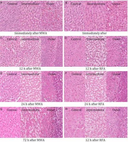
The comparison of microscopic findings follow -ing MWA and RFA was performed immediately and 12, 24, and 72 h after ablation. Immediately after ablation, no differences were observed between the lesions ablated via MWA and RFA with either H&E or AcP staining (Figures 1A, 1B and 2A, 2B). The central zone comprised cells that had maintained their structures but appeared slightly smaller and more
[image:4.612.89.525.71.469.2]ic than those in the intact liver (Figure 1A, 1B, left). The intermediate zone, which surrounded the central zone, comprised swollen hepato-cytes with blurred cell walls and/or nuclear loss (Figure 1A, 1B, middle). AcP activity was absent in the central and intermediate zones, indicat-ing that the cells were completely non-viable despite the normal shape of cells in the central zones. The outer zone, corresponding to the purple rim, was characterized by the dilation and congestion of sinusoids with abundant erythrocytes and the presence of intact
hepa-tocytes with distinct cell walls and normal nuclear morphology (Figure 1A, 1B, right). AcP activity was slightly attenuated in the outer zone compared with that in the intact liver (Figure 2A, 2B).
[image:5.612.93.523.71.512.2]However, the remodeling processes in the cen-tral zones induced by MWA and RFA evolved dif-ferently. After MWA, the cells in the central zone maintained their cell shapes, membranes, and nuclei until 72 h after ablation (Figure 1C, 1E and 1G, left). In contrast, cellular disruption
Comparison of MWA and RFA
was evident in some areas of the central zones of RFA samples at 12 h and disruption evolved progressively at 24 h. At 72 h, large number of
cells in this zone had liquefied and lost their cel -lular structures (Figure 1D, 1F and 1H, left). The intermediate and outer zones of the MWA and RFA lesions degenerated quickly in the
same manner. The major findings at 12 h
included interstitial edema, cell rupture and nuclear loss. More severe tissue degradation was observed at 24 and 72 h (Figure 1, mid-dle). At 12 h, the borderline between the ablat-ed and intact liver was well establishablat-ed by H&E staining in both MWA and RFA samples (Figure
1C, 1D, right). Neutrophils, macrophages, and
fibroblasts from the surrounding intact liver infiltrated the outer zones first and then to the
intermediate zone (Figure 1E-H, middle and right).
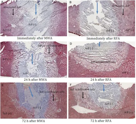
AcP activity was altered in the outer zones of both MWA and RFA lesions. In the outer zone hepatocytes, AcP activity present immediately after ablation had disappeared at subsequ- ent time points concomitantly with the
pres-ence of AcP activity in newly infiltrated cells.
[image:6.612.90.524.71.465.2]Consequently, a new pattern of positive AcP staining was observed beginning 24 h after ablation (Figure 2).
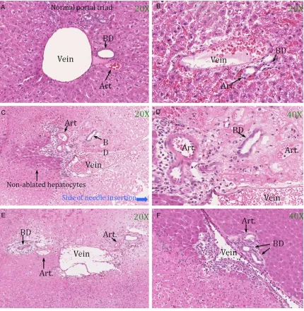
Assessment of vessel and bile duct injuries, Figure 3 shows the normal, immediately, 24, and 72 h after ablation hepatic portal triads (Figure 3A-E, respectively). Injuries of veins, arteries, and bile ducts occurred similarly fol-lowing MWA and RFA. Vessel injuries consisted of loss of endothelium causing extravasation of red blood cells, deformation or loss of nuclei, acidophilic cytoplasm, and sloughed off endo-thelium (Figure 3B-E). Injuries of bile duct cells were less severe with blurred cell wall and nor-mal or deformed cell nuclei. Structure of bile duct in the outer zones of ablated regions was preserved and no biloma was found in all sam-ples (Figure 3D, 3E). Neighboring ablated region bile ducts were well maintained their structure and cell shape (Figure 3F). The hepa-tocytes surrounding portal triad were complete-ly ablated in MWA samples whereas a number of non-ablated hepatocytes sometimes ap- peared on outer side of portal triad in RFA sam-ples (Figure 3C). The phenomenon was thought as a consequence of heat sink effect. In this
ablation model, the insufficiently ablated hepa -tocytes appeared beside vessels with diameter of 120 microns or larger and distance to the RFA needle of 3 mm or farther.
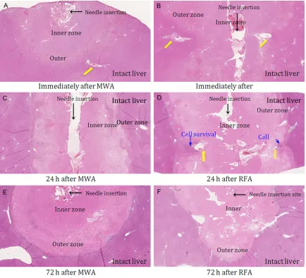
Observation at a low magnification (10×)
revealed differences in the margins and shapes of the regions ablated using MWA and RFA. MWA induced a sharp demarcation between the ablated and non-ablated regions, whereas RFA induced an irregular ablated margin (Figure 4A, 4C, 4E vs Figure 4B, 4D, 4F, respectively). Intralesional cell survival was sometimes observed in the outer zone of RFA samples, especially perivascular regions (Figure 4D). The presence of blood vessels in or around this zone affected the shape of the ablated region. This effect led to greater irregularity in the shape of RFA lesions compared with MWA lesions.
Discussion
AcP staining is reportedly useful for cell death detection and early delineation of ablated
regions, which are poorly defined by H&E stain -ing, particularly following MWA [18-20]. In this study, MWA and RFA induced similar alterations in AcP activity in the ablated region at every time point tested. Therefore, AcP staining allowed delineation of the lesions immediately
after ablation, but did not reveal any differenc-es between thdifferenc-ese two ablative techniqudifferenc-es. In addition, no differences between MWA and RFA in terms of macroscopic tissue changes and the ablation zone size were observed. However, differences in tissue changes were visible at a microscopic level. Tissue in RFA-induced region exhibited early damage and dis-ruption, whereas a portion of tissue around the microwave antenna comprised structurally sta-ble hepatocytes that resemsta-bled living cells until 3 day after ablation. Therefore, AcP
stain-ing is necessary for definstain-ing cell viability in the
MWA central zone. Although the cells in central zones of MWA and RFA lesions underwent dif-ferent remodeling processes, both were nega-tive for AcP activity indicating no evidence of viability.
In RFA lesions, the burned edge was less sharp-ly demarcated than that in MWA lesions, which were less affected byheat dissipation via blood vessels. These differences were potentially the
result of the specific heating mechanisms used
in each technique. Microwaves induce direct heating in a large volume of tissue around the antenna and the tissue within this volume is uniformly and consistently heated during micro-wave irradiation. This can propagate through tissue without depending on electric conductiv-ity [4, 7, 17, 21]. Consequently, tissue within the direct heating region undergoes homogenous cellular desiccation and protein coagulation, a
process known as microwave tissue fixation
[11, 19]. In contrast, tissue heating is inconsis-tent and inhomogeneous in RFA because the resistive heating induced by the electric current is modulated by the local tissue impedance and current density. Impedance depends on the ionic concentration of the tissue, which is attenuated quickly during RFA because of water vaporization, and the current density is inverse-ly proportional to the distance from the elec-trode. Furthermore, the tissue is primarily ablated by thermal conduction, which is
strong-ly influenced by the heat-sink effect [4].
In 1990, both McGahan et al. and Rossi et al.
independently published the first reports of
Comparison of MWA and RFA
has been wildly accepted during the past two decades, RFA currently remains the most popu-lar method of liver tumor ablation [6]. Comparison of MWA and RFA in both animal and clinical studies have revealed the superior-ity of MWA over RFA in terms of larger ablated zones, better lesion demarcation, reduced heat-sink effect, no limitations from tissue
charring, ability to exploit electromagnetic field
overlap and thus enhance heating by applying a multiple-antenna array, and no requirement for a ground pad to prevent the risk of skin burns [1, 4, 7, 12-16]. The key issue associated with MWA is the effective transmission of micro-wave power to tissues. Developments in the
field of MWA technology should focus on the
designs of microwave antennae and generators to address the major problems associated with
MWA such as microwave reflection, frequency
drift, and reduced generator effectiveness [21, 25].
In conclusion, tissue changes associated with
MWA included tissue fixation in the central
zone under direct heating and thermal conduc-tion with early tissue disrupconduc-tion in the outer
zone. In addition, MWA was less influenced by
heat-sink effect and could create a regularly shaped ablated lesion with a sharp burned edge. In contrast, the entire ablated region underwent a progressive damage process with RFA. The ablated lesion was irregularly shaped as a result of neighboring blood vessels and intralesional cell survival was sometimes evi-dent in the outer zone. This comparison revealed differences in tissue responses to the
resistive heating of RFA and magnetic field induction heating of MWA. These findings con -solidate the advantages of microwave technol-ogy relative to radiofrequency. MWA should therefore be completely developed to obtain the more reliable and controllable ablation zones and should be the recommended tech-nique option in future.
Disclosure of conflict of interest
None.
Address correspondence to: Dr. Nguyen Quoc Vinh, Department of Surgery, Shiga University of Medical Science, Seta-Tsukinowa, Otsu, Shiga 520-2192, Japan. Tel: 2238; Fax: +81-77-548-2240; E-mail: [email protected]
References
[1] Munireddy S, Katz S, Somasundar P and Espat NJ. Thermal tumor ablation therapy for colorec-tal cancer hepatic metastasis. J Gastrointest Oncol 2012; 3: 69-77.
[2] Crocetti L and Lencioni R. Thermal ablation of hepatocellular carcinoma. Cancer Imaging 2008; 8: 19-26.
[3] McDermott S and Gervais DA. Radiofrequency Ablation of Liver Tumors. Semin Interv Radiol 2013; 30: 49-55.
[4] Brace CL. Radiofrequency and Microwave Ab-lation of the Liver, Lung, Kidney, and Bone: What Are the Differences? Curr Probl Diagn Radiol 2009; 38: 135-143.
[5] Kim Y, Rhim H, Lim H, Choi D, Lee M and Park M. Coagulation Necrosis Induced by Radiofre-quency Ablation in the Liver: Histopathologic and Radiologic Review of Usual to Extremely Rare Changes. Radiographics 2011; 31: 377-390.
[6] Hong K and Georgiades C. Radiofrequency Ab-lation: Mechanism of Action and Devices. J Vasc Interv Radiol 2010; 21: S179-S186. [7] Simon CJ, Dupuy DE and Mayo-Smith WW.
Mi-crowave Ablation: Principles and Applications. Radiographics 2005; 25: S69-S83.
[8] Lubner MG, Brace CL, Hinshaw JL and Lee FT Jr. Microwave Tumor Ablation: Mechanism of Action, Clinical Results, and Devices. J Vasc In-terv Radiol 2010; 21: S192-S203.
[9] Goldberg S, Gazelle G, Compton C, Mueller P and Tanabe K. Treatment of intrahepatic ma-lignancy with radiofrequency ablation: radio-logic-pathologic correlation. Cancer 2000; 88: 2452-2463.
[10] Ohno T, Kawano K, Sasaki A, Aramaki M, Yo -shida T and Kitano S. Expansion of an ablated site and induction of apoptosis after micro-wave coagulation therapy in rat liver. J Hepato-biliary Pancreat Surg 2001; 8: 360-366. [11] Ozaki T, Mori I, Nakamura M, Utsunomiya H,
Tabuse K and Kakudo K. Microwave cell death: Immunohistochemical and enzyme histochem-ical evaluation. Pathol Int 2003; 53: 686-692. [12] Bhardwaj N, Strickland AD, Ahmad F, Atanesy-an L, West K Atanesy-and Lloyd DM. A comparative his-tological evaluation of the ablations produced by microwave, cryotherapy and radiofrequency in the liver. Pathology 2009; 41: 168-172. [13] Fan W, Li X, Zhang L, Jiang H and Zhang J.
Comparison of Microwave Ablation and Multi-polar Radiofrequency Ablation In Vivo Using Two Internally Cooled Probes. AJR Am J Roent-genol 2012; 198: W46-W50.
High-power Triaxial Antennas Create Larger Ab-lation Zones than Similarly Sized Internally Cooled Electrodes. J Vasc Interv Radiol 2009; 20: 1224-1229.
[15] Wright A, Sampson L, Warner T, Mahvi D and Lee F. Radiofrequency versus Microwave Abla-tion in a Hepatic Porcine Model. Radiology 2005; 236: 132-139.
[16] Zhang L, Wang N, Shen Q, Cheng W and Qian GJ. Therapeutic Efficacy of Percutaneous Ra -diofrequency Ablation versus Microwave Abla-tion for Hepatocellular Carcinoma. PLoS One 2013; 8: e76119.
[17] Andreano A and Brace C. A Comparison of Di-rect Heating During Radiofrequency and Micro-wave Ablation in Ex Vivo Liver. Cardiovasc Inter-vent Radiol 2013; 36: 505-511.
[18] Mukaisho K, Sugihara H, Tani T, Kurumi Y, Ka -mitani S, Tokugawa T and Hattori T. Effects of Microwave Irradiation on Rat Hepatic Tissue Evaluated by Enzyme Histochemistry for Acid Phosphatase. Dig Dis Sci 2002; 47: 376-379. [19] Yamaguchi T, Mukaisho K, Yamamoto H, Shio
-mi H, Kuru-mi Y, Sugihara H and Tani T. Disrup -tion of Erythrocytes Distinguishes Fixed Cells/ Tissues from Viable Cells/Tissues Following Microwave Coagulation Therapy. Dig Dis Sci 2005; 50: 1347-1355.
[20] Mukaisho K, Kurumi Y, Sugihara H, Naka S, Ka -mitani S, Tsubosa Y, Moritani S, Endo Y, Hana -sawa K, Morikawa S, Inubushi T, Hattori T and Tani T. CASE REPORT: Enzyme Histochemistry Is Useful to Assess Viability of Tumor Tissue Af-ter Microwave Coagulation Therapy (MCT): Metastatic Adenocarcinoma Treated by Lateral Segmentectomy After MCT. Dig Dis Sci 2002; 47: 2441-2445.
[21] Tabuse K. Basic knowledge of a microwave tis-sue coagulator and its clinical applications. Jo Hepatobiliary Pancreat Surgery 1998; 5: 165-172.
[22] McGahan J, Browning P, Brock J and Tesluk H. Hepatic ablation using radiofrequency electro-cautery. Invest Radiol 1990; 25: 267-270. [23] Rossi S, Fornari F, Pathies C and Buscarini L.
Thermal lesions induced by 480 KHz localized current field in guinea pig and pig liver. Tumori 1990; 76: 54-57.
[24] Seki T, Wakabayashi M, Nakagawa T, Itho T, Shiro T, Kunieda K, Sato M, Uchiyama S and Inoue K. Ultrasonically guided percutaneous microwave coagulation therapy for small hepa-tocellular carcinoma. Cancer 1994; 74: 817-825.