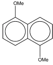
1,5-Dimethoxynaphthalene
Emmanuel Marfo-Owusua*‡ and Amber L. Thompsonb
aDepartment of Chemistry, University of Cape Coast, Cape Coast, Ghana, and bChemical Crystallography, Department of Chemistry, Chemistry Research
Labora-tory, University of Oxford, Mansfield Road, Oxford OX1 3TA, England Correspondence e-mail: [email protected]
Received 19 September 2013; accepted 28 September 2013
Key indicators: single-crystal X-ray study;T= 150 K; mean(C–C) = 0.001 A˚;
Rfactor = 0.034;wRfactor = 0.093; data-to-parameter ratio = 15.2.
The title compound, C12H12O2, lies across an inversion centre. The molecular structure suggests that the methoxy groups in the 1- and 5-positions of the naphthalene moiety do not significantly distort the planar conformation of the ring system, which has a maximum deviation of 0.0025 (9) A˚ . In the crystal, molecules pack in a herringbone arrangement in layers parallel to (100) and with chains propagating along [101] formed by very weak C—H O interactions.
Related literature
For details of the uses of 1,5-dimethoxynaphthalene, see: Ashtonet al.(1991); Amabilino & Veciana (2003); Kimet al.
(2008); Kato et al. (2003); Rawson et al. (2006). For related compounds, see: Allen & Kirby (1984); Beintema (1965); Belskii et al. (1990); Bolte & Bauch (1998); Cosmo et al.
(1990); Cruickshank (1957); Gaultier & Hauw (1967); Pawley & Yeats (1969); Rozycka-Sokolowska & Marciniak (2009); Rozycka-Sokolowska et al. (2004, 2005); Wiedenfeld et al.
(1999); Wilsonet al.(1996); Wilson (1997). For details of the low-temperature device used, see: Cosier & Glazer (1986). For details of the H-atom treatment, see: Cooperet al.(2010). For Cambridge Structural Database, see: Allen (2002).
Experimental
Crystal data
C12H12O2
Mr= 188.23
Monoclinic,P21=c
a= 7.0412 (3) A˚ b= 10.1058 (4) A˚ c= 6.5773 (2) A˚
= 95.509 (3)
V= 465.86 (3) A˚3
Z= 2
CuKradiation
= 0.73 mm1
T= 150 K
0.180.080.01 mm
Data collection
Oxford Diffraction SuperNova diffractometer
Absorption correction: multi-scan (CrysAlis PRO; Oxford Diffrac-tion, 2007)
Tmin= 0.59,Tmax= 1.00
7624 measured reflections 975 independent reflections 890 reflections withI> 2(I) Rint= 0.027
Refinement
R[F2> 2(F2)] = 0.034
wR(F2) = 0.093
S= 1.00 971 reflections
64 parameters
H-atom parameters constrained max= 0.22 e A˚
3
min=0.18 e A˚
[image:1.610.122.215.583.698.2]3
Table 1
Hydrogen-bond geometry (A˚ ,).
D—H A D—H H A D A D—H A
C7—H73 O6i
0.98 2.70 3.495 (1) 139
Symmetry code: (i)x;yþ1;zþ1.
Data collection: SUPERNOVA (Oxford Diffraction, 2007); cell refinement: CrysAlis PRO(Oxford Diffraction, 2007); data reduc-tion:CrysAlis PRO; program(s) used to solve structure:SUPERFLIP
(Palatinus & Chapuis, 2007); program(s) used to refine structure:
CRYSTALS (Betteridge et al., 2003); molecular graphics:
CAMERON(Watkinet al., 1996); software used to prepare material for publication:CRYSTALS.
EMO would like to thank the University of Cape Coast for assistance with travel and the Department of Chemistry, University of Oxford, for support.
Supplementary data and figures for this paper are available from the IUCr electronic archives (Reference: LH5655).
References
Allen, F. H. (2002).Acta Cryst.B58, 380–388.
Allen, F. H. & Kirby, A. J. (1984).J. Am. Chem. Soc.106, 6197–6200. Amabilino, D. B. & Veciana, J. (2003). InMagnetism, Molecules to Materials II,
edited by J. S. Miller & M. Drillon. New York: John Wiley & Sons, Inc.
Ashton, P. R., Brown, C. L., Chrystal, E. J. T., Goodnow, T. T., Kaifer, A. E., Parry, K. P., Phili, D., Slawin, A. M. Z., Spencer, N., Stoddart, J. F. & Williams, D. J. (1991).J. Chem. Soc. Chem. Commun.pp. 634–639. Beintema, J. (1965).Acta Cryst.18, 647–654.
Belskii, V. K., Kharchenko, E. V., Sobolev, A. N., Zavodnik, V. E., Kolomiets, N. A., Prober, G. S. & Oleksenko, L. P. (1990).Zh. Strukt. Khim.31, 116– 121.
Betteridge, P. W., Carruthers, J. R., Cooper, R. I., Prout, K. & Watkin, D. J. (2003).J. Appl. Cryst.36, 1487.
Bolte, M. & Bauch, C. (1998).Acta Cryst.C54, 1862–1863.
Cooper, R. I., Thompson, A. L. & Watkin, D. J. (2010).J. Appl. Cryst.43, 1100– 1107.
organic compounds
Acta Cryst.(2013). E69, o1655–o1656 doi:10.1107/S1600536813026731 Marfo-Owusu and Thompson
o1655
Acta Crystallographica Section EStructure Reports Online
ISSN 1600-5368
Cosmo, R., Hambley, T. W. & Sternhell, S. (1990). Acta Cryst.B46, 557– 562.
Cruickshank, D. W. J. (1957).Acta Cryst.10, 504–508. Gaultier, J. & Hauw, C. (1967).Acta Cryst.23, 1016–1024.
Kato, S., Suzuki, D. & Yoshiko, Y. S. (2003). US Patent No. US 6,656,328 B2, 2nd December.
Kim, Y. K., Jeonmin-dong, Y., Daejeon, L., Hyun, S., Eoeun-dong, Y., Daejeon, R., Mun, C., Jeonmin-dong, Y., Daejeon, C., Yong, S., Banseok-dong, Y., Daejeon, S., Gyu, Y. & Galma-Banseok-dong, S. (2008). Patent Number WO 2008/18645 A1.
Oxford Diffraction (2007).CrysAlis PRO. Oxford Diffraction Ltd, Abingdon, Oxfordshire, England.
Palatinus, L. & Chapuis, G. (2007).J. Appl. Cryst.40, 786–790.
Rawson, J. M., Alberola, A. & Whalley, A. (2006).J. Mater. Chem.16, 2560– 2575.
Rozycka-Sokolowska, E. & Marciniak, B. (2009).Acta Cryst.C65, o565–o568. Rozycka-Sokolowska, E., Marciniak, B. & Pavlyuk, V. (2004).Acta Cryst.E60,
o884–o885.
Rozycka-Sokolowska, E., Marciniak, B. & Pavlyuk, V. (2005).Acta Cryst.E61, o114–o115.
Watkin, D. J., Prout, C. K. & Pearce, L. J. (1996).CAMERON. Chemical Crystallography Laboratory, Oxford, England.
Wiedenfeld, D., Xiao, W. & Gravelle, P. W. (1999).J. Chem. Crystallogr.29, 955–959.
Wilson, C. C. (1997).Chem Commun.pp. 1281–1282.
supporting information
sup-1
Acta Cryst. (2013). E69, o1655–o1656
supporting information
Acta Cryst. (2013). E69, o1655–o1656 [doi:10.1107/S1600536813026731]
1,5-Dimethoxynaphthalene
Emmanuel Marfo-Owusu and Amber L. Thompson
S1. Comment
1,5-Dimethoxynaphthalene has numerous industrial applications and uses. It is employed in the synthesis of pesticides
(Kim et al., 2008) for the agriculture industry, involved in the preparation of polyhydric alcohols (Kato et al., 2003), as
well as a component in molecular magnetic devices (Ashton et al., 1991) for the electronics industry. It is also involved in
the synthesis of more complex paramagnetic supramolecular architectures including rotaxanes and catenanes (Amabilino
& Veciana, 2003, Rawson et al., 2006). Despite this, reports discussing single-crystal studies of naphthalene (Pawley et
al., 1969; Wilson et al., 1996; Wilson et al., 1997) and its analogues including naphthol (Rozycka-Sokolowska, et al.,
2004; Rozycka-Sokolowska et al., 2009; CSD (Allen, 2002) refcode NAPHOLO1), 1,4- and 1,5-dihydroxynaphthalene
(Gaultier, et al., 1967; CSD refcode NPHHQU10), Belskii et al., 1990; CSD refcode VOGRUE) and
1,4-dimethoxy-naphthalene (Wiedenfeld, et al., 1999; CSD refcode ALUJIA; Cosmo et al., 1990; CSD refcode MATFES) confirm that
the crystal structure of 1,5-dimethoxynaphthalene (I) is not known.
The colorless single-crystal of 1,5-dimethoxynaphthalene was crystallized while attemping to crystallize the rac
-1,1′-bi-2-naphthol/1,5-dimethoxynaphalene complex from a blend of methanol/ethylacetate solvents. It crystallizes in the
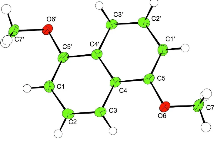
monoclinic space group P21/c with the molecule located on an inversion centre. The refined molecule and the labeling
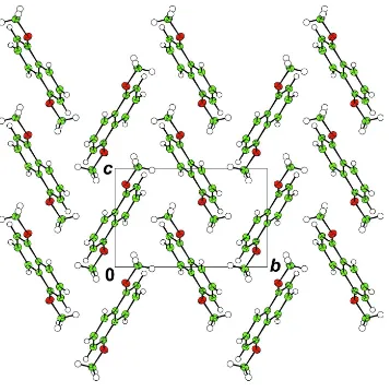
scheme are given in Fig. 1. It exhibits a herringbone packing motif and the molecules are arranged in layers parallel to the
lattice plane (100) as shown in Fig. 2. All bond distances and angles fall within expected ranges.
In 1,5-dimethylnaphthalene (Gaultier, et al., 1967; Belskii et al., 1990; Beintema, 1965) as well as those in
1,4-dimeth-oxynaphthalene (Wiedenfeld, et al., 1999), 1,8-dimethoxynaphthalene (Cosmo et al., 1990), the steric interactions of the
methyl groups cause a deviation from planarity in the naphthalene moiety. However, the ten-membered aromatic ring
formed by atoms C1–C10 in (I) is planar; the steric interactions of the methoxy and H atoms do not cause any significant
deviation from planarity. The exterior C4—C5—C4′ angle (122.13 (9)°; symmetry operator indicated by a prime is -x +
1, -y + 1, -z + 2) in the naphathalene moiety shows no evidence of distortion in the naphthalene core associated with
1,5-disubstitutions. This suggests that the methoxy group seems to be restrained in the packing structure as a result of steric
interaction between methoxy group and hydrogen atoms that reduce the propensity of the methoxy group to freely rotate
in the crystal structure.
The methoxy substituents point away from the centre of the naphthalene moiety and each one forms a weak hydrogen
bonded dimer with the neighbouring molecule. Since the molecule sits on an inversion centre, this leads to the formation
of chains in the [101] direction (Fig. 3) via the weak intermolecular C—H···O hydrogen bonds involving the methoxy
groups (with a C···O distance of 3.495 (1) Å).
In conclusion, the structure suggests that the methoxy groups in 1 and 5 positions around the naphthalene moiety do not
significantly distort the planar conformation of the naphthalene, and the size of the groups and their positions are not
The crystal of 1,5-dimethoxynaphthalene was obtained as a result of attemping to crystallize crystal complex of 1:1
mixture of rac-1,1′-bi-2-naphthol/1,5-dimethoxynaphalene from mixture of methanol and ethylacetate.
S3. Refinement
The H atoms were all located in a difference map, but those attached to carbon atoms were repositioned geometrically.
The H atoms were initially refined with soft restraints on the bond lengths and angles to regularize their geometry (C—H
in the range 0.93–0.98, O—H = 0.82 Å) and Uiso(H) (in the range 1.2–1.5 times Ueq of the parent atom), after which the
[image:4.610.128.486.211.446.2]positions were refined with riding constraints (Cooper et al., 2010).
Figure 1
The title compound with displacement ellipsoids drawn at the 50% probability level. H atoms are shown as spheres of
supporting information
sup-3
[image:5.610.130.487.67.426.2]Acta Cryst. (2013). E69, o1655–o1656
Figure 2
The packing in (I) viewed along [100] and showing the herringbone arrangement.
Figure 3
Intermolecular C—H···O hydrogen bonds forming chains that propagate along [101] (symmetry operator indicated by a
double prime is -x, -y + 1, -z + 1).
1,5-Dimethoxynaphthalene
Crystal data
C12H12O2
Mr = 188.23 Monoclinic, P21/c
Hall symbol: -P 2ybc a = 7.0412 (3) Å
b = 10.1058 (4) Å c = 6.5773 (2) Å β = 95.509 (3)° V = 465.86 (3) Å3
[image:5.610.124.487.463.554.2]Dx = 1.342 Mg m−3
Cu Kα radiation, λ = 1.54180 Å Cell parameters from 3622 reflections θ = 4–77°
T = 150 K
Plate, clear_pale_colourless 0.18 × 0.08 × 0.01 mm
Data collection
Oxford Diffraction SuperNova diffractometer
Graphite monochromator ω scans
Absorption correction: multi-scan
(CrysAlis PRO; Oxford Diffraction, 2007) Tmin = 0.59, Tmax = 1.00
7624 measured reflections
975 independent reflections 890 reflections with I > 2.0σ(I) Rint = 0.027
θmax = 76.7°, θmin = 6.3°
h = −8→7 k = −12→12 l = −8→8
Refinement
Refinement on F2
Least-squares matrix: full R[F2 > 2σ(F2)] = 0.034
wR(F2) = 0.093
S = 1.00 971 reflections 64 parameters 0 restraints
Primary atom site location: other
Hydrogen site location: difference Fourier map H-atom parameters constrained
Method = Modified Sheldrick w = 1/[σ2(F2) +
(0.06P)2 + 0.1P],
where P = (max(Fo2,0) + 2Fc2)/3
(Δ/σ)max = 0.0004116
Δρmax = 0.22 e Å−3
Δρmin = −0.18 e Å−3
Fractional atomic coordinates and isotropic or equivalent isotropic displacement parameters (Å2)
x y z Uiso*/Ueq
C1 0.46630 (14) 0.35777 (9) 1.31198 (15) 0.0246
C2 0.28469 (14) 0.36191 (10) 1.19978 (15) 0.0251
C3 0.25474 (13) 0.43124 (9) 1.02089 (14) 0.0226
C4 0.40796 (12) 0.50104 (9) 0.94482 (13) 0.0202
C5 0.38406 (14) 0.57492 (9) 0.75848 (14) 0.0219
O6 0.20285 (10) 0.57222 (7) 0.66380 (11) 0.0274
C7 0.16539 (15) 0.64994 (10) 0.48270 (16) 0.0296
H11 0.4837 0.3088 1.4350 0.0297*
H21 0.1819 0.3157 1.2519 0.0309*
H31 0.1296 0.4338 0.9479 0.0276*
H73 0.0325 0.6342 0.4324 0.0427*
H72 0.1849 0.7431 0.5170 0.0424*
H71 0.2476 0.6232 0.3793 0.0432*
Atomic displacement parameters (Å2)
U11 U22 U33 U12 U13 U23
C1 0.0273 (5) 0.0242 (5) 0.0220 (4) −0.0007 (4) 0.0001 (4) 0.0018 (3)
C2 0.0220 (5) 0.0258 (5) 0.0277 (5) −0.0029 (4) 0.0041 (4) 0.0001 (4)
C3 0.0179 (5) 0.0236 (5) 0.0260 (5) −0.0001 (3) −0.0002 (3) −0.0027 (3)
C4 0.0195 (5) 0.0186 (4) 0.0220 (4) 0.0012 (3) −0.0002 (4) −0.0031 (3)
supporting information
sup-5
Acta Cryst. (2013). E69, o1655–o1656
O6 0.0223 (4) 0.0314 (4) 0.0268 (4) −0.0028 (3) −0.0064 (3) 0.0060 (3)
C7 0.0298 (5) 0.0308 (5) 0.0261 (5) −0.0002 (4) −0.0077 (4) 0.0044 (4)
Geometric parameters (Å, º)
C1—C5i 1.3717 (14) C4—C4i 1.4236 (17)
C1—C2 1.4143 (13) C4—C5 1.4312 (13)
C1—H11 0.946 C5—O6 1.3653 (11)
C2—C3 1.3683 (14) O6—C7 1.4301 (11)
C2—H21 0.953 C7—H73 0.975
C3—C4 1.4197 (13) C7—H72 0.974
C3—H31 0.962 C7—H71 0.972
C5i—C1—C2 119.63 (9) C3—C4—C5 122.01 (9)
C5i—C1—H11 120.5 C4—C5—C1i 121.13 (9)
C2—C1—H11 119.9 C4—C5—O6 114.09 (8)
C1—C2—C3 121.39 (9) C1i—C5—O6 124.78 (9)
C1—C2—H21 118.6 C5—O6—C7 117.29 (8)
C3—C2—H21 120.0 O6—C7—H73 106.7
C2—C3—C4 119.86 (9) O6—C7—H72 109.1
C2—C3—H31 120.1 H73—C7—H72 110.2
C4—C3—H31 120.1 O6—C7—H71 110.8
C4i—C4—C3 119.90 (10) H73—C7—H71 109.5
C4i—C4—C5 118.09 (11) H72—C7—H71 110.5
Symmetry code: (i) −x+1, −y+1, −z+2.
Hydrogen-bond geometry (Å, º)
D—H···A D—H H···A D···A D—H···A
C7—H73···O6ii 0.98 2.70 3.495 (1) 139