Lehto J (1993) Ion exchange in the nuclear power industry. In: Dyer A, Hudson HG and Williams PA (eds) Ion Exchange Processes:Advances and Applications, p. 39. Cambridge, UK: The Royal Society of Chemistry. Lehto J and Harjula R (1997) Selective separation of
radionuclides from nuclear waste solutions with inor-ganic ion exchangers,React Funct Polym(in press).
Navratil JD (1989) Ion exchange technology in spent fuel reprocessing.Journal of Nuclear Sciences and Techno-logy, 26: 735.
Shultz WW, Wheelwright EJ, Godbee H, Mallory CW, Burney GA and Wallace RM (1984) Ion exchange and adsorption in nuclear chemical engineering. In:AIChE Symposium Series80(233): 96.
NUCLEIC ACIDS
Centrifugation
A. Marziali, University of British Columbia, Vancouver, Canada
Copyright^ 2000 Academic Press
Introduction
Centrifugation has been applied to nucleic acid isolation and puriRcation through numerous proto-cols which, at some level, contain elements of one or more of three basic techniques: isopycnic or density equilibrium separation, phenol}chloroform extrac-tion, and differential precipitation. Even if we consider only the protocols that are in current use, numerous variations on these appear in the literature. These variations result from the intended use of the product, the required purity from speciRc contaminants, the cost and through-put goals of the technique, and often the author’s personal preferences. This article will make no at-tempt to cover all variations but will instead illustrate by example the basic forms of centrifuge-based techniques for nucleic acid separation as they are presently used. A rough guide to these three basic techniques and their applications is contained in Table 1. Each of these will subsequently be described separately.
Recent demands imposed on nucleic acid puriR ca-tions by large scale DNA sequencing operaca-tions have led to the development, and increased use of R ltra-tion-based puriRcation methods for high through-put separations. Though the cost of the Rlter mem-branes required for these separations is much higher than the cost of centrifugation, the throughput and case of automation of the membrane based methods make them preferable in many situations. Recent developments in automation of centrifugation, dis-cussed in the last section of this article, may reverse this trend.
Isopycnic Separations
General Principle
Isopycnic separations rely on the balancing of the buoyant and centrifugal forces acting on a submerged sample during centrifugation. When a sample of den-sity s and effective volume V is placed in a me-dium of density min the presence of a centrifugal Reld a, the sample feels an upward buoyant force
Fb"mVa, and an opposing centrifugal force
Fc"sVa. Consequently, the sample will move ‘up’ toward the rotation axis if s(m and ‘down’ if
s'm. This motion terminates when the sample
reaches the boundary of the medium or when it enters a region of the medium weres"m. Based on this principle, if a sample container isRlled with a me-dium whose density increases gradually in the down-ward direction, a sample injected in this medium will migrate to the region of the medium that matches the sample density (provided such a region exists). This location is known as the isopycnic point of the sample.
Samples may therefore be separated based on their densities provided a medium is found that can be formed into a density gradient and whose density range includes that of the sample. One of the criteria in the selection of separation media for a speciRc sample is to ensure that this condition is met.
After a substantial migration period (often over a day), the sample fractions of different densities can be observed as bands within the medium. Extrac-tion of these bands is performed by puncturing the centrifuge tube with a hypodermic needle and with-drawing the desired band. The resolution provided by this method is a function of the separation medium and the relative density difference in the fractions to be separated.
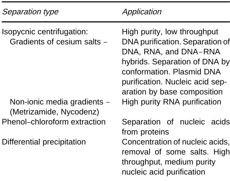
Table 1 Three common methods of nucleic acid separation employing centrifugation
Separation type Application
Isopycnic centrifugation: Gradients of cesium salts}
High purity, low throughput DNA purification. Separation of DNA, RNA, and DNA}RNA hybrids. Separation of DNA by conformation. Plasmid DNA purification. Nucleic acid sep-aration by base composition Non-ionic media gradients} High purity RNA purification (Metrizamide, Nycodenz)
Phenol}chloroform extraction Separation of nucleic acids from proteins
Differential precipitation Concentration of nucleic acids, removal of some salts. High throughput, medium purity nucleic acid purification
a centrifugal acceleration Reld they spontaneously form into a density gradient whose range can include that of DNA. RNA typically exhibits higher density than the maximum cesium gradient density and pel-lets at the bottom of the centrifuge tube. DNA may also be fractionated according to a number of vari-ables which affect its buoyant density. Single stranded DNA and double stranded DNA differ in their degree of hydration and therefore exhibit different buoyancies, allowing them to be separ-ated into two different bands in Cs2SO4 (or NaI) gradients. Also, the base composition (G#C con-tent) of DNA linearly affects its buoyancy allow-ing separation of DNA from different organisms, and, in some cases, even separation of DNA from different regions of the same eukaryotic genome (Figure 1).
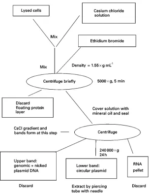
The addition of intercalating molecules such as ethidium bromide to the gradient may be used to separate DNA based on its conformation. Linear and relaxed circular DNA allow a larger amount of ethidium bromide to intercalate than supercoiled DNA, leading to decreased density and band separ-ation. In the example described below, this result is used to separate supercoiled plasmid DNA from genomic and nicked circular plasmid DNA.
Separation of RNA is difRcult to perform in cesium salt solutions because of its density and tend-ency to form a precipitate. Consequently, separation of RNA is now performed in nonionic media such as Metrazimide and Nycodenz.Table 2lists some com-monly used density gradient media and their asso-ciated use.
A common example of the use of cesium chloride gradients is illustrated in the isolation of plasmid DNA. Note that this is an abbreviated protocol: the
references at the end of this article should be consul-ted for further details.
Phenol Extraction for Separating
DNA/Proteins
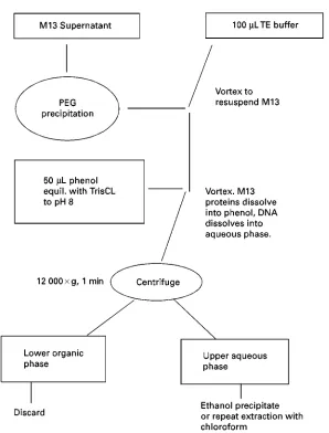
A common method for separating nucleic acids from proteins is extraction by phenol or phenol : chloro-form. In this technique, solutions containing protein and nucleic acids are combined with an equal part of phenol or phenol : chloroform and mixed into an emulsion. Since phenol and chloroform are solvents for denatured proteins while nucleic acids are soluble in the aqueous phase, centrifugation of the phases results in separation of nucleic acids from proteins. In some cases, multiple extractions may be required and may be followed by extractions in pure chloroform and by ethanol precipitation depending on the re-quired purity of the nucleic acid sample. A simple example of this technique is the puriRcation of DNA from M13 bacteriophage for purposes of DNA se-quencing. An abbreviated protocol is given in Figure 2.
Differential Precipitation Methods
Ethanol or Isopropanol Precipitation
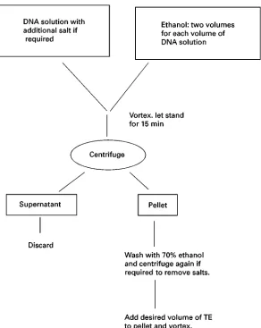
Perhaps the simplest way to concentrate nucleic acids by centrifugation is precipitation in ethanol or iso-propanol solutions. This technique takes advantage of the fact that nucleic acids can form a solid precipi-tate in these solutions when their negative charge is neutralized by the presence of monovalent cations. A common example of this is ethanol precipitation of DNA in which an aqueous DNA sample is mixed with ethanol and a small amount of salt (often so-dium acetate). After incubation, a solid precipitate of the sodium salt of DNA is formed which can be centrifuged into a pellet. Repeated washing of this pellet with 70%}80% ethanol solutions helps remove residual salts.
Though ethanol precipitation is not useful in separ-ating nucleic acids from many contaminants, this form of puriRcation is the Rnal step in many nucleic acid puriRcation schemes as it tends to both concen-trate the nucleic acid and remove any remaining salts or contaminants used in previous separations and extractions. In some cases, salt contaminants already present in the sample can be used to aid precipitation without further addition of sodium acetate (or other salts). The simplicity of this protocol has made it a cornerstone of high throughput nucleic acid puriR -cation.Table 3is a rough guide to the choice of salt used in the precipitation.
Figure 1 Plasmid DNA purification by CsCI}Ethidium bromide continuous gradient.
Table 2 Some density gradient media commonly used for nucleic acid separation
Separation medium Application
CsCI Isolation of plasmid DNA. Separation of DNA by conformation. Separation of DNA by base composition NaI Separation of single vs. double
stranded DNA Metrizamide, Nycodenz Fractionation of RNA
Precipitation of nucleic acids from buffers con-taining high concentrations of EDTA or phosphate ions may result in co-precipitation of these substan-ces. Also, precipitation of small nucleic acid strands ((100 nucleotides) may be improved by the addition of carriers such as glycogen, by the addition of MgCl2, or by increased duration and speed of the
centrifugation (100 000;g, 1}2 hours). Centrifu-gations for nucleic acid precipitations are typically carried out at 0}43C, though for substantial concen-trations ('40 ngL\) of long strands (such as DNA template for sequencing) incubation and centrifu-gation can also be carried out at room temperature. The generic protocol shown in Figure 3 is an example of ethanol precipitation forRnal concentra-tion of DNA from a plasmid or M13 preparaconcentra-tion.
[image:3.568.51.277.621.710.2]Figure 2 M13 DNA purification by phenol : chloroform extraction.
Table 3 Salt solutions used for nucleic acid precipitation
Salt Final
concentration
Advantage/application
Ammonium acetate 2.0}2.5M Reduces co-precipitation of dNTPs
Lithium chloride 0.8M Works with high concentra-tions of ethanol (as used in RNA precipitation) Sodium chloride 0.2M Allows SDS to remain
soluble in ethanol Used with samples containing SDS
Sodium acetate 0.3M Used for routine RNA and DNA precipitations After the initial precipitation, the pellet can be resuspended in TE buffer to the desired concen-tration, or, if particularly low salt concentration is
desired in theRnal product, a further ethanol wash can be performed. This is done by washing the pellet in 70% ethanol and centrifuging for a further Rve minutes before again discarding the ethanol.
Isopropanol may be used in place of ethanol. In this case, only one volume of isopropanol should be used per volume of DNA solution. This is usually less desirable as residual isopropanol is more difRcult to remove and more likely to cause coprecipitation of salts.
Precipitation of RNA is performed as for DNA except that 2.5 to 3 volumes of ethanol should be used per volume of RNA solution.
Plasmid Preparations by Differential Precipitation
[image:4.568.50.278.560.711.2]Figure 3 Ethanol precipitation of DNA.
acid molecules such as plasmid DNA from genomic DNA, RNA and protein contaminants. One example of this technique is the frequently used alkaline lysis preparation for the puriRcation of plasmid DNA from E. coli. The technique takes advantage of the fact that the large genomic DNA strands from lysed bacterial cells will precipitate much more easily than the smaller plasmid molecules. Consequently, a mix-ture can be generated in which the genomic DNA can be pelleted, allowing the plasmid DNA to be extrac-ted with the supernatant. The generic technique is shown inFigure 4.
Nucleic Acid Separation at
High Throughput
Present and future efforts in the development of novel nucleic acid puriRcation methods are likely to be aimed at satisfying the demand for inexpensive and high speed puriRcation of a large number of
samples simultaneously. This is particularly true in applications related to large scale DNA sequencing and analysis. Currently, large-scale sequencing labs are expanding their operations to reach sequencing rates of 50 000 to 100 000 DNA samples per day. Though density gradient separations such as CsCl have historically provided the highest purity DNA, they are far too laborious to be employed at this rate. Furthermore, because of the inherently parallel op-eration in these cases, any DNA puriRcation schemes must be compatible with standard microtitre plate formats.
Figure 4 Plasmid purification by differential precipitation.
microtitre plates (for bead separations) and microtitre membrane plates (now available with up to 384 sample wells per plate). Furthermore these two methods are easily automated, and numerous instru-ments now exist that can performRlter membrane or magnetic bead based separations with very little la-bour cost and at high throughput. The only drawback to these methods has typically been the cost asso-ciated with either the magnetic beads or the dispos-ableRlter membranes.
Precipitation based puriRcation methods, though inherently less expensive because of the lack of dis-posables involved, have been somewhat harder to adapt to large scale operation because of the human effort required to set-up and operate a conven-tional centrifuge. Though centrifugation of high density microtitre plates is routinely performed,
unat-tended automation of this process has been avoided until recently, and many large sequencing labs simply rely on manual execution of centrifuge based protocols.
Automation of Nucleic Acid Separation by Centrifugation
Two methods for performing high throughput auto-mated centrifugation have emerged which allow the construction of automated instruments for perform-ing centrifugation-based puriRcation protocols.
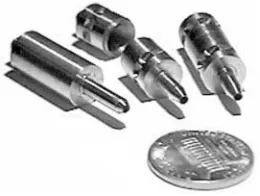
Figure 5 (See Colour Plate 108) Titanium belt driven and air driven rotors used in the arrayable flow-through centrifuge. A penny is shown for scale.
Figure 6 Principle of operation of arrayable flow-through centri-fuge: (A) Sample is injected through the upper axial orifice into the spinning rotor. The centrifugal field instantly presses the sample against the inside wall of the rotor preventing it from exiting through the bottom orifice. (B) Rapid spinning of the rotor separ-ates sample phases}any solid precipitate is pressed into the widest part of the rotor. (C) The rotor is stopped and the super-natant drips out the bottom orifice. The pellet can be re-sus-pended by injecting a small amount (100L) of buffer into the rotor and agitating the rotor through repeated clockwise and counterclockwise accelerations. This procedure can also be used to clean the rotor and prepare it for the next sample to be separated.
centrifuge. The difRculty involved with this method is that standard centrifuge rotors are not designed to stop at a repeatable indexed orientation. Consequently, the robotic arm which places and re-moves the samples from the samples from the centri-fuge, cannot know the location of the samples at the end of a run.
The solution to this problem is to index the rotor position by means of an electronic sensor or a mech-anical stop. One example of this type of solution is a plasmid preparation instrument developed at the Lawrence Berkeley National Laboratory (LBNL). This instrument consists of a robotic gantry equipped with a pipetting and gripping tool which can access a work surface that includes an indexing centrifuge. To prepare the centrifuge for robotic access, a pneu-matic actuator opens the lid while another actuator is extended to interfere with tabs attached to the rotat-ing rotor shaft. These tabs, when pressed against this actuator, deRne a well-indexed position for the rotor buckets. To ensure contact between these tabs and the actuator, the rotor is turned by an external fric-tion coupling which can slip once the tab is in contact with the actuator. With the rotor positioned in this fashion, the robotic tool can reliably enter the centri-fuge and locate the rotor bucket or sample plate.
With an instrument of this type, plasmid puriR ca-tion at the rate of 192 samples in 2.5}4 h can be performed. Final DNA puriRcation occurs by ethanol precipitation, automated within the centrifuge described.
Similar methods have been used to automate cen-trifugation in other instruments including the com-mercially available Autogen 740 and Autogen 850 instruments. These instruments also contain auto-mated centrifuges and are capable of various DNA and RNA puriRcations at rates up to 48 samples per 4}6 h.
Miniature, arrayable centrifuges A second ap-proach to automation of centrifugation for high throughput DNA puriRcations has recently been de-veloped at the Stanford DNA Sequencing and Tech-nology Center. The goal of this approach is to remove the inherent radial acceleration limit (&3500;g) im-posed on microtitre plate centrifugation by the struc-tural weakness of the sample plate. It is because of this strength limit that centrifugation of DNA sam-ples in microtitre plates typically requires 20}30 min per separation. By centrifuging the samples directly within a reusable, high strength rotor, accelerations of over 20 000;g can be reached, substantially de-creasing pelleting times. To implement this at high throughput, a large number of small rotors operating in parallel is required.
The Stanford group’s implementation of this con-cept consists of blocks of 96 individual, high speed rotors, arrayed on the same spacing as a standard 96 well microtitre plate. The rotors (Figure 5) can be spun at up to 70 000 rpm in both directions about their central axis by means of either compressed air or a motor driven belt.
[image:7.568.297.501.438.572.2]The small size of this Sow-through centrifuge allows highly parallel operation, smaller sedimenta-tion drift distances, and high angular velocities. These high velocities in turn translate to large sedimentation forces which, coupled with the short drift distance, lead to much shorter separation times than a conven-tional microtitre plate centrifuge. Calculations based onE.colisedimentation indicate a 40 fold decrease in pelleting time over a conventional centrifuge.
Applications of this technology to nucleic acid sep-aration are being exploited primarily in the area of plasmid DNA puriRcation for sequencing purposes. Instruments are being constructed based on this tech-nology which should be capable of purifying over 500 plasmid DNA samples from cell cultures within one hour using an alkaline lysis protocol. In this protocol, two separations are required, one to clear the cell lysate, followed by ethanol precipitation to collect the DNA. With this protocol in mind, a multi-stage version of thisSow-through centrifuge system (where the super-natant from one array of rotors drips into the input of a second array) is being developed at Stanford.
See Colour Plate 108.
See also: II/Centrifugation: Theory of Centrifugation.
Further Reading
Birnie GD and Rickwood D (1978)Centrifugal Separations in Molecular and Cell Biology. London: Butterworth. Ford TC and Graham JM (1991)An Introduction to
Cen-trifugation. Oxford: BIOS ScientiRc Publishers.
Marziali A, Willis TD and Davis RW (1999)An Arrayable Flow-Through Microcentrifuge for High Throughput Instrumentation. Proc. Natl. Acad. Sci., USA. 5 Jan. Parish JH (1972)Principles and Practice of Experiments
with Nucleic Acids. New York: John Wiley&Sons. Rickwood D (1984)Centrifugation(Second Edition).
Ox-ford: IRL Press.
Rickwood D, Ford TC and Steensgaard J (1994) Centrifu-gation}Essential Data. New York: John Wiley&Sons. Sambrook J, Fritsch EF and Maniatis T (1989)Molecular Clon ing.A Laboratory Manual.Second Edition. USA: Cold Spring Harbor Laboratory Press.
Sheeler P (1981) Centrifugation in Biology and Medical Science. New York: John Wiley&Sons.
Extraction
S. J. Walker and K. E. Vrana, Wake Forest University, Winston-Salem, NC, USA
Copyright^ 2000 Academic Press
Introduction
The recent explosion of information from recom-binant DNA technology and the human genome initi-ative has come about in response to a number of key technological advances. These include the discovery and characterization of restriction endonucleases, the development of plasmid and phage vectors, and the creation of high throughput DNA sequencing methodologies. Less dramatic, but no less important, has been the development and reRnement of proto-cols for extracting, purifying, and characterizing the various nucleic acids from complex biological mix-tures. The present chapter reviews these procedures with particular emphasis on the unique character-istics and methodological constraints involved in dealing with deoxyribonucleic acid (DNA) vs. ribonucleic acid (RNA).
Figure 1establishes the experimental hurdles to the isolation of puriRed nucleic acids. A eukaryotic cell contains a variety of biological macromolecules of
which the genetic material (nucleic acids) represents a minor component. The investigator is therefore faced with the daunting task of separating proteins, lipids, and nucleic acids from each other. Indeed, many applications require fractionating the genetic material into DNA and RNA and even subfractionat-ing the RNA into ribosomal and messenger RNA species. These last two tasks are complicated by the fact that chromosomal DNA is a fragile, double-stranded molecule of very high molecular weight (3 billion total base pairs). RNA, on the other hand, while much smaller (75}10 000 nucleotides), is a single-stranded molecule that is exquisitely sensitive to enzy-matic degradation. Fortunately, each biological fraction within a cell bears unique biophysical characteristics (charge, lipophilicity, chemical makeup, etc.) and these characteristics provide convenient mechanisms for re-solving the macromolecules from one another.