Original Article
Effect of social interaction on learning and
memory of morphine withdrawal mice
Xun Gou, Yanyou Liu, Zhou Jiang, Yuhui Wang, Zhengrong Wang
Key Laboratory of Chronobiology (Health and Family Planning Commission), School of Preclinical and Forensic Medicine, West China Medical Center, Sichuan University, Chengdu 610041, Sichuan, PR China
Received April 13, 2017; Accepted August 6, 2017; Epub October 15, 2017; Published October 30, 2017
Abstract: Drug abuse is characterized by its long term persistence which has physical and psychological depen-dence. Clinical treatment, psychological counselling and social intervention can help patients have a certain with-drawal from physical dependence, but psychological addiction remains a serious problem leading over 90% of relapse. Influence of social interaction on drug effects has a close relationship with psychological dependence. In order to study the enduring changes in long-term memory in brains of drug-withdrawal mice, we established a social interaction model to compared hippocampal and prefrontal cortex gene expression in drug abuse mice. Two groups of physical morphine-withdrawal mice were caged in morphine abuse mice and saline-treated mice for four weeks, respectively. Morris’ water maze was used for the detection of learning and memory, magnetic resonance spectroscopy (MRS) was used for the integrity of neurons in the brains between two groups. Expression GABAA and PKCα which mediated neurotransmission of GABA and glutamate in hippocampus and the prefrontal cortex were de-termined by immunohistochemistry and Western blot. The results showed that compared with morphine-withdrawal mice that social interacted with saline treated mice, the ones who lived with morphine abuse mice had a decreas-ing learndecreas-ing and memory ability and much more damaged neurons. Moreover, overexpression of mRNA and protein levels of GABAA and PKCα reveals the upregulation of GABA and downregulation of glutamate in neurons, enhancing the learning and memory in morphine withdrawal mice to trigger the reward pathway in morphine addiction at first. However, over activation of GABA and glutamate inside or outside of neurons had a negative feedback in learning and memory function. In conclusion, drug-withdrawal mice that social interacted with drug abuse mice had an in-creasing memory caused by environmental stimulus, but the over simulations may lead to neuron damages and a drop in functions of learning and memory through the over expression of GABAA and PKCα.
Keywords: Morphine, social interaction, GABAA, PKCα
Introduction
Opiate addiction is not only a chronic tolerance endangering people’s physical and mental health, the mortality caused by drug abuse is very high even after cardiovascular diseases and malignant tumors in some countries. One
significant characteristic of opiate addiction is
the physical and psychological dependence [1-3]. Clinical treatment, psychological counsel-ling and social intervention can help patients have a certain withdrawal from physical depen-dence, but psychological addiction is still a
worldwide problem. In the field of neuroscience,
brain function in addictive patient is altered in certain areas rather than nonaddicted brain, as the chronic administration of opiate is involved
in regulation of gene expression of genes in receptor regulation, signaling and transcription [4-6]. “Reward pathway” which including the ventral midbrain, nucleus accumbens and pre-frontal cortex in brain is the key in the develop-ment of addiction [7, 8]. Opiate inhibits the neu-ral activity of GABA in midbrain ventneu-ral tegmen-tal area to activate the DA neurons, therefore accelerates the release of dopamine in the nucleus accumbens to establish the reward cir-cuits [9, 10]. Hippocampus, another region of brain, associates with the effects of reward pathway in long time drug abuse and may be one factor in psychological dependence [11, 12]. Psychological addition in drug abuse
causes the relapse rate in detoxificated patients
“reward pathway”. Therefore, how to withdrawal from psychological addiction and help patients
return to normal life has been the key and diffi -culty in the study of drug withdrawal.
GABAergic neurons occupy about one fifth of
the central nervous system neurons and play a decisive role in the process of brain develop-ment [13]. As a kind of important inhibitory neu-rons, some mental disorders are often occurred by the impairment of GABAergic neurons with the excitability [10]. In the mammalian brain development, GABAA receptor is widely distrib-uted in hippocampus, prefrontal cortex and striatum, mediated with most of the GABAergic neuron activity [14, 15].
Protein kinase C (PKC) belongs to the multi-functional serine and threonine kinase, encod-ed by multi-gene families, molecular weight is about 67-83 KD [16]. It is widely distributed in a variety of tissues, organs and cells in mam-mals. PKC pathways play central roles in chang-es of neuronal physiology that involved in learn-ing and cognition [17]. PKCα is a kind of calcium dependent PKC sub-types, report shows that the over activation of it can inhibit the intake of glutamine by neurons to lead impairment of learning and memory [18, 19].
A number of animal studies showed that early psychologically harmful events can alter the normal development of brain, suggested the social environmental effects on brain function.
Moreover, social context influenced the affec -tive valence of drug abuse such as alcohol [20,
21]. The goal of this study was to find whether
the social factor was involved in the psychologi-cal dependence in drug relapse by establishing a social interaction model. In this model, mice in the period of morphine withdrawal were per-formed, and the environmental stimuli was to live with groups of mice that had morphine administration and saline as control. The exper-iments examined the psychological response in learning and memory to social interaction and the possible mechanisms involved in changes of GABAA and PKCα in prefrontal cortex and hip-pocampus, to provide a new aspect on drug addiction treatment.
Methods and materials
Animals
Male Bal B/C mice weighing 20-22 g was used in the study. Animals were housed in plastic
cages with free access to food and water under certain conditions (12 h/12 h light-dark cycle and temperature of 22±2°C). Experimental pro-cedures were approved by the Laboratorial Animal Care Committee of Sichuan University.
Animal procedure
40 mice were randomly divided into morphine abuse group (MA, n = 20) and morphine control group (MC, n = 20). Both MA and MC were exposed to consistent eight days of morphine injection. Morphine hydrochloride (Simopharm Group Sichuan Medicines Co., Ltd, Sichuan, China) dissolved in physiological saline was injected in constantly increasing doses accord-ing to Sukhotina (Day 1: 10 mg/kg; Day 2: 20 mg/kg; Day 3: 30 mg/kg till Day 8: 80 mg/kg) [22]. Physical morphine withdrawal was carried lasting for ten days since Day 9 in both groups. Another 40 mice were randomly divided intoso-cial interaction group (SI, n = 20) and social control group (SC, n = 20). SI and SC were treat-ed with 50 mg/kg morphine and same amount of saline water from Day 19 to Day 46, respec-tively. On Day 19, MA started to live with SI (in ratio of 1:1) and MC with SC (in ratio of 1:1) for 4 weeks until Day 46.
Morris’ water maze
The alternation of learning and memory in MA and MC mice was examined by Morris’ water maze (Chengdu Technology & Market Co., China) according to the procedure on day 47
and finished on day 53. The apparatus was a circular pool (120 cm diameter) fulfilled with
opaque water (24±1°C) added with milk. A cir-cular transparent platform around 12 cm diam-eter was submerged 1 cm under the surface of water in the third quadrant for testing learning and memory. Each mouse was placing into the tank at the edge of each quadrant and the latency was recorded for searching and locat-ing the platform. It would be guided to the plat-form and stayed for 10 s when 120 s of search-ing time had elapsed without locatsearch-ing. Three trials were conducted every day in seven con-secutive days. Probe trial was taken when the platform was removed. Percentage of times spent in the 3rd quarter and times of swimming
body (Santa Cruz Biotechnology, Inc, USA, 1:400) and rabbit anti-PKCα (Boster, Wuhan, China, 1:200), followed by the addition of goat anti-rabbit IgG secondary antibody (Boster, Wuhan, China, 1:200). Images were acquired from randomly four sections in different ratios (40×, 100×, 400×) and analyzed with Image Software (Nikon NIS-Elements D) by an observ-er blind to samples. Four grades wobserv-ere made according to the degree of expression as nega-tive (-), lightly posinega-tive (+), posinega-tive (++), strongly positive (+++).
Brain collection and Western blot
Another Ten mice in each group were sacrificed
and different parts of brains (the prefrontal cor-tex and hippocampus according to a mouse brain atlas) were dissected out immediately and frozen in liquid nitrogen for subsequent experiments. The dissected and frozen parts of brains (prefrontal cortex, hippocampus) in dif-ferent mice groups were thawed and homoge-nized in RIPA lysis buffer (1 mmol/L PMSF) according to the manufactures’ instruction (Beyotime Institute of Biotechnology, Jiangsu, China). Lysates were centrifuged at 14,000 g for 5 min, and the supernatant was collected.
Protein concentrations were quantified with
[image:3.612.92.372.82.305.2]BCA Protein Assay Kit (Beyotime Institute of Biotechnology, Jiangsu, China) at 562 nm. Sub- Figure 1. Time spend (latency) in acquisition experiment between MC and
MA group in Morris’ Water Maze. Note: Compared with MC mice, *P<0.05 in t-test.
was taken at Nanchong Cen- tral Hospital using a 1.5 T Signa scanner (GE Medical systems, USA).
Immunohistochemistry
Tenmice in MA and MC group were used for immunohisto-chemistry. Mice were anaesth- etized with 4% chloralic hydras (350 mg/kg), perfused with 0.1 mol/L phosphate-buffered saline (PBS, pH = 7.4) and th-
en fixed with 4% paraformal -dehyde (PFA) in 0.1 mol/L PBS. The brains were removed and stored overnight at 4°C. Subsequently, the tissues we- re given to College of Public Health (West China Center of Medical Sciences, Sichuan University) for immunohisto-chemistry. The antibodies us- ed were rabbit GABA anti-Table 1. Probe trial results between MC and
MA group in Morris’ water maze (_x±s) Groups n Time spent in Q3 (%) Times across platform MC 20 43.06±8.46 3.406±1.13 MA 20 28.48±9.21* 1.748±0.85* Note: Compared with MC mice, *P<0.05 in t-test.
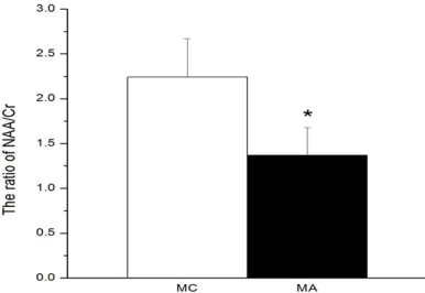
Figure 2. The ratio of NAA/Cr between MC and MA group in MRS experiment. Note: Compared with MC mice, *P<0.05 in t-test.
MRS examination
[image:3.612.91.287.389.440.2] [image:3.612.91.284.474.607.2]sequently, the protein sampl- es were prepared by mixing with loading buffer (0.1 mol/L Tris-HCl, pH = 6.8; 2% mercap-toethanol; 2% SDS; 0.01% bro-mophenol blue and 10% glyc-erol) followed by denaturation at 95°C water for 5 min and cooled to room temperature. Each sample was separated by 15% SDS-PAGE electroph- oresis using 150 V for 50 min. The durations of protein trans-ferring to PVDF membrane at 200 mA were different (GA- BAA-105 min; PKCα-150 min) depended on the size of pro-teins. The membranes were blocked with 5% milk (TBST buffer: 10 mmol/L Tris-HCl, pH 7.5; 0.2 mol/L NaCl; 0.01% Tween) for 1 h at room tem-perature followed by incuba-tion overnight at 4°C with primary antibodies. The dilu-tions of primary antibodies were: GABAA (1:1000) (Santa Cruz Biotechnology, Inc, USA); PKCα (1:500) (Boster, Wuhan,
China); β-actin (1:500)
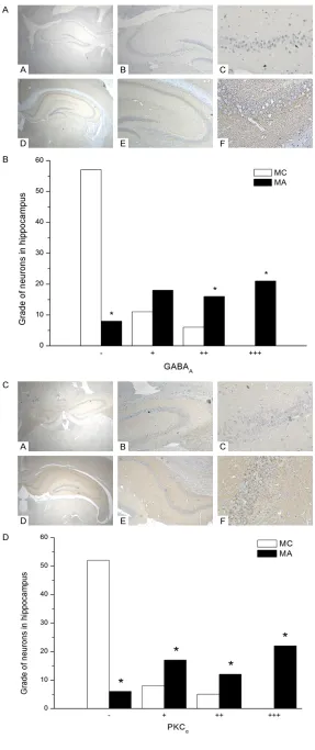
[image:4.612.90.377.64.747.2](Bei-jing Biosynthesis Biotechno- logy Co., LTD). The membranes were washed 3 times with TB- ST buffer before 1 h incubati- Figure 3. A: Immunohistochemi-cal detection of GABAA in hippo-campus. Representative photomi-crographs of hippocampus from MC and MA mice with different multiple are shown: A = MC group (40×); B = MC group (100×); C = MC group (400×); D = MA group (40×); E = MA group (100×); F = MA group (400×). B. The expres-sion of GABAA in hippocampus of mice between MC and MA mice. C. Immunohistochemical detec-tion of PKCα in hippocampus.
Rep-resentative photomicrographs of hippocampus from MC and MA mice with different multiple are shown: A = MC group (40×); B = MC group(100×); C = MC group (400×); D = MA group (40×); E = MA group (100×); F = MA group (400×). D. The expression of PKCα
on with conjugated goat anti-rabbit IgG second-ary antibody (Boster, Wuhan, China; 1:20,000)
and goat anti-rabbit β-actin (Beijing
Biosynth-esis Biotechnology Co., LTD; 1:20000), respec-tively. The blots were visualized by chemilumi-nescence using EasySee Western Blot kit
(TransGen Biotech, Beijing, China) under X-ray and analyzed with Image Lab (Bio-Rad, USA).
Statistical analysis
Data was expressed as mean ± SEM and
ana-lyzed by T-test to find the differences between
two groups (SPSS 16.0). Results
MA mice had worse performance on learning and memory
In Morris’ water maze, the results demonstrat-ed that the ability of learning and memory of the mice could be altered by the different living environments. Compared with MC, MA had a
significant worse performance on learning and
memory (Figure 1; Table1). The time spent in water maze searching for the platform in MA was much longer than that of MC at Morris’ water maze experimental at Day 1 to Day 7 (P<0.05, n = 20). The integrity of neurons in brains between MA and MC showed a differ-ence (Figure 2). In the MRS examination, NAA (n-acetyl-L-aspartic acid) is an amino acid locat-ed in neurons and its concentration corr- elates with neuronal mitochondrial function [23, 24]. The reduction of NAA concentration
reflects decline of both neuronal density and
integrity of neuronal mitochondria in several psychiatric disorders. Creatine is an important neurometabolite with dietary supplementation and used as a biomarker correlated with NAAin MRS. In the experiment, the ratio of NAA and Cr in MA was declined compared with that with MC, which had a more damaged in the integrity of neurons (P<0.05, n = 20) [25].
Expression of GABAA in MA mice was in-creased than MC mice
After living with drug-abuse mice, the level of GABAA in MA mice was significantly increased
[image:5.612.88.287.70.475.2]in both prefrontal cortex and hippocampus (Figures 3A, 3B, 4A, 4B). In the immunohisto-chemistry experiment, the expression of hippo-campal GABAA showed a much stronger expres-sion in MA than MC (P<0.05, n = 10). According to the blind test, the MC mice had more nega-tive expression (-) than MA, and less posinega-tive (++) and strongly positive (+++). In western blot, the ratio of GABAA/beta-actin in hippocampus was higher in MA rather than MC (P<0.05, n = Figure 4. A. Electrophoresis of Western blotting
prod-ucts of beta-actin, GABAA and PKCα in hippocampus
and PFC between MC and MA mice. Templates are total protein isolated from brain of BAL/B/C mice. Lane 1-isolated from PFC of MC mice, Lane 2-iso-lated from PFC of MA mice, Lane 3-iso2-iso-lated from Hip-pocampus of MC mice, Lane 4-isolated from Hippo-campus of MA mice. B. IOD ratio of western blotting products of GABAA and beta-actin in PFC between MC and MA mice. (*P<0.05). Note: Compared with MC mice, *P<0.05 in t-test. C. IOD ratio of western blot-ting products of PKCα and beta-actin in the PFC and
10). The expression of was also GABAA in MA was also higher in prefrontal cortex (P<0.05, n = 10).
Expression of PKCα in MA mice was increased than MC mice
The level of PKCα protein in morphine
withdraw-al mice showed a significant decrease com -pared with morphine control group in prefrontal cortex and hippocampus (Figures 3C, 3D, 4A, 4C). The expression of hippocampal PKCα show- ed a much stronger expression in MA than MC in the immunohistochemistry experiment (P< 0.05, n = 10). According to the blind test, the MC mice had more negative expression (-) than MA, and much less slightly positive (+), positive (++) and strongly positive (+++). In western blot, the ratio of PKCα/beta-actin in hippocampus was higher in MA rather than MC (P<0.05, n = 10). The expression of was also PKCα in MA was also higher in prefrontal cortex (P<0.05, n = 10).
Discussion
Many studies have suggested that the regula-tion of gene expression was involved in mor-phine administration and biological mechanis-
ms were specified in drug dependence for the
treatment of drug addiction [26-28]. However, few studies have been carried the treatment after the drug withdrawal, which over 90% of patients would have drug relapse in the next six months. Social environment was considered when the patient returned in the society after treatment. One characteristic of opiate addic-tion is long term memory consolidaaddic-tion that lead a strong desire for drug [29]. In this study, we established an animal model that mice had physical withdrawal treatment after acute mor-phine addiction in eight days, followed by a social interaction with mice which had mor-phine administration every day. Whether this stimulation of surrounding environmental fac-tor would alter the functions of brain, the mem-ory system in hippocampus and the reward pathway were examined.
The ability of learning and the formation of a stable spatial memory was established in Morris’ water maze, which is an experimental
study of the conditioned reflex. Studies have
shown that it was mainly involved in the regula-tion of neurons, neurotransmitters and recep-tors in regions of hippocampus, striatum, the
basal forebrain and cerebellum. The modula-tion in coordinamodula-tion of spatial learning ability in brain regions leads to different searching laten-cy. For example, the damage in hippocampus showed a longer time in exploring platform than control. In this study, result suggested that compared with the MC group, the learning and memory ability in MA mice had reduced after social interaction. This was different from that social interaction of morphine abuse mice stim-ulated the reward pathway of MA mice to sen-sation of morphine addiction. Reports have proved that even some exogenous non-drug dependence stimulus (such as food, sex) acti-vated the midbrain dopamine reward pathway, but the relative amount of dopamine release was very small, compared with drug addiction, and short duration [30]. We suspect that the environmental interaction stimulates the re- ward system which modulated in morphine administration period. However, the four weeks repeated social stimulation may lead to a chronic adaptive changes in nervous system and cause impairment of learning and memory. Further experiment in MRS also support the more serious damage in neurons in MA mice rather than MC mice. NAA (n-acetyl-L-aspartic acid) which mainly exists in the brain neurons and axons, is a biochemical indicator of neuron injury severity [24]. A study shows the decline of NAA in ischemic brain tissue indicates a novel metabolism in neuron system [23]. Creatine (Cr) is often used as a reference in the MRS examination for its relative stable concentra-tion in brain tissues [31, 32]. Therefore, the lower ratio of NAA and Cr in MA mice brains showed the relative neural functional damage compared with MC mice [25].
In our results, the morphine withdrawal and drug abuse social interaction caused an upreg-ulation in the expression of GABAA protein in both prefrontal cortex and hippocampus. Type A receptor belongs to Cl- ion channel receptor,
considered as one of the main factors in drug addiction. Hippocampus plays an important role in drug addiction since the consolidation of long-term memory for drug seeking and taking. The overexpression of GABAA in prefrontal cor-tex and hippocampus observed by western blot illustrated the social interaction with drug abuse mice increased the level of GABAA to alter the activities of reward pathway indrug seeking.
Glutamic acid (Glu) has been proved in promo-tion of learning and memory funcpromo-tion [37, 38]. Glu and NMDA receptors causing its Ca2+
chan-nels open, results in an increases of Ca2+
con-centration in the postsynaptic membrane, which in turn makes the membrane of the nature of the changes, cause long time history synaptic strengthening (long-term potentiation, LTP). LTP is an important indicator in learning and memory function at the synaptic level
reflects the process of information storage,
involved in the process of memory consolida-tion and the physiological activities of the neu-rons [11, 39]. Brain injection of Glu agonists also has the effect of memory enhancement [40]. Therefore, this experiment, PKCα protein level in prefrontal cortex and hippocampus of
MA mice were significantly increased, not only
can prompt the inhibitory neurons on the absorption of Glu, lead to abnormal high con-centration of Glu to form pathological LTP, therefore the disorder of learning and memory. Moreover, by combination of the expression of GABAA, high ratio of Glu/GABA in MA mice showed that a decreased cognition in morphine withdrawal mice social interacted with mor-phine abuse mice.
Disclosure of conflict of interest None.
Address correspondence to: Dr. Zhengrong Wang, Key Laboratory of Chronobiology (Health and Family Planning Commission), School of Preclinical and Forensic Medicine, West China Medical Center, Sichuan University, Chengdu 610041, Sichuan, PR China. Tel: 0086-18908099005; E-mail: [email protected]
References
[1] Jamali A, Soleimanjahi H, Moin M, Mahdavi M, Hashemi H, Sabahi F, Hassan ZM and Bamdad T. Withdrawal from morphine reduces
cell-me-diated immunity against herpes simplex virus generated by natural immunization. Neuroim- munomodulation 2012; 19: 229-34.
[2] Bates ML, Emery MA, Wellman PJ and Eitan S.
Social housing conditions influence morphine dependence and the extinction of morphine place preference in adolescent mice. Drug Alcohol Depend 2014; 142: 283-9.
[3] Umathe SN, Mundhada YR and Bhutada PS.
Differential effects of acute morphine, and chronic morphine-withdrawal on obsessive-compulsive behavior: inhibitory influence of CRF receptor antagonists on chronic mor-phine-withdrawal. Neuropeptides 2012; 46: 217-21.
[4] Thiel KJ, Okun AC and Neisewander JL. Social reward-conditioned place preference: a model revealing an interaction between cocaine and social context rewards in rats. Drug Alcohol Depend 2008; 96: 202-12.
[5] Wan L, Su L, Xie Y, Liu Y, Wang Y and Wang Z.
Protein receptor for activated C kinase 1 is involved in morphine reward in mice. Neuro- science 2009; 161: 734-42.
[6] Balter RE and Dykstra LA. The effect of envi-ronmental factors on morphine withdrawal in C57BL/6J mice: running wheel access and group housing. Psychopharmacology (Berl) 2012; 224: 91-100.
[7] Kedia G, Mussweiler T and Linden DE. Brain mechanisms of social comparison and their influence on the reward system. Neuroreport 2014; 25: 1255-65.
[8] Ernst M and Luciana M. Neuroimaging of the dopamine/reward system in adolescent drug use. CNS Spectr 2015; 20: 427-41.
[9] Lee JS, Jung S, Park IH and Kim JJ. Neural ba-sis of anhedonia and amotivation in patients with schizophrenia: the role of reward system.
Curr Neuropharmacol 2015; 13: 750-9. [10] Paine TA, Swedlow N and Swetschinski L.
Decreasing GABA function within the medial prefrontal cortex or basolateral amygdala de-creases sociability. Behav Brain Res 2016; 317: 542-552.
[11] Jacquet M, Lecourtier L, Cassel R, Loureiro M, Cosquer B, Escoffier G, Migliorati M, Cassel JC, Roman FS and Marchetti E. Dorsolateral stria-tum and dorsal hippocampus: a serial contri-bution to acquisition of cue-reward associa-tions in rats. Behav Brain Res 2013; 239: 94-103.
[12] Bunzeck N, Doeller CF, Dolan RJ and Duzel E.
Contextual interaction between novelty and re-ward processing within the mesolimbic sys-tem. Hum Brain Mapp 2012; 33: 1309-24. [13] Cabral A, Ruggiero RN, Nobre MJ, Brandao ML
withdrawal-potentiated startle from acute morphine. Prog Neuropsychopharmacol Biol Psychiatry 2009; 33: 334-44.
[14] Feng J, Cai X, Zhao J and Yan Z. Serotonin re-ceptors modulate GABA(A) receptor channels through activation of anchored protein kinase C in prefrontal cortical neurons. J Neurosci 2001; 21: 6502-11.
[15] Ma X, Bao W, Wang X, Wang Z, Liu Q, Yao Z, Zhang D, Jiang H and Cui S. Role of spinal GABAA receptor reduction induced by stress in rat thermal hyperalgesia. Exp Brain Res 2014; 232: 3413-20.
[16] Zhang GR, Wang X, Kong L, Lu XG, Lee B, Liu M, Sun M, Franklin C, Cook RG and Geller AI.
Genetic enhancement of visual learning by ac-tivation of protein kinase C pathways in small groups of rat cortical neurons. J Neurosci 2005; 25: 8468-81.
[17] Hyde R, Corkins ME, Somers GA and Hart AC.
PKC-1 acts with the ERK MAPK signaling path-way to regulate caenorhabditis elegans mech-anosensory response. Genes Brain Behav 2011; 10: 286-98.
[18] Shu Q, Zhang J, Ma W, Lei Y and Zhou D.
Orexin-A promotes Glu uptake by OX1R/ PKCalpha/ERK1/2/GLT-1 pathway in astro-cytes and protects co-cultured astroastro-cytes and neurons against apoptosis in anoxia/hypogly-cemic injury in vitro. Mol Cell Biochem 2017; 425: 103-112.
[19] Wang K, Jin YH, Yu QL, Liu L and Zhao CX.
[Effects of perfluorooctane sulfonate on Glu, PKC and PKA activities in mouse brain].
Zhonghua Yu Fang Yi Xue Za Zhi 2007; 41: 466-70.
[20] Hostetler CM and Ryabinin AE. Social partners prevent alcohol relapse behavior in prairie voles. Psychoneuroendocrinology 2014; 39: 152-7.
[21] Hamilton DA, Magcalas CM, Barto D, Bird CW, Rodriguez CI, Fink BC, Pellis SM, Davies S and Savage DD. Moderate prenatal alcohol expo-sure and quantification of social behavior in adult rats. J Vis Exp 2014.
[22] Sukhotina IA. Morphine withdrawal-facilitated aggression is attenuated by morphine-condi-tioned stimuli. Pharmacol Biochem Behav 2001; 68: 93-8.
[23] Mazzoccoli C, Ruggieri V, Tataranni T, Agriesti F, Laurenzana I, Fratello A, Capitanio N and Piccoli C. N-acetylaspartate (NAA) induces neu-ronal differentiation of SH-SY5Y neuroblasto-ma cell line and sensitizes it to chemothera-peutic agents. Oncotarget 2016; 7: 26235-46. [24] Paslakis G, Traber F, Roberz J, Block W and
Jessen F. N-acetyl-aspartate (NAA) as a corre-late of pharmacological treatment in psychiat-ric disorders: a systematic review. Eur Neuro- psychopharmacol 2014; 24: 1659-75.
[25] Liu W, Yu H, Jiang B, Pan B, Yu S, Li H and Zheng L. The predictive value of baseline NAA/ Cr for treatment response of first-episode schizophrenia: a (1)H MRS study. Neurosci Lett 2015; 600: 199-205.
[26] Addolorato G, Leggio L, Hopf FW, Diana M and Bonci A. Novel therapeutic strategies for alco-hol and drug addiction: focus on GABA, ion channels and transcranial magnetic stimula-tion. Neuropsychopharmacology 2012; 37: 163-77.
[27] Holmes D. Prescription drug addiction: the treatment challenge. Lancet 2012; 379: 17-8. [28] Leshner AI. Addiction is a brain disease, and it
matters. Science 1997; 278: 45-7.
[29] Harris AC and Gewirtz JC. Elevated startle dur-ing withdrawal from acute morphine: a model of opiate withdrawal and anxiety. Psychoph- armacology (Berl) 2004; 171: 140-7.
[30] Rogers PJ. Food and drug addictions: simil- arities and differences. Pharmacol Biochem Behav 2017; 153: 182-190.
[31] Muench F, Retel J, Jeuthe S, D Oh-I, van Rossum B, Wassilew K, Schmerler P, Kuehne T, Berger F, Oschkinat H and Messroghli DR.
Alterations in creatine metabolism observed in experimental autoimmune myocarditis using ex vivo proton magic angle spinning MRS. NMR Biomed 2015; 28: 1625-33.
[32] Sarkar BK, Chakraborty C, Sharma AR, Bae KJ, Sharma G, Doss GP, Dutta D, Ding S, Ganbold B, Nam JS and Lee SS. Novel biomarker for prostate cancer diagnosis by MRS. Front Biosci (Landmark Ed) 2014; 19: 1186-201.
[33] Fritschy JM and Panzanelli P. GABAA receptors and plasticity of inhibitory neurotransmission in the central nervous system. Eur J Neurosci 2014; 39: 1845-65.
[34] Paydar A, Lee B, Gangadharan G, Lee S, Hwang EM and Shin HS. Extrasynaptic GABAA recep-tors in mediodorsal thalamic nucleus modu-late fear extinction learning. Mol Brain 2014; 7: 39.
[35] Abdallah CG, Jackowski A, Sato JR, Mao X, Kang G, Cheema R, Coplan JD, Mathew SJ and Shungu DC. Prefrontal cortical GABA abnor-malities are associated with reduced hippo-campal volume in major depressive disorder.
Eur Neuropsychopharmacol 2015; 25: 1082-90.
[36] Waschkies CF, Bruns A, Muller S, Kapps M, Borroni E, von Kienlin M, Rudin M and Kun- necke B. Neuropharmacological and neurobio-logical relevance of in vivo (1)H-MRS of GABA and glutamate for preclinical drug discovery in mental disorders. Neuropsychopharmacology 2014; 39: 2331-9.
anesthe-sia in rats. Int J Clin Exp Med 2015; 8: 12365-73.
[38] Wang S, Pan DX, Wang D, Wan P, Qiu DL and Jin QH. Nitric oxide facilitates active avoidance learning via enhancement of glutamate levels in the hippocampal dentate gyrus. Behav Brain Res 2014; 271: 177-83.
[39] Subramaniyan S, Hajali V, Scherf T, Sase SJ, Sialana FJ, Groger M, Bennett KL, Pollak A, Li L, Korz V and Lubec G. Hippocampal receptor complexes paralleling LTP reinforcement in the spatial memory holeboard test in the rat.
Behav Brain Res 2015; 283: 162-74.
[40] Lennox R, Porter DW, Flatt PR and Gault VA.