Original Article
Effect of miR-506 on the biological behavior of PC12
cells by regulating BACE1 gene
Jia-Li Jin1*, Kai-Yao Hua2*, Yu-Xing Ge1, Min Fang1, Xue-Yuan Liu1, Yan-Xin Zhao1
1Department of Neurology, Shanghai Tenth People’s Hospital, School of Medicine, Tongji University, Shanghai
200072, China; 2Department of General Surgery, Shanghai Tenth People’s Hospital, Tongji University, Shanghai,
200072, China. *Equal contributors.
Received October 29, 2015; Accepted January 18, 2016; Epub February 15, 2016; Published February 29, 2016
Abstract: Background and purpose: The amyloid β-protein (Aβ) aggregation is widely recognized as the main molecu-lar mechanisms of Alzheimer’s disease. However, regulation of BACE1 (β-site amyloid precursor protein-cleaving en-zyme 1), the rate-limiting enen-zyme of Aβ, remains to be fully elucidated. In this study, we observed the effect of miR-506 on the function of rat adrenal gland PC12 cells by targeting the predicted gene BACE1. Methods: miRNA-miR-506 mimics, miRNA-506 inhibitor and NC group was respectively transfected with LipofectamineTM2000 in Rat PC12
cells to influence the BACE1 expression. The effect of miR-506 on cell proliferation was assessed by MTT assay and colony formation assay. Cell cycle and apoptosis assay were also used to explore the potential function on cell cycle and apoptosis. Quantitative real time-Polymerase chain reaction (qRT-PCR) and Western blot analysis were carried out to confirm the successful suppression of BACE1 gene and protein by miR-506. Finally, luciferase reporter as-says were performed to validate the targeted binding of miR-506 to BACE1 gene. Results: As MTT assay and colony formation assay showed, the proliferation of PC12 cells was successfully enhanced by miR-506 mimics group com-pared with negative control while the miR-506 inhibitor group was not significantly changed. Besides, we found that percentage of cells in miR-506 mimics group at G0/G1 phase decreased than other two groups. Luciferase assays revealed that miR-506 directly bound to the 3’-untranslated region (3’-UTR) of BACE1. Western blot analysis verified that miR-506 regulated expression of BACE1 at protein level. Conclusion: miR-506 can promote cellular growth and regulate cell cycle by targeting BACE1.
Keywords: Alzheimer’s disease, miR-506, BACE1, cellular function
Introduction
Alzheimer’s disease (AD) is a neurodegenera-tive disease with progressive cognineurodegenera-tive dysfunc-tion and behavioral impairment. To date, the most generally acknowledged pathology of AD consists of amyloid-β deposition and neurofi-brillary tangles. Still no efficient therapeutic strategies can reverse the cognitive dysfunc-tion. The exact mechanisms underlying AD has not been fully elucidated.
β-site amyloid precursor protein-cleaving enzy- me 1 (BACE1), known as a membrane-bound aspartate protease, catalyses the initial step in the amyloidogenic metabolism, releasing a sol-uble amyloid precursor protein (sAPPβ) and C-terminal fragment consisting of 99 amino acids (CTF99). Then is further processed by
γ-secretase enzyme which generates AT pep-tide [1, 2]. The identification of Aβ, as basis of neurofibrillary tangles and progressive insolu-ble senile plaques, is strongly implicated in the pathogenesis of AD. Thus, interruption of ‘amy-loid cascade’ at BACE1 site potentially becomes a promising target to alleviate the amyloid bur-den. However, the regulation mechanisms of BACE1 expression have not been established firmly.
physiologi-cal and pathologiphysiologi-cal processes as apoptosis, proliferation, differentiation. What’s more, ab- normal miRNA expression has been generally observed in the development of neurodegener-ative diseases including AD [3]. The presump-tion that variapresump-tion in miRNAs networks in the brain lead to neurodegenerative disease is attractive. Studies are as follows: elevated miR-146a targets complement factor H (CFH) and IL-1 receptor-associated kinase-1 (IRAK1) [4]; miR-103a targets actin-binding protein cofilin [5]; miR-29a targets neuronnavigator 3 (NAV3), which has been recognized to correlate with AD [6]. Moreover, several miR-clusters such as miR-29a/b [7], miR-107 [8], miR-298, miR-328 [9], miR-485-5P [10], miR-124 [11], miR-195, miR-135a, etc. have been reported to signifi-cantly decrease in AD patients, with a correla-tion with aberrantly high BACE1 protein. There- fore, miRNAs has shown an emerging new look of regulating BACE1 levels. Deregulated miR-NAs might play important roles in the pathogen-esis of AD. However, the mechanism for miR-NAs in the progression of AD has not been fully elucidated.
In present study, we investigated the effect of miR-506 on the function of rat PC12 cells. Then we elucidated the expression alteration of one of its potential downstream targets, BACE1, and the profiles of cell death and survival fol-lowing treatments with mimics or inhibitor of miR-506.
Materials and methods
Cell culture and extraction
The rat PC12 cell line was purchased from Chinese of Sciences in Shanghai. Cells were maintained in Dulbecco’s Modified Eagle’s Medium (DMEM) (Gibco, USA) supplemented with 10% Fetal Bovine Serum (FBS) (Gibco), penicillin (100 units/ml) and streptomycin (100 μg/ml) (Enpromise, China). Cells were incubat-ed at 37°C in a humidifiincubat-ed chamber supple-mented with 5% CO2.
Firstly, execute the pregnant SD rats on embry-onic day 15 by cervical dislocation. Dissociate the brain cortex and wash it in Hank’s buffered saline solution. Then cut it into small pieces, digest with trypsin, isolate with pipette, and centrifuge to separate un-dissociated tissue. Make sure the extracted cells were single with
the 150-200 mesh filter. At last, the neurons were resuspended in serum-free neurobasal medium and plated into 6-well plates at a cul-ture density of 1 × 10⁶ cells/mL at 37°C supple-mented with 5% CO2. Cytosine arabinoside was added at 48 h to inhibit the excessive growth of non neuronal cells such as Glial cells and few fibroblasts.
miRNA transfection
Select the cells of exponential growth phase; add cells (1 × 10⁵) into each well of a 6-well plate and culture them with DMEM medium off with serum and antibiotics. Culture the con-fluency of rat adrenal gland PC12 cells to 30-50%. Meanwhile miR-506 mimics, miRNA-506 inhibitor and lipofect were respectively diluted to 250 μl at the ratio of 1 μg: 1 μg: 3 μl by DMEM medium, and incubated for 5 min at room temperature. MiR-506 mimics and miRNA-506 inhibitor respectively were softly mixed with lipofect and incubated for 25 min. Then 500 μl of 50 nM were added to each well. After 4-6 h of incubation, replace the DMEM medium with DMEM with 10% FBS, and incu-bate these cells at 37°C in a CO2 incubator for 48 h before further tests.
RNA extraction and quantitative reverse-tran-scription PCR (qRT-PCR)
Total RNA was extracted from the PC12 cells using TRIzol (Invitrogen, Carlsbad, CA, USA) as to the manufacturer’s protocol, then analyzed by ultraviolet spectrophotometer. To detect the miR-506 expression, primer design and qRT-PCR were experimented as to the manufactur-er’s directions. The primers were as followed: miR-506 forward, 5’-TAAGGCACCCTTCTGAGTA- GA-3’, reverse, 5’-GCGAGCACAGAATTAATACG- AC-3’; U6 forward, 5’-AGAGCCTGTGGTGTCCG-3’, reverse, 5’-CATCTTCAAAGCACTTCCCT-3’; BAC- E1 forward, 5’-AGCTGGATTATGGTGGCCTGAG-3’, reverse, 5’-CCTGCAGCTTTCAGGGTCTTC-3’; β- actin forward, 5’-CGTCTTCCCCTCCATCGT-3’, re- verse, 5’-GAAGGTGTGGTGCCAGATTT-3’.
45 sec at 65°C and 45 sec at 72°C . The fold-change of mRNA expression was calculated by the relative quantification equation, 2-ΔΔCT. Each sample was experimented in triplicate.
Western blot analysis
Cells were extracted after 48 h transfection. Total cell protein was extracted by RIPA lysis buffer. The concentration was quantified by bicinchoninic acid assay (Pierce, USA). Then, denature 20 μg protein samples with 5X sodi-um dodecyl sulfate (SDS) loading buffer at 95°C for 5 min. Next, separate the total cell protein by 10% SDS-polyacrylamide gel electro-phoresis (SDS-PAGE) and transfer onto 0.45-μm nitrocellulose membranes (Beyotime). Afterwards, block membranes in 5% fat-free milk and incubate with the BACE1 antibody (1:1,000) and the β-actin antibody (1:1,000) (both from Cell Signaling Technology, USA) over-night at 4°C. Wash the protein blots and then incubate for 1 h with specific secondary anti-bodies. Washed by PBST for 3 times again, immune-reactive protein bands were detected using the odyssey scanning system (LI-COR, Lincoln, NE, USA). The experiment was per-formed three times in triplicate. Representative photographs are shown.
Cell viability assay
Select the PC12 cells of Logarithmic growth phase; add cells (2 × 10³) into each well of a 96-well plate (BD Biosciences, USA) and cul-ture them with DMEM medium. Transfect PC12 cells with 50 nM miR-506 mimics, miR-506
inhibitor and NC control. We evaluated the cell multiplication at 24, 48, 72 and 96 h post-transfection by the MTT assay. In short, add 20 μl (5 mg/ml) MTT (Sigma, USA) solution to each well. After a 4-h incubation at 37°C, the super-natant was replaced with 100 μl dimethylsulf-oxide (DMSO; Sigma) and vortexed for 10 min. At last, the optical density (OD) of each well was analyzed using a microplate spectrophotome-ter at 490 nm. Data was collected from three independent experiments.
Colony formation assay
Five hundred cells of each group (miR-506 mimics, miR-506 inhibitor and NC) were added in a 6-well plate in complete medium. After 8-10 days, or when the colonies were visible by the naked eye, the culture was terminated. Then wash the plates twice in phosphate buff-ered saline (PBS) after removing the complete medium. The colonies were fixed in 95% etha-nol for 15 min, dried and stained with 0.1% crystal violet solution for 15 min. Finally, each plate was washed twice with flowing water. Colonies less than 2 mm in diameter and faintly stained were neglected, the number of colonies was counted under a microscope in 10 random view fields. The experiment was repeated for three times.
Cell cycle analysis
To investigate if miR-506 can lead to apoptosis of PC12 cells, PC12 cells were transfected with 50 nM of miR-506 mimics, inhibitor or NC for 48 h. Post-transfected PC12 cells were trypsin-ized and centrifuged at 1,000 rpm for 5 min, then subsequently washed by precooled PBS twice. Add 3.0 ml ice-cold ethanol and fix the cells for two hours. 250 μl 0.05 g/L Propidium iodide (PI) staining solution was added into each sample and incubated for 30 min in dark at room temperature. Finally, analyze the cells on a flow cytometer (FACSCantoTM II, BD Biosciences). The experiment was performed three times in triplicate.
Cell apoptosis analysis
[image:3.612.90.285.73.246.2]Post-transfected PC12 cells till 48 h were tryp-sinized and centrifuged at 1,000 rpm for 5 min, then subsequently washed by precooled PBS twice. Double-stained with fluorescein (FITC)-conjugated Annexin V, cells were added with propidium iodide (FITC-Annexin V/PI) (BD
Biosciences, San Diego, CA, USA) and then ana-lyzed on a FACSCalibur flow cytometer (BD Biosciences) to assess rate of apoptosis. The experiment was repeated for three times.
Luciferase assays
Amplifie the 3’-untranslated region (3’UTR) of mRNA sequence of BACE1 containing predict-ed miR-506 binding site by PCR following the direction of Primer star kit (Takara). ClonePCR products into the Xho I site in the 3’-UTR of Renilla luciferase of psiCHECK-2 reporter vec-tor (Promega, USA). Reporter plasmids (200 ng psiCHECK-2 reporter vector containing BACE1 3’UTR) and 100 nM miR-506 mimics were cotransfected into 293T cells (80-90% conflu-ence) using Lipofectamine 2000 (Invi-trogen, USA), control groups include cells cotransfect-ed with psiCHECK-2/BACE1 3’-UTR (200 ng) and NC (100 nM), psiCHECK-2/BACE1 3’-UTR mutant (200 ng) and miR-506 mimics (100 nM) as well as psiCHECK-2/BACE1 3’-UTR mutant (200 ng) and NC (100 nM). After 48 h, lyse the cells and detect the reporter activity using Dual-luciferase report assay system (Promega, USA). Firefly luciferase values were normalized to Renilla, and the ratio of firefly/renilla was shown. All experiments were performed three times.
Statistical analysis
Each vitro experiment was performed in tripli-cate. Data were expressed as the means ±
standard deviation (SD). One-way ANOVA was used for comparisons. P values < 0.05 were defined statistically significant between groups. Results
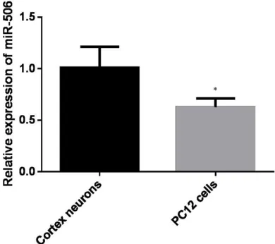
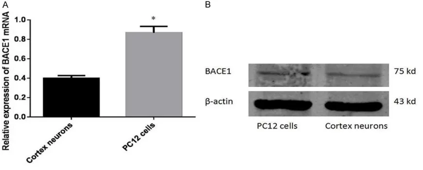
Expression levels of miR-506 and BACE1 in PC12 cells
[image:4.612.91.526.74.249.2]In order to determine the expression level of miR-506 and BACE1 in PC12 cell, we compared it with primary cortical neurons at both mRNA and protein level. As shown in Figure 1, PC12 cells had less expression of miR-506 in com-parison with cortical neurons (P < 0.05). Contrary to the low level of miR-506, endoge-nous BACE1 was up-regulated in PC12 cells (Figure 2).
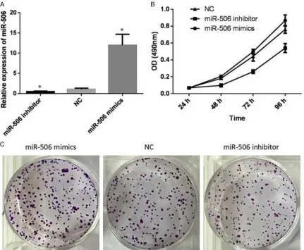
Over-expression of miR-506 promotes prolif-eration of PC12 cells
To examine the effect of miR-506 on the prolif-eration of PC12 cells, 506 mimics, miR-506 inhibitor and NC were transfected into PC12 cells at the final concentrations of 50 nM. We evaluated cell proliferation at 24, 48, 72 and 96 h post-transfection by MTT assays. In short, inhibition rate was calculated as follow-ing: inhibition rate (%) = (OD value of the control group-OD value of experimental group)/OD value of control group iR-506. In comparison with the NC group, miR-506 mimics group was promoted significantly (P < 0.05, Figure 3B) in time dependent manner. Conversely, cell
eration was strongest inhibited when cells were interfered with 50 nM miR-506 inhibitor for 96 h (P < 0.05, Figure 3B). Colony formation assays also showed much more colony forma-tion in the group transfected with miR-506 mimics. The colony number decreased when transfected with miR-506 inhibitors (P < 0.05, Figure 3C). On the whole, these data suggest-ed that over-expression of miR-506 promotsuggest-ed proliferation of PC12 cells.
Over-expression of miR-506 can change the PC12 cells cycle distribution
Forty-eight hours after the transfection, flow cytometry analysis indicated that the percent-age of G0/G1 phase (57.44%) dramatically de-
creased in the miR-506 mimics group, when compared with that of the NC group (64.19%) and miR-506 inhibitor group (65.19%) (P < 0.05, Figure 4), while there were no statistical difference between the NC group and miR-506 inhibitor group (P > 0.05). These findings revealed that miR-506 can reduce G0/G1 phase arrest. Upregulation of miR-506 expres-sion could lead to increase of S-phase and G2/M phase cells (Figure 4).
Over-expression of miR-506 has no significant effect on apoptosis of PC12 cells
[image:5.612.93.524.74.429.2]Apoptotic cell death was assessed using flow cytometric analysis of Annexin V-FITC/PI stain-ing. As presented in Figure 5, the percentage of
the early and late apoptotic cells was a little higher in the miR-506 inhibitor group compared with the miRNA mimics group. But we found no statistical difference between these three groups (P > 0.05). These results indicated that miR-506 had no significant effect on PC12 cell apoptosis in vitro.
miR-506 can specifically target BACE1 gene and inhibit BACE1 at protein levels
In the preliminary experiment, we found that contrary to the low expression of miR-506, endogenous BACE1 mRNA and protein
expres-sion were up-regulated in PC12 cells compared with primary cortical neurons. These results supported our previous hypothesis.
[image:6.612.95.520.74.239.2]To validate that miR-506 can bind to the pre-dicted site BACE1, we performed a luciferase reporter assay in the 293T cell line. As present-ed in Figure 6, the luciferase activity signifi-cantly decreased after co-transfection with psi-CHECK-2/BACE1 3’-UTR and miR-506 mimics in comparison with NC. However, this signifi-cant action of miR-506 was abolished following co-transfection of psiCHECK-2/BACE1 3’-UTR mutant and miR-506 mimics. Briefly, this result
Figure 4. Cell cycle distribution was analyzed by flow cytometry 48 h after transfection of PC12 cells with 50 nM miR-506 mimics or inhibitor or NC. Flow cytometry analysis indicated that the percentage of G0/G1 phase cells (57.44%) dramatically decreased in the miR-506 mimics group, when compared with that of the NC group (64.19%) and miR-506 inhibitor group (65.79%) (P < 0.05).
[image:6.612.97.518.319.484.2]apeutic agents can effectively reverse its pro-gression. In search for non-invasive biomarkers for Alzheimer’s disease (AD) diagnosis and prognosis, circulating miRNAs are emerging as a promising candidates these decades. What’s more, increasing evidence indicates that miR-NAs may affect cancer pathogenesis. Dys- regulation of miRNAs is related to initiation and progression of AD since they may affect the amyloid production as well as neurofibrillary tangles. For example, miR-29 expression con-trarily correlated with BACE1 (miR-29) whereas it had been presented to increase amyloid pro-duction in vitro [7]; miR-34, expressed highly in the hippocampus of patients with AD, regulated the p53 associated to tau phosphorylation [12]; additionally, miR-107 [13], reported to be down-regulated in temporal cortex at an early stage of AD had a converse correlation with BACE1 and plaque density [14]. In the present study, we are interested in the role of miR-506 in AD. The role of miR-506 in cell differentiation and senescence was recently demonstrated in some tumour types, including neuroblastoma [15], lung [16], breast, ovarian [17] and cervical
indicated that miR-506 spe-cifically binds to the 3’-UTR of BACE1 mRNA.
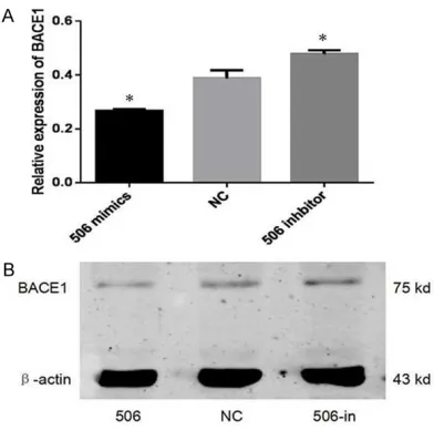
To discover whether miR-506 regulates BACE1, we trans-fected miR-506 inhibitors or mimics (50 nM) into PC12 cells, and the levels of BACE1 protein were detected.
As shown in Figure 7, we found that at the protein lev-els, BACE1 expression clearly decreased in the miR-506 mimics group in comparison with NC group and miR-506 inhibitor group. Together, th- ese results validate our previ-ous hypothesis that BACE1 is a novel target of miR-506. Discussion
Alzheimer’s disease (AD) is a progressive neurodegenera-tive disorder and its most common characteristic is defined as cognitive dysfunc-tion. To date, no existing
[image:7.612.93.375.74.308.2]ther-Figure 6. MiR-506 can specifically target BACE1 gene. A. The binding site for miR-506 in the 3’-UTR of BACE1 mRNA. B. The relative luciferase activ-ity was analyzed in PC12 cells after co-transfection of the BACE1 3’-UTR or BACE1 3’-UTR mutant luciferase construct with either miR-506 mimics or NC. As shown above, the relative BACE1 expression was significantly down-regulated by BACE1 3’-UTR luciferase construct with miR-506 mimics com-pared with NC group (P < 0.05). However, no significant difference was found between BACE1 3’-UTR mutant luciferase construct with miR-506 mimics and NC control.
[image:7.612.91.287.428.622.2]cancer [18] etc. The previous studies showed that miR-506, effectively reduced the tumor burden and inhibited invasive growth and metastasis. Therefore, miR-506 has been con-ceived as a promising new therapeutic agent that can be carried to suppress cancer progres-sion. But no connection has been discovered with the neurodegenerative disease.
We performed qRT-PCR to assess the expres-sion level of miR-506 in primary cortical neu-rons and PC12 cell lines. We further investigat-ed the biological impact of miR-506 and the molecular mechanisms by which miR-506 mod-ulates the behavior of PC12 cells.
Firstly, we investigated the expression levels of miR-506 in primary cortical neurons and PC12 cell lines. Intriguingly, we found that the expression levels of miR-506 were obviously decreased in PC12 cells compared with corti-cal neurons extracted from SD fetal rats. What’s more, we further tested the gain-or-loss effects of miR-506. MiR-506 mimics, miR-506 inhibitor and NC were transfected into PC12 cells to adjust its own expression. The exoge-nous over-expression of miR-506 regulating by miR-506 mimics strikingly promoted prolifera-tion and colony formaprolifera-tion ability of PC12 cells as evaluated by MTT and colony formation assays, respectively. We also found that miR-506 distinctly decreased percentage of cells at the G0/G1 phase in the cell cycle assay.
To determine how miR-506 caused these effects on the cell function, we tested assumed targets of miR-506 and identified BACE1, which is the most critical protease in the amyloido-genic metabolism. In previous research, BACE1, levels of which have been shown to be increased in the common late-onset sporadic AD [19]. Considered as a potential target for therapies against AD, BACE1 is characterized by accumu-lation of plaques formed of amyloid precursor protein [20-22]. Furthermore, Willem M found BACE1 was important in the formation of myelin sheaths in peripheral nerve cells [23]. Recently, BACE1 level has shown to be regulated by microRNAs, such as 29a, 29b-1, miR-29c [24], miR-107 and miR-124 [25] etc. in vitro.
In our study, we performed a luciferase report-er assay to detreport-ermine that miR-506 can bind to
the predicted site BACE1. We identified BACE1 as a direct target of miR-506 in PC12 cells. As shown above, BACE1 decreased when trans-fected with miR-506 mimics but increased when transfected with miR-506 inhibitors. In a word, these results show that enforced expres-sion of miR-506 in PC12 cells triggered an evi-dent inhibitory effect on BACE1 expression. On the whole, this experiment is carried out in the transient transfection of miR-506 with LipofectamineTM2000, and the inhibition of gene is not stable enough. In addition, we have not clarified the role of miR-506 in vivo but only in PC12 cells. Related research shall be further expanded.
To conclude, our findings indicate that miR-506 is down-regulated in PC12 cells and over-expression of miR-506 is able to promote cel-lular proliferation as well as disrupt the cell cycle via direct regulation of BACE1. Further- more, its expression has a negative correlation with BACE1, which is highly direct correlation with amyloid production, neuritic plaque densi-ty, as well as neurofibrillary tangles. Just as the emerging role for miRNA in AD, miR-506 may also act as a potential therapeutic target spot for AD.
Acknowledgements
This study was supported by grants from the National Natural Science Foundation of China (No. 81171163, No. 81371212).
Disclosure of conflict of interest
None.
Address correspondence to: Xue-Yuan Liu and Yan-Xin Zhao, Department of Neurology, Shanghai Tenth People’s Hospital, Tongji University, Middle Yan- chang Rd. 301#, Zhabei District, Shanghai, China. Tel: 86-21-66302582; E-mail: 1053793158@qq. com (XYL); Tel: 86-21-66301167; E-mail: zhao_ [email protected] (YXZ)
References
[1] Hunt CE, Turner AJ. Cell biology, regulation and inhibition of beta-secretase (BACE-1). FEBS J 2009; 276: 1845-59.
[3] Provost P. Interpretation and applicability of microRNA data to the context of Alzheimer’s and age-related diseases. Aging 2010; 2: 166-9.
[4] Lukiw WJ, Zhao Y, Cui JG. An NF-kappaB-sensitive micro RNA-146a-mediated inflamma-tory circuit in Alzheimer disease and in stressed human brain cells. J Biol Chem 2008; 283: 31315-22.
[5] Yao J, Hennessey T, Flynt A, Lai E, Beal MF, Lin MT. MicroRNA-related cofilin abnormality in Alzheimer’s disease. PLoS One 2010; 5: e15546.
[6] Shioya M, Obayashi S, Tabunoki H, Arima K, Saito Y, Ishida T, Satoh J. Aberrant microRNA expression in the brains of neurodegenerative diseases: miR-29a decreased in Alzheimer dis-ease brains targets neurone navigator 3. Neuropathol Appl Neurobiol 2010; 36: 320-30.
[7] Hebert SS, Horre K, Nicolai L, Papadopou- lou AS, Mandemakers W, Silahtaroglu AN, Kauppinen S, Delacourte A, De Strooper B. Loss of microRNA cluster miR-29a/b-1 in spo-radic Alzheimer’s disease correlates with in-creased BACE1/beta-secretase expression. Proc Natl Acad Sci U S A 2008; 105: 6415-20. [8] Wang WX, Rajeev BW, Stromberg AJ, Ren N,
Tang G, Huang Q, Rigoutsos I, Nelson PT. The expression of microRNA miR-107 decreases early in Alzheimer’s disease and may acceler-ate disease progression through regulation of beta-site amyloid precursor protein-cleaving enzyme 1. J Neurosci 2008; 28: 1213-23. [9] Boissonneault V, Plante I, Rivest S, Provost P.
MicroRNA-298 and microRNA-328 regulate expression of mouse beta-amyloid precur- sor protein-converting enzyme 1. J Biol Chem 2009; 284: 1971-81.
[10] Faghihi MA, Zhang M, Huang J, Modarresi F, Van der Brug MP, Nalls MA, Cookson MR, St-Laurent G 3rd, Wahlestedt C. Evidence for natural antisense transcript-mediated inhibi-tion of microRNA funcinhibi-tion. Genome Biol 2010; 11: R56.
[11] Lagos-Quintana M, Rauhut R, Yalcin A, Meyer J, Lendeckel W, Tuschl T. Identification of tissue-specific microRNAs from mouse. Curr Biol 2002; 12: 735-9.
[12] Hooper C, Meimaridou E, Tavassoli M, Melino G, Lovestone S, Killick R. p53 is upregulated in Alzheimer’s disease and induces tau phos-phorylation in HEK293a cells. Neurosci Lett 2007; 418: 34-7.
[13] Nelson PT, Wang WX. MiR-107 is reduced in Alzheimer’s disease brain neocortex: valida-tion study. J Alzheimers Dis 2010; 21: 75-9.
[14] Goodall EF, Heath PR, Bandmann O, Kirby J, Shaw PJ. Neuronal dark matter: the emerging role of microRNAs in neurodegeneration. Front Cell Neurosci 2013; 7: 178.
[15] Zhao Z, Ma X, Hsiao TH, Lin G, Kosti A, Yu X, Suresh U, Chen Y, Tomlinson GE, Pertsemlidis A, Du L. A high-content morphological screen identifies novel microRNAs that regulate neu-roblastoma cell differentiation. Oncotarget 2014; 5: 2499-512.
[16] Zhao Y, Liu H, Li Y, Wu J, Greenlee AR, Yang C, Jiang Y. The role of miR-506 in transformed 16HBE cells induced by anti-benzo[a]pyrene-trans-7, 8-dihydrodiol-9, 10-epoxide. Toxicol Lett 2011; 205: 320-6.
[17] Liu G, Sun Y, Ji P, Li X, Cogdell D, Yang D, Parker Kerrigan BC, Shmulevich I, Chen K, Sood AK, Xue F, Zhang W. MiR-506 suppresses prolifera-tion and induces senescence by directly target-ing the CDK4/6-FOXM1 axis in ovarian cancer. J Pathol 2014; 233: 308-18.
[18] Wen SY, Lin Y, Yu YQ, Cao SJ, Zhang R, Yang XM, Li J, Zhang YL, Wang YH, Ma MZ, Sun WW, Lou XL, Wang JH, Teng YC, Zhang ZG. miR-506 acts as a tumor suppressor by directly target-ing the hedgehog pathway transcription factor Gli3 in human cervical cancer. Oncogene 2015; 34: 717-25.
[19] Li Q, Chen M, Liu H, Yang L, Yang G. Expression of APP, BACE1, AChE and ChAT in an AD model in rats and the effect of donepezil hydrochlo-ride treatment. Mol Med Rep 2012; 6: 1450-4. [20] Hussain I, Powell D, Howlett DR, Tew DG, Meek
TD, Chapman C, Gloger IS, Murphy KE, Southan CD, Ryan DM, Smith TS, Simmons DL, Walsh FS, Dingwall C, Christie G. Identification of a novel aspartic protease (Asp 2) as beta-secre-tase. Mol Cell Neurosci 1999; 14: 419-27. [21] Sinha S, Anderson JP, Barbour R, Basi GS,
Caccavello R, Davis D, Doan M, Dovey HF, Frigon N, Hong J, Jacobson-Croak K, Jewett N, Keim P, Knops J, Lieberburg I, Power M, Tan H, Tatsuno G, Tung J, Schenk D, Seubert P, Suomensaari SM, Wang S, Walker D, Zhao J, McConlogue L, John V. Purification and cloning of amyloid precursor protein beta-secretase from human brain. Nature 1999; 402: 537-40. [22] Yan R, Bienkowski MJ, Shuck ME, Miao H, Tory
MC, Pauley AM, Brashier JR, Stratman NC, Mathews WR, Buhl AE, Carter DB, Tomasselli AG, Parodi LA, Heinrikson RL, Gurney ME. Membrane-anchored aspartyl protease with Alzheimer’s disease beta-secretase activity. Nature 1999; 402: 533-7.
nerve myelination by the beta-secretase BACE1. Science 2006; 314: 664-6.
[24] Lei X, Lei L, Zhang Z, Zhang Z, Cheng Y. Downregulated miR-29c correlates with in-creased BACE1 expression in sporadic Alzheimer’s disease. Int J Clin Exp Pathol 2015; 8: 1565-74.