shown that some new metabolic parameters of 18F-FDG (Fluor-18-fluorodeoxyglucose, 18 F-FDG) PET/CT, including MTV (Metabolic Tumor Volume), TLG (Total Lesion Glycolysis), HF (Intra-tumoral Heterogeneity Factor) have value for the assessment of prognosis of malignant tumors [2-8]. Therefore, whether 18F-FDG PET/ CT metabolic parameters can be used to pre-dict the regional lymph node metastasis of esophageal cancer deserved further investiga -tion. The current study aimed to investigate potential correlation between metabolic param-eters and lymph node metastasis, diagnostic value of metabolic parameters on lymph node metastasis of esophageal cancer, and the scope of lymph node dissection during opera -tion based on preoperative metabolic parame -ters of 18F-FDG PET/CT and postoperative pathology results.
Introduction
Esophageal cancer is a common malignant tumor with an increasingly high incidence. In China, the incidence of new esophageal can-cer was about 47.79% and the mortality rate was about 37.5% in 2015 [1]. Accurate staging and assessment of the range of lesions is the basis for comprehensive diagnosis and sur -gical treatment of esophageal cancer. In addi -tion, an accurate assessment of the existence of regional lymph node metastasis of esopha -geal cancer is of great significance to select therapeutic regimens and guide subsequent treatments. At present, lymph node metastasis is mainly identified by imaging techniques such as CT, intracavitary ultrasound and Positron Emission Tomography-Computed Tomography (PET/CT). In recent years, some studies have
Original Article
Correlation between
18F-FDG PET/CT metabolic
parameters and lymph node metastasis
of esophageal cancer
Zhiyu Huang1, Shaoxing Chen2, Jiancheng Li1
1Department of Radiation Oncology, Fujian Cancer Hospital Affiliated to Fujian Medical University, Fuzhou, PR
China; 2Department of Radiation Oncology, Provincial Clinical College of Fujian Medical University, Fuzhou, PR
China
Received June 2, 2018; Accepted August 4, 2018; Epub March 15, 2019; Published March 30, 2019
Abstract: Objective: The goal of this study was to investigate the correlation between lymph node metastasis and
18F-FDG PET/CT metabolic parameters in primary esophageal patients. Materials and Methods: A total of 82 esoph
-ageal cancer patients that were pathologically confirmed and received initial treatment in our hospital from October 2011 to September 2016 were retrospectively analyzed. All patients underwent systemic 18F-FDG PET/CT
exami-nation within two weeks before operation but without receiving any anti-tumor treatment before operation. The metabolic parameters of primary lesions and postoperative pathological lymph node metastasis of esophageal cancer patients were recorded. Results: There was strong correlation between metabolic parameters of primary lesion (including SUVmax, SUVmean, MTV, TLG, HF) and lymph node metastasis of esophageal cancer, and TLG of primary lesion had a certain diagnostic value for the lymph node metastasis of esophageal cancer. In addition, ROC curve analysis showed that the minimum TLG for diagnosis of lymph node metastasis of esophageal cancer at the upper and middle segment was 23.06, while the thoracic esophageal cancer at the lower segment was not statisti
-cally significant. Conclusion: 18FDG-PET/CT metabolic parameters (SUVmax, SUVmean, MTV, TLG, HF) are closely
related to lymph node metastasis of esophageal cancer. The TLG has a certain diagnostic value for the lymph node metastasis of esophageal cancer.
Materials and methods
Subjects
Esophageal cancer patients treated in our hos -pital from October 2011 to September 2016 were collected. There were 82 cases meeting inclusion criteria, and all of them were diag -nosed with squamous cell carcinoma, including 70 males and 12 females, aged between 41 and 78 years, and medium age of 59 years. None of the patients had a medical history of a tumor, and they underwent systematic 18F-FDG PET/CT examination two weeks before the operation.
The present study was conducted in accor -dance with the Declaration of Helsinki and was approved by the Ethics Committee of Fujian Provincial Cancer Hospital. Written informed consent was obtained from all participants.
18F-FDG PET/CT equipment and method
[image:2.612.87.299.96.272.2]The Gemini TF 64 PET/CT instrument (Philips, the Netherlands) was used. 18F-FDGT was gen -erated from Sumitomo Corporation HM-10 cyclotron, with the radiochemical purity grea-ter than 95%. Before the examination, pa-tients were fasted for more than 6 hours, and blood glucose levels were controlled within 3.9~7.5 mmol/L. After intravenous injection of
Table 1. MTVs corresponding to different percentages of SUVmax in a patient
40%
SUVmax SUVmax50% SUVmax60% SUVmax70% SUVmax80% MTV (cm3) 32.817 25.777 18.792 11.295 4.334
Figure 1. Linear relationship based on Table 1. HF value was the slope of the liner function. The slope was negative because of negative correlation be
-tween them. In this study, the absolute value was taken, i.e. the HF of the patient was 0.715 (HF =
0.715).
the skull base to the upper end of femur at a rate of 1 min/person. The acquisition of imag-es was performed by a three-dimensional mo-del, then attenuation correction was made by CT data. The fusion of PET and CT images was performed on EBW2.0 post-processing workstation, to get the PET, CT and PET/CT fusion images of cross-section, sagittal, and coronal plane. The maximum standard uptake (SUVmax) was measured by ROI (region of inter -est, ROI).
Acquisition of metabolic parameters
SUVmax refers to the maximum SUV in ROI. SUVmean refers to the mean SUV in ROI. MTV refers to the volume of high metabolic tumor tissue. The volume of lesions was divided according to different bottom-line thresholds, and the MTV values were different. At present, the commonly used thresholds were SUV = 2.5, 40% SUVmax, 50% SUVmax, etc. [9]. The PET Cancer Imaging Guide of 2010 Annual Con-ference of European Society of Nuclear Me-dicine recommended 40% SUVmax as the low-er threshold of ROI [10]. Thlow-erefore, in this st-udy, 40% SUVmax was used as a threshold to measure the images, and the system automati -cally calculated SUVmean, MTV, TLG = MTV × SUVmean [11].
Measurement of metabolic heterogeneity fac-tor (HF)
Tumor metabolic heterogeneity was determin-ed by primary lesion metabolic volume and volume of adjacent normal tissue [12]. A differ -ent perc-entage of SUVmax had a correspond-ing MTV, and there was a linear relationship between both of them, and through conver-sion, the HF value was obtained. It has been reported that when converting HF, the SUVmax that was less than 40% and greater than 80% should be ruled out [12]. For example, the MTVs corresponding to different percentages of SUVmax in a patient are shown in Table 1
and Figure 1.
Univariate analysis of PET/CT metabolic pa-rameters and lymph node metastasis
As shown in Table 2, the PET/CT metabolic parameters were divided into two groups, including SUVmax (cutoff value 7.350, divided into two groups: SUVmax ≥ 7.350, SUVmax < 7.350), SUVmean (cutoff value 4.399, divided into two groups: SUVmean ≥ 4.399, SUVmean < 4.399), MTV (cutoff value 5.375, divided into two groups: MTV ≥ 5.375, MTV < 5.375), TLG (cutoff value 24.080, divided into two gro-ups: TLG ≥ 24.080, TLG < 24.080), and HF (cut -off value 0.1385, divided into two groups: HF ≥ 0.1385, HF < 0.1385). The univariate analysis of PET/CT metabolic parameters and lymph node metastasis was performed by logistic binary regression and the results showed that, all metabolic parameters were statistically sig -nificant (P = 0.001, P = 0.001, P = 0.001, P < 0.001, P = 0.001, respectively), suggesting that there was a correlation between metabo- lic parameters and lymph node metastasis of esophageal cancer.
Multivariate analysis of PET/CT metabolic parameters and lymph node metastasis
The above univariate analysis on PET/CT me-tabolic parameters showed that SUVmax,
Statistical analysis
Data analysis was performed by SPSS for Windows 17.0 software. ROC curve analysis was conducted to calculate the AUC, sensitivity, specificity. Logistic binary regression analysis was performed, and P < 0.05 was considered statistically significant.
Results
Correlation of PET/CT metabolic parameters and lymph node metastasis: ROC curve analy-sis
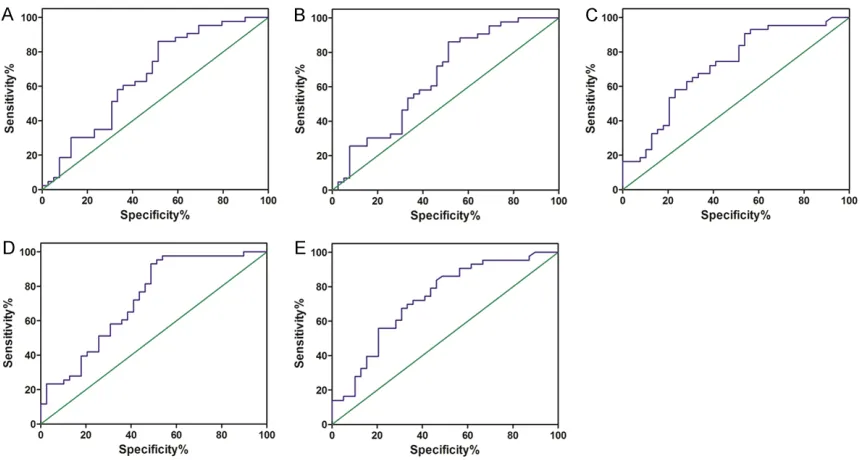
[image:3.612.91.524.70.300.2]The correlation between metabolic parame- ters and lymph node metastasis was investi -gated by ROC (Receiver Operating Charac-teristic Curve, ROC) analysis. As shown in Fi- gure 2, the AUC of SUVmax was 0.657, cutoff value was 7.350, P = 0.014. The AUC of SUVmean was 0.655, cutoff value was 4.399, P = 0.016. The AUC of MTV was 0.719, cutoff value was 5.375, P = 0.001. The AUC of TLG was 0.722, cutoff value was 24.080, P = 0.001. The AUC of HF was 0.727, cutoff value was 0.1385, P = 0.001. These data suggested th-at there was correlth-ation between metabolic parameters and lymph node metastasis of esophageal cancer.
Figure 2. ROC analysis for the correlation of PET/CT metabolic parameters and lymph node metastasis. A. Relation
diagnosis of lymph node metastasis of esopha -geal cancer at the upper and middle segment was 23.06.
Discussion
Esophageal cancer is a common malignant tumor, and current treatment is mainly focused on the comprehensive treatment. The exis -tence of lymph node metastasis plays a deci -sive role for the choice of surgical approaches and the extent of lymph node dissection, so it is important to assess lymph node metastas-is before surgery. The commonly-used lymph node examination methods such as CT, MRI, intracavity ultrasound belong to anatomical structure imaging with some limitations. It was found that, the PET/CT diagnosis of esophageal cancer lymph node metastasis was more ac-curate than the enhanced CT [11]. In addition, another research study by van Vliet and his group [13] conducted the meta-analysis with the results showing that, both sensitivity and specificity of PET/CT for diagnosis of regional lymph node of esophageal cancer were superi -or to CT and intra-cavity ultrasound. At present, the PET/CT diagnosis of lymph node metasta -sis of esophageal cancer should be subject to the locally high metabolic lesions in the lym -phatic drainage area observed by naked eyes. A number of lymph node metastases were found after operations that were not found before operation. The PET/CT metabolic param-eters were correlated with the lymph node metastasis, but whether it can be used to pre -dict lymph node metastasis needs to be further studied.
Accurate preoperative staging played an impor -tant role in the development of surgical proce -SUVmean, MTV, TLG, and HF were statistically
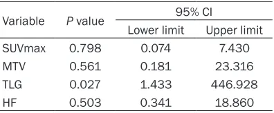
[image:4.612.97.522.84.243.2]significant, then we conducted multivariate analysis on all parameters (SUVmax, SUVmean, MTV, TLG, HF) and found that only TLG was statistically significant, P = 0.027 (Table 3). The correlation between TLG and lymph node metastasis was analyzed by ROC curve. Its AUC was greater than 0.7. When the cutoff value was 24.080, the sensitivity was 93.0%, speci -ficity was 51.3%, and Youden index was 0.443, P = 0.001, suggesting that TLG of primary lesion has diagnostic value for lymph node metastasis of esophageal cancer, and the mini -mum TLG for diagnosis of lymph node metasta -sis of esophageal cancer was 24.08. In addi -tion, according to different sites of primary lesion of esophageal cancer, ROC curve analy -sis on TLG of the thoracic esophageal cancers at the upper and middle segment and the lower segment was performed, and it was found that the thoracic esophageal cancers at the upper and middle segment were statistically signifi -cant. When TLG > 23.06, its AUC was 0.755, the sensitivity was 96.7%, specificity was 51.7%, and Youden index was 0.484, P = 0.001, while the thoracic esophageal cancer at the lower segment was not statistically signifi -cant, suggesting that the minimum TLG for
Table 2. Correlation between lymph node metastasis and PET parameters
Variable Range lymph node metastasisNumber of cases with Number of cases without lymph node metastasis P value 95% CI Lower limit Upper limit
SUVmax ≥ 7.350 37 20 0.001 2.016 17.027
< 7.350 6 19
SUVmean ≥ 4.399 37 20 0.001 2.016 17.027
< 4.399 6 19
MTV ≥ 5.375 39 21 0.001 2.501 27.923
< 5.375 4 18
TLG ≥ 24.080 40 20 < 0.001 3.348 47.927
< 24.080 3 19
HF ≥ 0.1385 36 18 0.001 2.152 16.732
< 0.1385 7 21
Table 3. Multivariate analysis of lymph node metastasis and PET parameters
Variable P value 95% CI
Lower limit Upper limit
SUVmax 0.798 0.074 7.430
MTV 0.561 0.181 23.316
TLG 0.027 1.433 446.928
[image:4.612.91.289.289.371.2]diameter of tracheal esophageal bypass, peri -cardium and abdominal lymph node long-axis diameter > 0.5 cm on the CT images. The PET/ CT metabolic parameters are mainly used to evaluate whether tumors are benign or malig -nant. Currently, at SUVmax > 2.5, malignancy is suspected. It was difficult to identify the region -al lymph node metastasis of esophage-al can -cer by the SUVmax value [18]. For other param -eters, a small number of studies have suggest -ed a correlation with the lymph node metasta -sis of esophageal cancer [17-21], but its diag -nostic value on lymph node metastasis of esophageal cancer was rarely mentioned. TLG is the product of SUVmean and MTV, which combines two factors namely SUV and MTV, so theoretically its application value is higher than other metabolic parameters. In this study, the logistic univariate regression analysis showed that, there was a correlation between PET/CT metabolic parameters (SUVmax, SUVmean, MTV, TLG and HF) and lymph node metastasis of esophageal cancer, all P values were less than 0.01. The further logistic multivariate regression analysis on each metabolic param -eter found that, TLG was statistically signifi -cant, P = 0.027, suggesting that TLG had diag -nostic value for the lymph node metastasis of esophageal cancer. In addition, by studying the TLG and lymph node metastasis of esophageal cancer through ROC curves, it was found that when TLG = 24.08, its AUC was 0.722, the sen -sitivity was 93.0%, the specificity was 51.3%, P = 0.001. The results show that TLG had a cer -tain diagnostic value for lymph node metasta -sis of esophageal cancer, and the minimum TLG was 24.08 for the diagnosis. According to the different sites of primary lesions of esopha -geal cancer, ROC curve analysis was conducted for TLG of the thoracic esophageal cancers at the upper and middle segment and the lower segment. The results show that the thoracic esophageal cancers at the upper and middle segment were statistically significant. When TLG > 23.06, its AUC was 0.755, the sensitivity was 96.7%, specificity was 51.7%, P = 0.001, while the thoracic esophageal cancer at the lower segment was not statistically significant, suggesting that the minimum TLG for diagnosis of lymph node metastasis of esophageal can -cer at the upper and middle segment was 23.06.
At present, lymph node dissection of esopha -geal cancer surgery mainly includes chest and dures for esophageal cancer. It was found that,
for patients receiving surgery of esophageal cancer, the 5-year survival rate of patients with lymph node metastasis was less than 15% (< 15%), while the 5-year survival rate of patients without lymph node metastasis was more than 40% (> 40%) [14]. Because PET/CT examina -tion is expensive, it is mainly used for clinical examination of esophageal cancer with distant metastasis, and the CT examination mainly used for of esophageal cancer without lymph node metastasis. At present, fewer studies on the correlation between PET metabolic param-eters and lymph node metastasis have been reported. In earlier studies, for example, retro -spective analysis on 49 patients with esopha -geal squamous cell carcinoma examined by PET/CT before operation showed that, SUVmax and MTV were correlated with lymph node metastasis [15]. Another retrospective analys-is was conducted on 51 esophageal cancer patients by preoperative PET/CT examination and found that there was correlation between metabolic parameters (SUVmax, MTV, TLG, HF) and lymph node metastasis, and further multi -variate analysis showed that only HF was sta-tistically significant [16]. It was also found that MTV, SUVmax were correlated with the lymph node metastasis of esophageal cancer [17]. 18F-FDG PET/CT metabolic parameters were proportional to the glucose metabolism of tis -sues, and most tumors were highly metabolic [9]. The larger the metabolic parameters, the more advanced the tumor, and the higher de-gree of malignancy, and therefore, the higher possibility the occurrence of regional lymph node metastasis. In this study, PET/CT meta -bolic parameters and lymph node metastasis were studied using ROC curve, and all P values were less than 0.05, suggesting that there was a correlation between PET/CT metabolic param-eters (including SUVmax, SUVmean, MTV, TLG, HF) and lymph node metastasis of esophageal cancer. Then logistic univariate regression an-alysis also suggested that there was such correlation, in which, SUVmax (P = 0.001), SUVmean (P = 0.001), MTV (P = 0.001), TLG (P < 0.001), and HF (P = 0.001).
with esophageal cancers at upper and middle thoracic segments.
Disclosure of conflict of interest
None.
Address correspondence to: Dr. Jiancheng Li, De-partment of Radiation Oncology, Fujian Cancer Hospital Affiliated to Fujian Medical University, 420 Fuma Road, Fuzhou 350014, Fujian, PR China.
Tel: +86-13906900190; Fax: +86-591-83928767;
E-mail: [email protected]
References
[1] Chen W, Zheng R, Baade PD, Zhang S, Zeng H, Bray F, Jemal A, Yu XQ and He J. Cancer statis
-tics in China, 2015. CA Cancer J Clin 2016; 66:
115-132.
[2] Chang S and Kim SJ. Prediction of recurrence and mortality of locally advanced esophageal cancer patients using pretreatment F-18 FDG PET/CT parameters: intratumoral heterogene
-ity, SUV, and volumetric parameters. Cancer
Biother Radiopharm 2016; 31: 1-6.
[3] Liu XQ, Zhou HZ and Du J. Prognostic value of metabolic tumor volume measured by 18F-FDG
PET/CT imaging in preoperative breast cancer
patients. J Clin Med Pract 2014; 18: 55-59. [4] Chung MK, Jeong HS, Park SG, Jang JY, Son YI,
Choi JY, Hyun SH, Park K, Ahn MJ, Ahn YC, Kim HJ, Ko YH and Baek CH. Metabolic tumor vol
-ume of [F-18]-Fluorodeoxyglucose positron emission tomography/computed tomography predicts short-term outcome to radiotherapy with or without chemotherapy in pharyngeal
cancer. Clin Cancer Res 2009; 15: 5861-5868.
[5] Hyun SH, Choi JY, Shim YM, Kim K, Lee SJ, Cho YS, Lee JY, Lee KH and Kim BT. Prognostic val
-ue of metabolic tumor volume measured by 18F-fluorodeoxyglucose positron emission to
-mography in patients with esophageal carci
-noma. Ann Surg Oncol 2010; 17: 115-122.
[6] Xie YT, Zheng R, Wu N, Zhang WJ, Liang Y, Liu Y, Li XM, Liang M and Xiao ZF. Prognostic value
of FDG PET-CT metabolic parameters before
treatment in patients with esophageal cancer receiving radiotherapy. Chin J Radiat Oncol
2016; 25: 452-456.
[7] Yildirim F, Yurdakul AS, Ozkaya S, Akdemir UO and Ozturk C. Total lesion glycolysis by 18F-FDG
PET/CT is independent prognostic factor in pa
-tients with advanced non-small cell lung can -cer. Clin Respir J 2017; 11: 602-611.
[8] Son SH, Kim DH, Hong CM, Kim CY, Jeong SY, Lee SW, Lee J and Ahn BC. Prognostic implica
-stomach, or neck, chest, abdomen. There is no consensus on prognosis of the two surgical pro -cedures. It was found that, the 1-year, 2-year and 5-year survival rates of esophageal cancer patients who underwent three-field lymph node dissection were higher than those who under -went two-field lymph node dissection [21]. But it was also reported that there was no statisti-cal difference in the postoperative survival time between the two kinds of surgical procedures [20]. Due to the complex anatomical structure around the cervical lymph nodes, it was easy to cause injury of recurrent laryngeal nerves, anastomotic fistula and respiratory complica -tions during dissection. Therefore, not all eso-phageal cancer patients should choose the neck, chest, and abdominal lymph node dissec -tion, instead, we should conduct assessment on the lymph node metastasis before opera-tion and select appropriate surgical proce -dures. For patients who were highly suspicious of lymph node metastasis with high TLG of pri -mary lesions found in preoperative PET/CT examination, the possibility of metastasis was high, and neck, chest, abdominal lymph node dissection should be performed to achieve better prognosis. For patients who were suspi -cious of lymph node metastasis with low TLG of primary lesions found in preoperative PET/ CT examination, the possibility of metastasis was low. Therefore, considering tumor sites, we should avoid the neck, chest, abdominal lymph node dissection to reduce the surgical complications.
Conclusion
[16] Kim SJ, Pak K and Chang S. Determination of regional lymph node status using (18)F-FDG PET/CT parameters in oesophageal cancer pa
-tients: comparison of SUV, volumetric parame
-ters and intratumoral heterogeneity. Br J Radiol
2016; 89: 20150673.
[17] I HS, Kim SJ, Kim IJ and Kim K. Predictive value of metabolic tumor volume measured by
18F-FDG PET for regional lymph node status in
patients with esophageal cancer. Clin Nucl
Med 2012; 37: 442-446.
[18] Tangoku A, Yamamoto Y, Furukita Y, Goto M and Morimoto M. The new era of staging as a key for an appropriate treatment for esopha
-geal cancer. Ann Thorac Cardiovasc Surg
2012; 18: 190-199.
[19] Jung JH, Kim CY, Son SH, Kim DH, Jeong SY, Lee SW, Lee J and Ahn BC. Preoperative pre
-diction of cervical lymph node metastasis us
-ing primary tumor SUVmax on 18F-FDG PET/CT
in patients with papillary thyroid carcinoma. PLoS One 2015; 10: e0144152.
[20] Igaki H, Tachimori Y and Kato H. Improved sur
-vival for patients with upper and/or middle me
-diastinal lymph node metastasis of squamous
cell carcinoma of the lower thoracic
esopha-gus treated with 3-field dissection. Ann Surg
2004; 239: 483-490.
[21] Soydal C, Yuksel C, Kucuk NO, Okten I, Ozkan E and Doganay Erdogan B. Prognostic value of metabolic tumor volume measured by 18F-FDG
PET/CT in esophageal cancer patients. Mol Imaging Radionucl Ther 2014; 23: 12-15. tion of intratumoral metabolic heterogeneity in
invasive ductal carcinoma of the breast. BMC
Cancer 2014; 14: 585.
[9] Hu N, Wu YG, Xiao LZ, Tan YL and Wang YH.
Metabolic parameters of 18F-FDG PET/CT and
their application in lymphoma. Int J Radiat
Med Nucl Med 2015; 39: 342-347.
[10] Boellaard R, O’Doherty MJ, Weber WA, Mottaghy FM, Lonsdale MN, Stroobants SG, Oyen WJ, Kotzerke J, Hoekstra OS, Pruim J, Marsden PK, Tatsch K, Hoekstra CJ, Visser EP, Arends B, Verzijlbergen FJ, Zijlstra JM, Comans EF, Lammertsma AA, Paans AM, Willemsen AT, Beyer T, Bockisch A, Schaefer-Prokop C, Delbeke D, Baum RP, Chiti A and Krause BJ. FDG PET and PET/CT: EANM procedure guide
-lines for tumour PET imaging: version 1.0. Eur J Nucl Med Mol Imaging 2010; 37: 181-200.
[11] Tan R, Zhu RJ, Ge QX, Lu BY, Yao SZ, Li X and Liu QW. The application of 18F-FDG PET/CT and
contrast enhanced MSCT in detecting meta
-static lymph nodes in esophageal cancer. Chin
J Nucl Med 2007; 27: 356-359.
[12] Kidd EA and Grigsby PW. Intratumoral meta
-bolic heterogeneity of cervical cancer. Clin
Cancer Res 2008; 14: 5236-5241.
[13] van Vliet EP, Heijenbrok-Kal MH, Hunink MG, Kuipers EJ and Siersema PD. Staging investi
-gations for oesophageal cancer: a meta-analy -sis. Br J Cancer 2008; 98: 547-557.
[14] Tanaka T, Matono S, Mori N, Shirouzu K and Fujita H. T1 squamous cell carcinoma of the esophagus: long-term outcomes and prognos
-tic factors after esophagectomy. Ann Surg
Oncol 2014; 21: 932-938.
[15] Zhu WQ, Yu JM, Sun XR, Xing LG, Xie P, Sun XD, Guo HB, Yang GR and Kong L. The prognostic value of metabolic tumor volume in FDG PET/ CT evaluation of post-operative survival in pa
-tients with esophageal squamous cell cancer.