organic papers
o236
Saminathan and Sivakumar C8H7NC6H3N3O7 doi:10.1107/S1600536806052512 Acta Cryst.(2007). E63, o236–o238 Acta Crystallographica Section E
Structure Reports
Online
ISSN 1600-5368
Indole–picric acid (1/1)
Kolandaivelu Saminathan and Kandasamy Sivakumar*
Department of Physics, Anna University, Chennai 600 025, India
Correspondence e-mail: [email protected]
Key indicators
Single-crystal X-ray study
T= 293 K
Mean(C–C) = 0.009 A˚ Disorder in main residue
Rfactor = 0.069
wRfactor = 0.271
Data-to-parameter ratio = 11.0
For details of how these key indicators were automatically derived from the article, see http://journals.iucr.org/e.
Received 20 September 2006 Accepted 4 December 2006
#2007 International Union of Crystallography All rights reserved
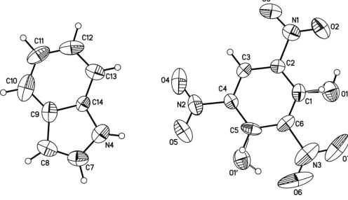
In the title molecular complex, C8H7NC6H3N3O7, the picric
acid molecule is disordered, corresponding to a rotation about one H—C C—N axis and resulting in two sites for the hydroxyl group. In the crystal structure, the indole and picric acid molecules are stacked in columns parallel to the b axis with significant –interactions. In addition, intermolecular hydrogen bonds link molecules in a zigzag fashion along thea
axis, giving ring patterns with graph-set motifs R22(8) and
R2 2
(7).
Comment
Picric acid acts not only as an acceptor forming various -stacking arrangements but also as an acid forming salts through electrostatic or hydrogen-bonding interactions (Inet al., 1997; Shrineret al., 1980). Indole derivatives form donor– acceptor (charge-transfer) complexes with a variety of aromatic electron acceptors (Gartlandet al., 1974). The indole molecule, with a lone pair of electrons on the N atom, does not act like a base as do amines or anilines, because the lone pair is delocalized and contributes to the aromatic system. Hence, very strong acids are needed to protonate a substantial amount of indole. Our attempts to co-crystallize picric acid and indole resulted in the formation of the 1:1 complex (I) and not the salt form, as expected.
The asymmetric unit of (I) is shown in Fig. 1. The positional disorder of the hydroxyl group observed in the picric acid molecule is similar to that in the structure of the indole-3-acetic acid–picric acid complex (Nagata et al., 1995). The essentially planar indole group and the aromatic ring of the picric acid are almost parallel to each other with a dihedral angle of 2.47 (1)and are packed alternately in columns along the b axis. This arrangement gives significant – stacking interactions with aCg1 Cg2(1x,1
2+y, 1
2x) distance of
3.507 (3) A˚ (whereCg1 andCg2 are the centroids defined by the rings N4/C7–C9/C14 and C1–C6, respectively) and a perpendicular distance of 3.35 A˚ .
two essentially equal to each other and one showing the nitro group to be almost in the plane of the benzene ring, may explain why the rotational disorder is about the C6 C3 axis so that both disorder components of the hydroxyl group of at positions C1 and C-5 have similar environments. The C—N distances show varying lengths, with C6—N3 having more single bond character (see Table 1). The C7—N4 and C14—N4 bond lengths in the indole molecule show clear bond fixation, indicating the absence of electron delocalization within the pyrole ring.
In the crystal structure, picric acid and indole molecules are linked by N—H O and C—H O hydrogen bonds to form chains of alternatingR2
2(8) andR 2
2(7) rings (Bernstein et al.,
1995) propagating in thea-axis direction (Table 2 and Fig. 2). In addition, intramolecular O1—H1 O2 and O10—H10 O6 hydrogen bonds form six-membered rings, both with a graph-set motif ofS(6).
Experimental
The title compound was prepared by dissolving equimolar amounts (1:1) of picric acid and indole in ethanol. Slow evaporation of the solution resulted in the formation of transparent red needle-shaped single crystals.
Crystal data C8H7NC6H3N3O7
Mr= 346.26
Monoclinic,P21=c
a= 13.4586 (19) A˚
b= 6.723 (3) A˚
c= 16.158 (4) A˚
= 94.912 (19)
V= 1456.7 (8) A˚3
Z= 4
Dx= 1.579 Mg m
3
MoKradiation
= 0.13 mm1
T= 293 (2) K Needle, red 0.40.20.2 mm
Data collection Enraf–Nonius CAD-4
diffractometer
!–2scans
Absorption correction: scan (Northet al., 1968)
Tmin= 0.989,Tmax= 1.000
2659 measured reflections
2546 independent reflections 816 reflections withI> 2(I)
Rint= 0.015
max= 25.0
2 standard reflections every 100 reflections intensity decay: 1%
Refinement Refinement onF2
R[F2> 2(F2)] = 0.069
wR(F2) = 0.271
S= 0.91 2546 reflections 231 parameters
H-atom parameters constrained
w= 1/[2
(Fo2) + (0.1531P)2]
whereP= (Fo2+ 2Fc2)/3
(/)max< 0.001
max= 0.40 e A˚
3
min=0.24 e A˚
3
Table 1
Selected bond lengths (A˚ ).
N1—C2 1.480 (6) N2—C4 1.450 (6) N3—C6 1.514 (7) C1—O1 1.286 (7)
C5—O10
[image:2.610.314.563.73.237.2]1.298 (7) N4—C7 1.353 (7) N4—C14 1.400 (6)
Table 2
Hydrogen-bond geometry (A˚ ,).
D—H A D—H H A D A D—H A
N4—H4 O5 0.86 2.50 3.346 (7) 168 C13—H13 O4 0.93 2.56 3.423 (8) 155 C7—H7 O2i
0.93 2.69 3.524 (8) 150 C8—H8 O3i
0.93 2.70 3.391 (7) 132 O1—H1 O2 0.82 1.90 2.590 (6) 142 O10—H10 O6 0.82 1.53 2.302 (6) 156
Symmetry code: (i)x1;y;z.
The hydroxyl group of the picric acid molecule is disordered over two sites at positions C1 and C5, as are the H atoms bonded to atoms C1 and C5. The relative occupancies of all disordered atoms were initially refined but were fixed at 0.5 in the final cycles of refinement. All H atoms were located in difference Fourier maps, and then refined as riding on their parent atoms, with C—H = 0.93 A˚ , O—H = 0.82 A˚ and N—H = 0.86 A˚, and withUiso(H) = 1.2Ueq(C,N) and 1.5Ueq(O). The crystal used was very small and diffracted very poorly, resulting in a structure with lower than normal precision, as reflected in the higher than normal weightedRfactor of 0.271 and the very low proportion of significant observed data.
organic papers
Acta Cryst.(2007). E63, o236–o238 Saminathan and Sivakumar C
[image:2.610.46.294.75.218.2]8H7NC6H3N3O7
o237
Figure 1The asymmetric unit of (I), showing 30% probability displacement ellipsoids with the atom-numbering scheme. Both disorder components
are shown. Figure 2
[image:2.610.312.566.521.596.2]Data collection: CAD-4 EXPRESS (Enraf–Nonius, 1994); cell refinement: CAD-4 EXPRESS; data reduction:XCAD4 (Harms & Wocadlo, 1995); program(s) used to solve structure: SHELXS97
(Sheldrick, 1997); program(s) used to refine structure:SHELXL97
(Sheldrick, 1997); molecular graphics:ORTEP-3(Farrugia, 1997) and
PLATON (Spek, 2003); software used to prepare material for publication:SHELXL97.
The authors thank Dr Babu Varghese, SAIF, Indian Insti-tute of Technology, Chennai, for the single-crystal data collection.
References
Bernstein, J., Davis, R. E., Shimoni, L. & Chang, N.-L. (1995).Angew. Chem. Int. Ed. Engl.34, 1555–1573.
Enraf–Nonius (1994). CAD-4 EXPRESS. Version 5.0/1.2. Enraf–Nonius, Delft, The Netherlands.
Farrugia, L. J. (1997).J. Appl. Cryst.30, 565.
Gartland, G. L., Freeman, G. R. & Bugg, C. E. (1974).Acta Cryst.B30, 1841– 1849.
Harms, K. & Wocadlo, S. (1995). XCAD4. University of Marburg, Ger-many.
In, Y., Nagata, H., Doi, M., Ishida, T. & Wakahara, A. (1997).Acta Cryst.C53, 367–369.
Nagata, H., In, Y., Doi, M., Ishida, T. & Wakahara, A. (1995).Acta Cryst.B51, 1051–1058.
North, A. C. T., Phillips, D. C. & Mathews, F. S. (1968).Acta Cryst.A24, 351– 359.
Sheldrick, G. M. (1997). SHELXS97 and SHELXL97. University of Go¨ttingen, Germany.
Shriner, R. L., Fuson, R. C., Curtin, D. Y. & Morrill, T. C. (1980).Qualitative Identification of Organic Compounds, 6th ed., pp. 236–237. New York: Wiley.
Spek, A. L. (2003).J. Appl. Cryst.36, 7–13.
organic papers
o238
Saminathan and Sivakumar Csupporting information
sup-1 Acta Cryst. (2007). E63, o236–o238
supporting information
Acta Cryst. (2007). E63, o236–o238 [https://doi.org/10.1107/S1600536806052512]
Indole
–
picric acid (1/1)
Kolandaivelu Saminathan and Kandasamy Sivakumar
Indole–picric acid (1/1)
Crystal data
C8H7N·C6H3N3O7 Mr = 346.26 Monoclinic, P21/c Hall symbol: -P 2ybc a = 13.4586 (19) Å b = 6.723 (3) Å c = 16.158 (4) Å β = 94.912 (19)° V = 1456.7 (8) Å3 Z = 4
F(000) = 712 Dx = 1.579 Mg m−3
Mo Kα radiation, λ = 0.71073 Å Cell parameters from 25 reflections θ = 15–20°
µ = 0.13 mm−1 T = 293 K Needle, red 0.4 × 0.2 × 0.2 mm
Data collection
Enraf–Nonius CAD-4 diffractometer
Radiation source: fine-focus sealed tube Graphite monochromator
ω–2θ scans
Absorption correction: ψ scan (North et al., 1968)
Tmin = 0.989, Tmax = 1.000 2659 measured reflections
2546 independent reflections 816 reflections with I > 2σ(I) Rint = 0.015
θmax = 25.0°, θmin = 2.5° h = 0→15
k = 0→7 l = −19→19
2 standard reflections every 100 reflections intensity decay: 1%
Refinement
Refinement on F2 Least-squares matrix: full R[F2 > 2σ(F2)] = 0.069 wR(F2) = 0.271 S = 0.91 2546 reflections 231 parameters 1 restraint
Primary atom site location: structure-invariant direct methods
Secondary atom site location: difference Fourier map
Hydrogen site location: inferred from neighbouring sites
H-atom parameters constrained w = 1/[σ2(F
o2) + (0.1531P)2] where P = (Fo2 + 2Fc2)/3 (Δ/σ)max < 0.001
Δρmax = 0.40 e Å−3 Δρmin = −0.24 e Å−3
Special details
Geometry. All e.s.d.'s (except the e.s.d. in the dihedral angle between two l.s. planes) are estimated using the full
supporting information
sup-2 Acta Cryst. (2007). E63, o236–o238
Refinement. Refinement of F2 against ALL reflections. The weighted R-factor wR and goodness of fit S are based on F2,
conventional R-factors R are based on F, with F set to zero for negative F2. The threshold expression of F2 > σ(F2) is used only for calculating R-factors(gt) etc. and is not relevant to the choice of reflections for refinement. R-factors based on F2 are statistically about twice as large as those based on F, and R- factors based on ALL data will be even larger.
Fractional atomic coordinates and isotropic or equivalent isotropic displacement parameters (Å2)
x y z Uiso*/Ueq Occ. (<1)
O2 0.9954 (3) 0.6384 (8) 0.3538 (3) 0.132 (2)
O3 0.9088 (3) 0.5860 (8) 0.2402 (3) 0.1146 (17)
O4 0.5585 (4) 0.5859 (12) 0.2248 (4) 0.187 (3)
O5 0.4805 (4) 0.6338 (11) 0.3266 (4) 0.180 (3)
O6 0.6683 (7) 0.6280 (10) 0.6028 (4) 0.186 (4)
O7 0.8263 (6) 0.6326 (9) 0.6088 (3) 0.164 (3)
N1 0.9188 (4) 0.6158 (7) 0.3144 (3) 0.0759 (12)
N2 0.5562 (4) 0.6100 (9) 0.2968 (4) 0.0960 (17)
N3 0.7418 (8) 0.6266 (9) 0.5726 (3) 0.119 (2)
C1 0.8306 (3) 0.6201 (7) 0.4414 (3) 0.0608 (13)
H5′ 0.8919 0.6209 0.4727 0.073* 0.50
O1 0.9103 (5) 0.6204 (14) 0.4914 (5) 0.0961 (17) 0.50
H1 0.9593 0.6254 0.4646 0.144* 0.50
C2 0.8259 (3) 0.6157 (7) 0.3572 (3) 0.0512 (11)
C3 0.7366 (3) 0.6136 (6) 0.3097 (3) 0.0515 (11)
H3 0.7349 0.6108 0.2520 0.062*
C4 0.6500 (3) 0.6158 (7) 0.3481 (3) 0.0536 (11)
C5 0.6509 (4) 0.6177 (8) 0.4336 (4) 0.0698 (15)
H5 0.5921 0.6154 0.4599 0.084* 0.50
O1′ 0.5742 (5) 0.6155 (15) 0.4771 (5) 0.0961 (17) 0.50
H1′ 0.5932 0.6105 0.5266 0.144* 0.50
C6 0.7447 (5) 0.6233 (7) 0.4791 (3) 0.0664 (14)
N4 0.2733 (4) 0.6127 (6) 0.1969 (3) 0.0819 (14)
H4 0.3211 0.6082 0.2357 0.098*
C7 0.1746 (5) 0.6148 (9) 0.2074 (4) 0.0861 (17)
H7 0.1496 0.6126 0.2593 0.103*
C8 0.1178 (5) 0.6204 (8) 0.1379 (4) 0.0781 (16)
H8 0.0485 0.6226 0.1312 0.094*
C9 0.1872 (4) 0.6226 (7) 0.0726 (4) 0.0733 (15)
C10 0.1728 (6) 0.6264 (8) −0.0118 (5) 0.110 (3)
H10 0.1094 0.6302 −0.0397 0.132*
C11 0.2606 (8) 0.6241 (10) −0.0542 (4) 0.100 (2)
H11 0.2541 0.6246 −0.1120 0.120*
C12 0.3545 (6) 0.6212 (9) −0.0144 (4) 0.094 (2)
H12 0.4095 0.6213 −0.0456 0.113*
C13 0.3690 (4) 0.6181 (7) 0.0700 (4) 0.0744 (15)
H13 0.4324 0.6156 0.0978 0.089*
supporting information
sup-3 Acta Cryst. (2007). E63, o236–o238
Atomic displacement parameters (Å2)
U11 U22 U33 U12 U13 U23
O2 0.059 (2) 0.184 (5) 0.154 (4) −0.002 (3) 0.007 (3) 0.001 (4)
O3 0.106 (3) 0.138 (5) 0.107 (3) −0.001 (3) 0.049 (3) 0.001 (3)
O4 0.117 (4) 0.294 (10) 0.139 (5) 0.003 (5) −0.055 (4) −0.019 (6)
O5 0.062 (3) 0.299 (9) 0.178 (5) 0.012 (5) −0.001 (3) −0.001 (6)
O6 0.346 (11) 0.112 (4) 0.124 (5) −0.030 (6) 0.152 (6) −0.017 (4)
O7 0.296 (8) 0.118 (4) 0.068 (3) 0.028 (5) −0.038 (4) −0.010 (3)
N1 0.074 (3) 0.063 (3) 0.092 (3) 0.007 (3) 0.017 (3) 0.006 (3)
N2 0.067 (3) 0.105 (4) 0.112 (4) −0.007 (4) −0.016 (3) 0.009 (4)
N3 0.245 (9) 0.043 (3) 0.067 (4) 0.003 (6) 0.004 (5) 0.006 (4)
C1 0.066 (3) 0.041 (3) 0.072 (3) −0.004 (3) −0.012 (3) 0.002 (3)
O1 0.084 (3) 0.089 (4) 0.112 (4) 0.005 (4) −0.008 (3) 0.011 (5)
C2 0.057 (3) 0.041 (3) 0.057 (3) 0.000 (3) 0.009 (2) 0.002 (3)
C3 0.071 (3) 0.033 (2) 0.050 (2) 0.008 (3) 0.001 (2) 0.002 (3)
C4 0.054 (3) 0.037 (3) 0.070 (3) −0.003 (3) 0.003 (2) 0.002 (3)
C5 0.078 (3) 0.041 (3) 0.099 (4) −0.003 (3) 0.050 (3) 0.001 (4)
O1′ 0.084 (3) 0.089 (4) 0.112 (4) 0.005 (4) −0.008 (3) 0.011 (5)
C6 0.120 (5) 0.034 (3) 0.045 (3) 0.000 (4) 0.005 (3) 0.003 (3)
N4 0.116 (4) 0.058 (3) 0.071 (3) 0.008 (3) −0.002 (3) −0.001 (3)
C7 0.107 (5) 0.061 (4) 0.095 (5) 0.012 (4) 0.039 (4) 0.006 (4)
C8 0.081 (4) 0.042 (3) 0.116 (5) −0.003 (3) 0.035 (4) 0.001 (4)
C9 0.089 (4) 0.030 (3) 0.097 (4) 0.002 (3) −0.017 (3) −0.001 (4)
C10 0.161 (7) 0.038 (3) 0.119 (6) 0.000 (5) −0.054 (5) 0.002 (4)
C11 0.183 (7) 0.061 (4) 0.058 (4) −0.001 (6) 0.024 (5) −0.004 (4)
C12 0.147 (6) 0.049 (3) 0.096 (5) 0.000 (5) 0.066 (4) 0.001 (4)
C13 0.079 (3) 0.048 (3) 0.100 (5) 0.000 (3) 0.027 (3) −0.006 (4)
C14 0.071 (3) 0.036 (2) 0.054 (3) 0.009 (3) −0.002 (2) 0.001 (3)
Geometric parameters (Å, º)
O2—N1 1.174 (5) C5—H5 0.9300
O3—N1 1.211 (6) O1′—H1′ 0.8200
O4—N2 1.178 (7) N4—C7 1.353 (7)
O5—N2 1.175 (7) N4—C14 1.400 (6)
O6—N3 1.141 (10) N4—H4 0.8600
O7—N3 1.234 (9) C7—C8 1.303 (8)
N1—C2 1.480 (6) C7—H7 0.9300
N2—C4 1.450 (6) C8—C9 1.468 (7)
N3—C6 1.514 (7) C8—H8 0.9300
C1—O1 1.286 (7) C9—C10 1.361 (8)
C1—C6 1.353 (7) C9—C14 1.392 (7)
C1—C2 1.358 (6) C10—C11 1.417 (10)
C1—H5′ 0.9300 C10—H10 0.9300
O1—H1 0.8200 C11—C12 1.369 (9)
C2—C3 1.369 (6) C11—H11 0.9300
supporting information
sup-4 Acta Cryst. (2007). E63, o236–o238
C3—H3 0.9300 C12—H12 0.9300
C4—C5 1.380 (7) C13—C14 1.379 (6)
C5—O1′ 1.298 (7) C13—H13 0.9300
C5—C6 1.406 (7)
O2—N1—O3 125.2 (5) C1—C6—C5 121.8 (4)
O2—N1—C2 119.0 (5) C1—C6—N3 123.0 (6)
O3—N1—C2 115.7 (5) C5—C6—N3 115.1 (6)
O5—N2—O4 121.4 (6) C7—N4—C14 107.8 (5)
O5—N2—C4 120.3 (7) C7—N4—H4 126.1
O4—N2—C4 118.3 (6) C14—N4—H4 126.1
O6—N3—O7 126.5 (8) C8—C7—N4 113.7 (6)
O6—N3—C6 121.6 (9) C8—C7—H7 123.2
O7—N3—C6 111.9 (8) N4—C7—H7 123.2
O1—C1—C6 114.6 (6) C7—C8—C9 104.9 (5)
O1—C1—C2 126.5 (6) C7—C8—H8 127.6
C6—C1—C2 118.9 (4) C9—C8—H8 127.6
C6—C1—H5′ 120.5 C10—C9—C14 120.0 (6)
C2—C1—H5′ 120.5 C10—C9—C8 132.4 (6)
C1—O1—H1 109.5 C14—C9—C8 107.6 (5)
C1—C2—C3 121.7 (4) C9—C10—C11 115.6 (6)
C1—C2—N1 119.9 (4) C9—C10—H10 122.2
C3—C2—N1 118.3 (4) C11—C10—H10 122.2
C4—C3—C2 119.1 (4) C12—C11—C10 123.2 (6)
C4—C3—H3 120.4 C12—C11—H11 118.4
C2—C3—H3 120.4 C10—C11—H11 118.4
C3—C4—C5 121.4 (4) C13—C12—C11 121.3 (6)
C3—C4—N2 118.3 (5) C13—C12—H12 119.4
C5—C4—N2 120.3 (5) C11—C12—H12 119.4
O1′—C5—C4 127.1 (6) C12—C13—C14 115.6 (5)
O1′—C5—C6 115.9 (6) C12—C13—H13 122.2
C4—C5—C6 117.0 (5) C14—C13—H13 122.2
C4—C5—H5 121.5 C13—C14—C9 124.4 (5)
C6—C5—H5 121.5 C13—C14—N4 129.5 (5)
C5—O1′—H1′ 109.5 C9—C14—N4 106.1 (5)
O1—C1—C2—C3 179.3 (7) C4—C5—C6—C1 −2.2 (8)
C6—C1—C2—C3 −0.3 (8) O1′—C5—C6—N3 −0.2 (8)
O1—C1—C2—N1 −1.6 (10) C4—C5—C6—N3 179.6 (5)
C6—C1—C2—N1 178.9 (5) O6—N3—C6—C1 −179.5 (7)
O2—N1—C2—C1 −7.0 (8) O7—N3—C6—C1 2.3 (9)
O3—N1—C2—C1 170.9 (5) O6—N3—C6—C5 −1.4 (10)
O2—N1—C2—C3 172.1 (6) O7—N3—C6—C5 −179.6 (6)
O3—N1—C2—C3 −10.0 (8) C14—N4—C7—C8 0.5 (7)
C1—C2—C3—C4 −0.1 (7) N4—C7—C8—C9 −0.1 (7)
N1—C2—C3—C4 −179.2 (4) C7—C8—C9—C10 179.4 (6)
C2—C3—C4—C5 −0.8 (7) C7—C8—C9—C14 −0.3 (6)
supporting information
sup-5 Acta Cryst. (2007). E63, o236–o238
O5—N2—C4—C3 −171.6 (7) C8—C9—C10—C11 −179.0 (6)
O4—N2—C4—C3 6.6 (9) C9—C10—C11—C12 −0.9 (10)
O5—N2—C4—C5 10.3 (10) C10—C11—C12—C13 0.7 (11)
O4—N2—C4—C5 −171.6 (7) C11—C12—C13—C14 −0.2 (9)
C3—C4—C5—O1′ −178.4 (7) C12—C13—C14—C9 0.0 (8)
N2—C4—C5—O1′ −0.3 (11) C12—C13—C14—N4 178.7 (5)
C3—C4—C5—C6 1.9 (8) C10—C9—C14—C13 −0.2 (8)
N2—C4—C5—C6 180.0 (5) C8—C9—C14—C13 179.5 (5)
O1—C1—C6—C5 −178.1 (7) C10—C9—C14—N4 −179.1 (5)
C2—C1—C6—C5 1.5 (8) C8—C9—C14—N4 0.6 (6)
O1—C1—C6—N3 −0.1 (9) C7—N4—C14—C13 −179.5 (5)
C2—C1—C6—N3 179.5 (5) C7—N4—C14—C9 −0.6 (6)
O1′—C5—C6—C1 178.0 (8)
Hydrogen-bond geometry (Å, º)
D—H···A D—H H···A D···A D—H···A
N4—H4···O5 0.86 2.50 3.346 (7) 168
C13—H13···O4 0.93 2.56 3.423 (8) 155
C7—H7···O2i 0.93 2.69 3.524 (8) 150
C8—H8···O3i 0.93 2.70 3.391 (7) 132
O1—H1···O2 0.82 1.90 2.590 (6) 142
O1′—H1′···O6 0.82 1.53 2.302 (6) 156