(1
H
-1,2,4-Triazol-1-yl)methyl
2-(2,4-dichlorophenoxy)acetate
Yun-Man Xie* and Yu-Min Li
Henan Chemical Industry Research Institute Co. Ltd, Zhengzhou 450052, People’s Republic of China
Correspondence e-mail: [email protected]
Received 30 March 2010; accepted 2 April 2010
Key indicators: single-crystal X-ray study;T= 293 K; mean(C–C) = 0.003 A˚;
Rfactor = 0.036;wRfactor = 0.096; data-to-parameter ratio = 13.1.
In the title compound, C11H9Cl2N3O3, the triazole and
benzene rings are roughly parallel to one another [dihedral angle = 4.99 (2)] because the C—O—C—C—O chain that links the two rings is folded [O—C—C—O = 8.60 (2)] rather than fully extended. In the crystal, weak intermolecular C— H N and C—H O interactions are present, and – interactions are indicated by the short distances [3.749 (3) A˚ ] between the centroids of the triazole and benzene rings.
Related literature
For details of the biological activities of triazole-containing compounds, see: Xuet al.(2002). For bond-length data, see: Allenet al.(1987).
Experimental
Crystal data
C11H9Cl2N3O3 Mr= 302.11
Monoclinic,P21=c
a= 10.814 (2) A˚
b= 6.4514 (13) A˚
c= 18.698 (4) A˚
= 101.05 (3)
V= 1280.2 (4) A˚3
Z= 4
MoKradiation
= 0.51 mm1
T= 293 K
0.740.220.05 mm
Data collection
Rigaku R-AXIS RAPID IP area-detector diffractometer Absorption correction: multi-scan
(ABSCOR; Higashi, 1995)
Tmin= 0.702,Tmax= 0.976
2253 measured reflections 2253 independent reflections 1816 reflections withI> 2(I)
Rint= 0.000
Refinement
R[F2> 2(F2)] = 0.036
wR(F2) = 0.096
S= 1.02 2253 reflections
172 parameters
H-atom parameters constrained
max= 0.24 e A˚
3
min=0.34 e A˚
[image:1.610.313.565.326.408.2]3
Table 1
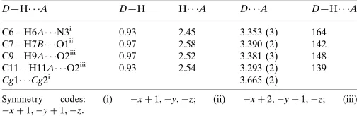
Hydrogen-bond geometry (A˚ ,).
Cg1 andCg2 are the centroids of the triazole ring (C10/C11/N1–N3) and the benzene ring (C1–C6), respectively.
D—H A D—H H A D A D—H A
C6—H6A N3i
0.93 2.45 3.353 (3) 164 C7—H7B O1ii
0.97 2.58 3.390 (2) 142 C9—H9A O2iii
0.97 2.52 3.381 (3) 148 C11—H11A O2iii 0.93 2.54 3.293 (2) 139
Cg1 Cg2i
3.665 (2)
Symmetry codes: (i) xþ1;y;z; (ii) xþ2;yþ1;z; (iii)
xþ1;yþ1;z.
Data collection:RAPID-AUTO (Rigaku, 2004); cell refinement: RAPID-AUTO; data reduction:RAPID-AUTO; program(s) used to solve structure: SHELXTL (Sheldrick, 2008); program(s) used to refine structure: SHELXTL; molecular graphics: SHELXTL; soft-ware used to prepare material for publication:SHELXTL.
Supplementary data and figures for this paper are available from the IUCr electronic archives (Reference: HG2669).
References
Allen, F. H., Kennard, O., Watson, D. G., Brammer, L., Orpen, A. G. & Taylor, R. (1987).J. Chem. Soc. Perkin Trans. 2, pp. S1–19.
Higashi, T. (1995).ABSCOR. Rigaku Corporation, Tokyo, Japan. Rigaku (2004).RAPID-AUTO.Rigaku Corporation, Tokyo, Japan. Sheldrick, G. M. (2008).Acta Cryst.A64, 112–122.
Xu, L. Z., Zhang, S. S. & Li, H. J. (2002).J. Chem. Res. Chin. Univ.18, 284–286.
Acta Crystallographica Section E Structure Reports
Online
supporting information
Acta Cryst. (2010). E66, o1122 [https://doi.org/10.1107/S1600536810012456]
(1
H
-1,2,4-Triazol-1-yl)methyl 2-(2,4-dichlorophenoxy)acetate
Yun-Man Xie and Yu-Min Li
S1. Comment
Compounds containing the triazole ring system are well known as efficient fungicides in agriculture, where they act by
inhibiting the biosynthesis of ergosterol (Xu et al., 2002). In order to search for new triazole compounds with higher
bioactivity, the title compound was synthesized and its structure is reported here.
In title compound, all bond lengths in the molecular are normal (Allen et al., 1987). Triazole ring (C10/C11/N1—N3)
and benzene ring (C1—C6) are essentially parallel to one another (dihedral angle of 4.99 (2)°) because the C—O—C—C
—O chain that links the two rings is folded [O—C—C—O torsion angle = 8.60 (2)°] rather than fully extended. π—π
interactions are indicated by the short distance (Cg1···Cg1 distance of 3.749 (3) Å, symmetry code: 1-x,-y,-z) between the
centroids of the triazole rings (C10/C11/N1—N3) (Cg1) and benzene rings C1—C6 (Cg2) (Table 1). There are weaker C
—H···N, C—H···O intermolecular interactions, which stabilized the structure (Table 1).
S2. Experimental
To a 100 ml flask were added 10 mmol of (1H-1,2,4-triazol-1-yl)methanol and 11 mmol of triethylamine in 20 ml of
dryed acetone, to which 10 mmol of 2-(2,4-dichlorophenoxy)acetyl chloride in 10 ml of acetone was then dropwise
added with stirring on ice-cold water bath within 0.5 h. The reaction took place immediately, and a lot of white solid
appeared. The mixture was heated and refluxed for 2 h, and then cooled to room temperature. After filtering and distilling
in reduced pressure, a crude product was obtained and purified by flash column chromatography (silicagel, using ethyl
ethanoate: cyclohexane = 1:3 as eluent) to afford the title compound. Single crystals suitable for X-ray measurements
were obtained by recrystallization from ethyl acetate at room temperature.
S3. Refinement
H atoms were positioned geometrically and refined using a riding model, with C—H = 0.93 or 0.97 Å and with Uiso(H) =
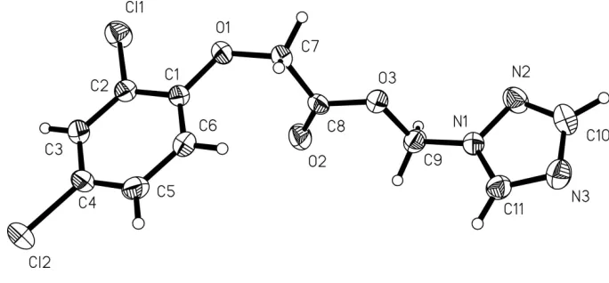
Figure 1
The molecular structure of (I), with atom labels and 40% probability displacement ellipsoids for non-H atoms.
(1H-1,2,4-Triazol-1-yl)methyl 2-(2,4-dichlorophenoxy)acetate
Crystal data
C11H9Cl2N3O3
Mr = 302.11
Monoclinic, P21/c
Hall symbol: -P 2ybc a = 10.814 (2) Å b = 6.4514 (13) Å c = 18.698 (4) Å β = 101.05 (3)° V = 1280.2 (4) Å3
Z = 4
F(000) = 616 Dx = 1.567 Mg m−3
Mo Kα radiation, λ = 0.71073 Å Cell parameters from 11868 reflections θ = 1.9–27.4°
µ = 0.51 mm−1
T = 293 K
Thin platelet, colorless 0.74 × 0.22 × 0.05 mm
Data collection
Rigaku R-AXIS RAPID IP area-detector diffractometer
Radiation source: Rotating Anode Graphite monochromator
ω Oscillation scans
Absorption correction: multi-scan (ABSCOR; Higashi, 1995) Tmin = 0.702, Tmax = 0.976
2253 measured reflections 2253 independent reflections 1816 reflections with I > 2σ(I) Rint = 0.000
θmax = 25.0°, θmin = 1.9°
h = 0→12 k = 0→7 l = −22→21
Refinement
Refinement on F2
Least-squares matrix: full R[F2 > 2σ(F2)] = 0.036
wR(F2) = 0.096
S = 1.02 2253 reflections 172 parameters 0 restraints
Primary atom site location: structure-invariant direct methods
Secondary atom site location: difference Fourier map
Hydrogen site location: inferred from neighbouring sites
H-atom parameters constrained w = 1/[σ2(F
o2) + (0.0589P)2 + 0.0748P]
where P = (Fo2 + 2Fc2)/3
(Δ/σ)max < 0.001
Δρmax = 0.24 e Å−3
Special details
Geometry. All esds (except the esd in the dihedral angle between two l.s. planes) are estimated using the full covariance matrix. The cell esds are taken into account individually in the estimation of esds in distances, angles and torsion angles; correlations between esds in cell parameters are only used when they are defined by crystal symmetry. An approximate (isotropic) treatment of cell esds is used for estimating esds involving l.s. planes.
Refinement. Refinement of F2 against ALL reflections. The weighted R-factor wR and goodness of fit S are based on F2,
conventional R-factors R are based on F, with F set to zero for negative F2. The threshold expression of F2 > σ(F2) is used
only for calculating R-factors(gt) etc. and is not relevant to the choice of reflections for refinement. R-factors based on F2
are statistically about twice as large as those based on F, and R- factors based on ALL data will be even larger.
Fractional atomic coordinates and isotropic or equivalent isotropic displacement parameters (Å2)
x y z Uiso*/Ueq
Cl2 0.79559 (6) 0.99712 (12) 0.32117 (3) 0.0704 (2) Cl1 1.04334 (5) 0.97477 (8) 0.09915 (3) 0.04955 (18) O3 0.66872 (11) 0.28902 (19) −0.04910 (7) 0.0376 (3) O2 0.66425 (13) 0.6226 (2) −0.01578 (8) 0.0476 (4) N1 0.49666 (14) 0.1167 (2) −0.11561 (8) 0.0339 (3) N2 0.52884 (17) 0.0126 (3) −0.17284 (9) 0.0438 (4) N3 0.39084 (16) −0.1726 (3) −0.12151 (9) 0.0464 (4) C1 0.87849 (16) 0.6753 (3) 0.12208 (9) 0.0344 (4) C6 0.79694 (17) 0.5867 (3) 0.16172 (10) 0.0415 (5) H6A 0.7583 0.4613 0.1466 0.050* C4 0.82796 (18) 0.8697 (4) 0.24517 (10) 0.0425 (5) C5 0.77190 (18) 0.6821 (4) 0.22373 (10) 0.0451 (5) H5A 0.7179 0.6202 0.2506 0.054* C3 0.91140 (18) 0.9612 (3) 0.20703 (10) 0.0399 (5) H3B 0.9496 1.0869 0.2223 0.048* C2 0.93678 (16) 0.8624 (3) 0.14596 (9) 0.0347 (4) O1 0.90954 (12) 0.5941 (2) 0.06039 (7) 0.0449 (4) C7 0.84682 (18) 0.4108 (3) 0.02987 (11) 0.0427 (5) H7A 0.8396 0.3144 0.0687 0.051* H7B 0.8964 0.3449 −0.0018 0.051* C8 0.71716 (17) 0.4595 (3) −0.01307 (9) 0.0340 (4) C9 0.54632 (17) 0.3176 (3) −0.09501 (10) 0.0385 (4) H9A 0.4909 0.3915 −0.0687 0.046* H9B 0.5539 0.3974 −0.1379 0.046* C11 0.41456 (18) 0.0036 (3) −0.08681 (11) 0.0388 (4) H11A 0.3792 0.0446 −0.0475 0.047* C10 0.4627 (2) −0.1587 (3) −0.17314 (11) 0.0470 (5) H10A 0.4651 −0.2639 −0.2068 0.056*
Atomic displacement parameters (Å2)
U11 U22 U33 U12 U13 U23
N1 0.0339 (8) 0.0340 (8) 0.0343 (8) −0.0040 (7) 0.0081 (6) −0.0048 (7) N2 0.0538 (10) 0.0439 (9) 0.0368 (9) −0.0011 (8) 0.0166 (7) −0.0047 (7) N3 0.0482 (10) 0.0400 (9) 0.0501 (10) −0.0091 (8) 0.0077 (8) −0.0015 (8) C1 0.0277 (9) 0.0454 (10) 0.0295 (9) 0.0023 (8) 0.0040 (7) −0.0079 (8) C6 0.0319 (10) 0.0476 (11) 0.0447 (11) −0.0079 (9) 0.0063 (8) −0.0108 (9) C4 0.0337 (10) 0.0625 (13) 0.0319 (9) 0.0053 (9) 0.0077 (7) −0.0116 (9) C5 0.0304 (10) 0.0656 (13) 0.0406 (10) −0.0045 (9) 0.0107 (8) −0.0032 (10) C3 0.0365 (10) 0.0439 (11) 0.0379 (10) 0.0012 (8) 0.0037 (8) −0.0105 (8) C2 0.0309 (9) 0.0391 (10) 0.0336 (9) 0.0012 (8) 0.0053 (7) −0.0015 (8) O1 0.0374 (7) 0.0564 (8) 0.0436 (7) −0.0089 (7) 0.0145 (6) −0.0223 (7) C7 0.0360 (10) 0.0464 (11) 0.0457 (11) −0.0004 (9) 0.0075 (8) −0.0202 (9) C8 0.0350 (10) 0.0394 (10) 0.0312 (9) −0.0045 (8) 0.0155 (7) −0.0090 (8) C9 0.0328 (10) 0.0351 (9) 0.0468 (11) −0.0031 (8) 0.0058 (8) −0.0030 (8) C11 0.0361 (10) 0.0435 (10) 0.0380 (10) −0.0053 (8) 0.0108 (8) −0.0006 (8) C10 0.0603 (13) 0.0379 (10) 0.0420 (11) −0.0002 (10) 0.0074 (9) −0.0081 (9)
Geometric parameters (Å, º)
Cl2—C4 1.7346 (19) C6—H6A 0.9300 Cl1—C2 1.7336 (19) C4—C5 1.378 (3) O3—C8 1.343 (2) C4—C3 1.385 (3) O3—C9 1.445 (2) C5—H5A 0.9300 O2—C8 1.194 (2) C3—C2 1.380 (3) N1—C11 1.339 (2) C3—H3B 0.9300 N1—N2 1.364 (2) O1—C7 1.427 (2) N1—C9 1.427 (2) C7—C8 1.509 (3) N2—C10 1.316 (3) C7—H7A 0.9700 N3—C11 1.310 (3) C7—H7B 0.9700 N3—C10 1.353 (3) C9—H9A 0.9700 C1—O1 1.366 (2) C9—H9B 0.9700 C1—C6 1.380 (3) C11—H11A 0.9300 C1—C2 1.395 (3) C10—H10A 0.9300 C6—C5 1.384 (3)
C3—C4—Cl2 118.25 (16) N1—C9—H9B 110.2 C4—C5—C6 119.20 (19) O3—C9—H9B 110.2 C4—C5—H5A 120.4 H9A—C9—H9B 108.5 C6—C5—H5A 120.4 N3—C11—N1 110.84 (18) C2—C3—C4 118.78 (18) N3—C11—H11A 124.6 C2—C3—H3B 120.6 N1—C11—H11A 124.6 C4—C3—H3B 120.6 N2—C10—N3 116.03 (17) C3—C2—C1 121.00 (18) N2—C10—H10A 122.0 C3—C2—Cl1 118.77 (15) N3—C10—H10A 122.0
C11—N1—N2—C10 −0.5 (2) C6—C1—O1—C7 5.1 (3) C9—N1—N2—C10 −179.25 (16) C2—C1—O1—C7 −175.59 (16) O1—C1—C6—C5 179.94 (17) C1—O1—C7—C8 78.8 (2) C2—C1—C6—C5 0.7 (3) C9—O3—C8—O2 2.2 (3) C3—C4—C5—C6 −1.9 (3) C9—O3—C8—C7 −177.47 (15) Cl2—C4—C5—C6 178.27 (16) O1—C7—C8—O2 −8.3 (3) C1—C6—C5—C4 1.1 (3) O1—C7—C8—O3 171.40 (15) C5—C4—C3—C2 0.9 (3) C11—N1—C9—O3 98.2 (2) Cl2—C4—C3—C2 −179.26 (14) N2—N1—C9—O3 −83.4 (2) C4—C3—C2—C1 0.9 (3) C8—O3—C9—N1 −167.03 (15) C4—C3—C2—Cl1 −178.88 (14) C10—N3—C11—N1 −0.4 (2) O1—C1—C2—C3 178.97 (17) N2—N1—C11—N3 0.6 (2) C6—C1—C2—C3 −1.7 (3) C9—N1—C11—N3 179.20 (17) O1—C1—C2—Cl1 −1.2 (2) N1—N2—C10—N3 0.3 (2) C6—C1—C2—Cl1 178.12 (15) C11—N3—C10—N2 0.0 (2)
Hydrogen-bond geometry (Å, º)
D—H···A D—H H···A D···A D—H···A
C6—H6A···N3i 0.93 2.45 3.353 (3) 164
C7—H7B···O1ii 0.97 2.58 3.390 (2) 142
C9—H9A···O2iii 0.97 2.52 3.381 (3) 148
C11—H11A···O2iii 0.93 2.54 3.293 (2) 139
Cg1···Cg2i 3.665 (2)