Original Article
Diffuse hyperplastic mesothelial cells in multiple lymph
nodes: case report with review of the literature
Libo Peng1,2*, Qin Shen1*, Xia Liu3, Jiandong Wang1,2, Shanshan Shi1, Bo Yu1, Xiaojun Zhou1,2
Department of Pathology, 1Jinling Hospital, Nanjing University School of Medicine, 2Clinical Medical School of
Southern Medical University, Nanjing, China; 3Department of Pathology, Xuzhou Central Hospital, Xuzhou, China. *The authors contributed equally.
Received March 15, 2013; Accepted March 30, 2013; Epub April 15, 2013; Published April 30, 2013
Abstract: We report a case of diffuse hyperplastic mesothelial cells in multiple lymph nodes. Microscopically, the lymph nodes had a normal follicular pattern. The lymphatic sinus was extremely expanded, within the sinuses the epithelial-like cells proliferated actively in the form of sheets and clusters. Epithelioid-like cells had eosinophilic cyto-plasm, prominent nucleoli and vesicular nuclei. Mitotic figures were rarely observed. These cells were immunoposi-tive for Calretinin, CK5/6, D2-40, MC and Ckpan and immunonegaimmunoposi-tive for S-100, HMB45, MelanA, TTF-1, CDX-2, Villin, ALK, CD30, CD20, CD3, CD1a and CD68. In addition, during a 22 months follow-up period failed to identify any malignant neoplasms, thus confirming the benign nature of these cells. It is the first reported case of diffuse hyperplastic of mesothelial cells mainly in the cervical lymph nodes associated with systemic multiple lymph node involvement. Awareness of this event is important for the pathologist in preventing the misdiagnosis of malignancy.
Keywords: Hyperplastic mesothelial cell, lymph nodes, serous effusions, differential diagnosis
Introduction
The presence of hyperplastic mesothelial cells (HMCs) in lymph nodes is extremely rare, and there is no unified naming convention for it. Adenopathy is more common in the thoracic, abdominal, and pelvic. Occurrence of superfi-cial lymph node enlargement is rare. The pres-ence of multiple diffuse hyperplasia mesotheli-al cells in the lymph nodes can be easily missed in daily practice and often erroneously diag-nosed as malignant. We report a case of dif-fuse hyperplastic of mesothelial cells mainly in the cervical lymph nodes associated with sys-temic multiple lymph node involvement. To fur-ther understand the essence of such lesions and explore the pathogenesis, clinicopatholog-ic features as well as differential diagnosis by review of the literature.
Case presentation
Clinical information
A 12-year-old man, previously healthy, was admitted to the hospital with a 3-months
his-tory of right neck mass gradually enlarged. Physical examination revealed bilateral cervical and axillary adenopathy, maximum diameter from 0.5 cm to 2.0 cm, medium to texture, no tenderness and no abnormal changes in the skin surface. CT venograms revealed thrombo-sis of the right internal jugular vein. Whole body PET-CT showed bilateral neck, the medial edge of the sternocleidomastoid, the bilateral poste-rior triangle, the bilateral supraclavicular regions and bilateral axillary, upper mediasti-num, around the abdominal aorta with multiple nodules with an increase in FDG metabolism; pelvic fluid was also identified. The patient was diagnosed as having metastatic carcinoma of unknown primary origin and underwent the biopsy of the cervical lymph node.
Pathologic findings
927 Int J Clin Exp Pathol 2013;6(5):926-931 within the sinuses the epithelial-like cells
prolif-erated actively in the form of sheets and clus-ters. These cells showed no cytologic features of malignancy. Medullary sinuses were expand-ed by clusters of round, polygonal epithelial cells with eosinophilic cytoplasm, no pleomor-phism. These cells showed very poor cell adhe-sion, clear membrane borders, clear cytoplasm, round or oval nuclei, lightly stained nuclear, without nuclear atypia, nucleoli visible, Mitotic activity was not appreciated. Hyperplastic cells occupy the lymphatic sinus quietly and don’t cause damage to surrounding tissue. And there was no stromal reaction (Figure 1A and 1B). All immunohistochemical stains were repeated.
[image:2.612.91.526.70.236.2]The large cells demonstrated strong staining with Calretinin, CK5/6, WT1, Ckpan, EMA, CK7, MC, D2-40 (Figure 2A and 2B). While CK20, TTF-1, CDX-2, Villin, ALK, CD20, CD3, CD30, S-100, HMB45, MelanA, CD1a and CD68 immunohistochemical stains were all negative. The patient was misdiagnosed as metastasis carcinoma, mesothelioma and sinus histiocyto-sis in different hospitals. A final diagnohistiocyto-sis of dif-fuse hyperplastic mesothelial cells in multiple lymph nodes was rendered eventually. The patient’s clinical evaluations during a 22 months period failed to identify any malignant neoplasms, thus confirming the benign nature of these nodal HMCs.
Figure 1. A. Low-power view showing the lymphatic sinus were extremely expanded, within the sinuses the epithelial-like cells proliferated actively in the form of sheets and clusters. (Hematoxylin-eosin, x100). B. High-power view of mesothelial cells. Magnification medullary sinuses were expanded by clusters of round, polygonal mesothelial cells with eosinophilic cytoplasm. These cells showed clear cytoplasm, round or oval nuclei, without nuclear atypia, nucleoli visible. Mitotic figures were rarely observed. (Hematoxylin-eosin, x400).
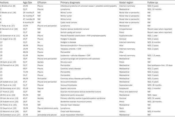
[image:2.612.93.524.320.485.2]Table 1. Clinical features of previously reported cases with benign mesothelial cells in Lymph Node
Authors Age/Sex Effusion Primary diagnosis Nodal region Follow-up
1 Brooks et al. [8] 23/M Pleura Infectious syndrome of unknown cause + possible cardiomyopathy Internal mammary NED, 3 years
2 52/F Pleural Lymphoma Mediastinal NED, 10 months
3 Weeks et al. [16] 24 months/F NM Wilms’ tumor Renal hilar or periaortic NM
4 42 months/F NM Wilms’ tumor Renal hilar or periaortic NM
5 47 months/M NM Wilms tumor Renal hilar or periaortic NM
6 8 months/M NM Cystic renal tumors Renal hilar or periaortic NM
7 Rutty et al. [17] 23/M Pleural and pericardial Lymphoma Mediastinal NM
8 Clement et al. [18] 59/F NM Ovarian serous borderline tumors Intraperitoneal Recent case (when reported)
9 21/F NM Sertoli-Leydig cell tumor Pelvic Recent case (when reported)
10 Groisman et al. [11] 41/M Pleura Pleural Possible tuberculosis + PVP lymphadenopathy Supraclavicular NED, 1 year
11 Argani et al. [9] 25/F Pleura Hodgkin’s disease Cervical NED, 2 years
12 32/F No Hodgkin’s disease Internal mammary NED, 8 months
13 38/M Pleura Glomerulonephritis + Pneumoconiosis Hilar NED, 2 years
14 14/M Pleura Takayasu arteritis + CHF Internal mammary NED, 2 years
15 48/M Pleura Pericarditis Hilar
16 61/M Ascites Leukemia + valvular disease + CHF Internal mammary NED, 1.5 years
17 19/F Pleural and pericardial Lymphoma/large cell lymphoma with sclerosis Mediastinal NM
18 Cohn et al. [19] 52/F Ascites Struma ovarii Pelvic NM
19 Parkash et al. [20] 52/F Pleura Pericarditis Mediastinal Died postopera- tion, 15 days
20 69/F Pleura Coronary artery disease Mediastinal NED, 2 years
21 70/M Pleura Pericarditis Mediastinal NED, 6 years
22 55/F Pleura Pericarditis Mediastinal NED, 3 years
23 68/M Pericardial Coronary artery disease pericarditis Mediastinal NED, 3 years
24 Isotalo et al. [14] 47/F Pleural and pericardial Venous thrombosis Mediastinal NED, 6 years
25 Paull et al. [12] 51/F Pleural Venous thrombosis Supraclavicular NED, 16 months
26 SionVardy et al. [21] 65/M Pleural Gastric carcinoma Subpleural NED, 2 months
27 Kiret al. [22] 32/F NM Ovarian microinvasive serous borderline tumors Pelvic and abdominal NM
28 Kim et al. [23] 65/M NM Abdominal aortic aneurysm Mediastinal NED, 1year
29 Van der et al. [13] 42/F Ascites Primary infertility ovarian hyperstimulation syndrome Cervical NED, 31 weeks
30 Acikalin et al. [24] 26/F No Borderline ovarian mucinous tumors Pelvic NED, 18 months
31 Pelosi et al. [25] 74/M NM Valvular heart disease Mediastinal NM
32 Goyal et al. [10] 16/M Pleural abdominal cavity Lymphoma Neck NM
33 Moonim et al. [26] 19/F NM Chest wall fibromatosis Axillary NED, 6 months
34 Colebatch et al. [27] 37/M pericardial and pleural acute myocardial infarction Mediastinal NM
929 Int J Clin Exp Pathol 2013;6(5):926-931 Discussion and review
Benign inclusions in lymph nodes may be glan-dular. The most well-known intranodal inclusion is typically glandular endometrium [1], parotid gland [2], thyroid [3], breast [4] and pancreas [5], the non-glandular components such as nevus cells [6] and decidual [7] are rare, and mesothelial cells in the lymph nodes is uncom-mon and under-recognized. The findings of benign mesothelial cells in mediastinal lymph nodes were first described by Brooks et al. in 1990 [8]. To the best of our knowledge, only 34 cases has been reported since (Table 1), 16 males and 18 females, the average age at pre-sentation was 39 years (range, 8–74 years). It often occur at one certain site, mesothelial cell inclusions frequently located in the mediastinal lymph node groups [8]. Successively in abdomi-nal, pelvic, renal hilar and periaortic lymph nodes, rarely in superficial lymph nodes (such as the neck, internal mammary and axillary). Only five cases occurrence in the neck [9-13]. We present a case of benign epithelial inclu-sions within the bilateral cervical lymph nodes associated with axillary, mediastinal and intra-peritoneal multiple lymph node involvement. Most of these patients have occurred concur-rently with serosal effusions (21/23). 10 cases with tumor, 9 cases with cardiovascular dis-ease, 2 cases with secondary venous thrombo-sis [12, 14]. In all cases, no evidence of a pri-mary malignant mesothelioma was identified. From the literature, HMCs usually associated with either inflammation, infection, mechanical causes or tumour. A feature common to most of the reported cases was a background of chron-ic mesothelial irritation caused by inflammatory or neoplastic process. Mesothelial cells gain access to the lymph nodes through the expan-sion lymphatic channels [8]. Mesothelial cells are thought to be transported to the lymph nodes through lymphatics system, thus there were mesothelial cell clusters identified within nodal lymphatics [14]. The present case of HMCs within mediastinal lymph nodes had pel-vic effusion secondary to venous thrombosis, which was similar to the previously described cases. Mesothelial reactions are thought to dis-rupt mesothelial stomata, allowing dislodged mesothelial cells access to sub-mesothelial lymphatics, lead to presence of the clusters of mesothelial cells in the lymph nodes.
The histopathological of HMCs need immuno-histochemistry and ultrastructural studies to
negative for epithelial and mesothelial mark-ers. (5) Metastatic malignant mesothelioma: often with a definite primary lesion, serosal sur-face blockbuster fusion of nodules, with high-grade atypia, with formation of nipple and microtubules, convinced destructive stromal infiltrates and invasion was necessary to con-firm the diagnosis of malignant. The homozy-gous deletion of p16 detected by FISH was a reliable way to distinguish the benign/reactive and malignant mesothelial proliferations. The majority of mesothelioma cases were positive for the p16 gene deletion, whereas none of the benign/reactive cases were positive the dele-tion [15].
To our knowledge, diffuse hyperplasia of meso-thelial cells in lymph multiple nodes have never been described. The potential problem of mis-diagnosis as mesothelioma can be avoided by an awareness of these conclusions, supported by clinical course and immunohistochemical results. Over 22-months follow-up period, the patient is free of disease. However, we wonder whether such diffuse proliferative mesothelial cells will last ‘static, benign’. Careful clinical follow-up and many more cases are required for depth of investigation.
Address correspondence to: Dr. Xiaojun Zhou, Department of Pathology, Jinling Hospital, Nanjing University School of Medicine, Nanjing, 210002, China. Tel: 80860192; Fax: +86-25-80860191; E-mail: [email protected]
References
[1] Javert CT. The spread of benign and malignant endometrium in the lymphatic system with a note on coexisting vascular involvement. Am J Obstet Gynecol 1952; 64: 780-806.
[2] Brown RB, Gaillard RA and Turner JA. The sig-nificance of aberrant or heterotopic parotid gland tissue in lymph nodes. Ann Surg 1953; 138: 850-856.
[3] Meyer JS and Steinberg LS. Microscopically be-nign thyroid follicles in cervical lymph nodes. Serial section study of lymph node inclusions and entire thyroid gland in 5 cases. Cancer 1969; 24: 302-311.
[4] Turner DR and Millis RR. Breast tissue inclu-sions in axillary lymph nodes. Histopathology 1980; 4: 631-636.
[5] Murayama H, Kikuchi M, Imai T, Yamamoto Y and Iwata Y. A case of heterotopic pancreas in lymph node. Virchows Arch A Pathol Anat Histol 1978; 377: 175-179.
[6] Epstein JI, Erlandson RA and Rosen PP. Nodal blue nevi. A study of three cases. Am J Surg Pathol 1984; 8: 907-915.
[7] Burnett RA and Millan D. Decidual change in pelvic lymph nodes: a source of possible diag-nostic error. Histopathology 1986; 10: 1089-1092.
[8] Brooks JS, LiVolsi VA and Pietra GG. Mesothe-lial cell inclusions in mediastinal lymph nodes mimicking metastatic carcinoma. Am J Clin Pathol 1990; 93: 741-748.
[9] Argani P and Rosai J. Hyperplastic mesothelial cells in lymph nodes: report of six cases of a benign process that can stimulate metastatic involvement by mesothelioma or carcinoma. Hum Pathol 1998; 29: 339-346.
[10] Goyal M, Kodandapani S, Sharanabasappa SN and Palanki SD. Mesothelial cell inclusions mimicking adenocarcinoma in cervical lymph nodes in association with chylous effusion. In-dian J Med Paediatr Oncol 2010; 31: 62-64. [11] Groisman GM, Amar M, Weiner P and Zamir D.
Mucicarminophilic histiocytosis (benign signet-ring cells) and hyperplastic mesothelial cells: two mimics of metastatic carcinoma within a single lymph node. Arch Pathol Lab Med 1998; 122: 282-284.
[12] Paull G and Mosunjac M. Fine-needle aspira-tion biopsy and intraoperative cytologic smear findings in a case of benign mesothelial-cell inclusions involving a lymph node: case report and review of the literature. Diagn Cytopathol 2003; 29: 163-166.
[13] van der Weiden RM, Meijers CJ and Hegt VN. Ectopic mesothelial cell proliferation in cervi-cal lymph nodes after severe ovarian hyper-stimulation syndrome. Fertil Steril 2005; 83: 739-741.
[14] Isotalo PA, Veinot JP and Jabi M. Hyperplastic mesothelial cells in mediastinal lymph node sinuses with extranodal lymphatic involve-ment. Arch Pathol Lab Med 2000; 124: 609-613.
[15] Monaco SE, Shuai Y, Bansal M, Krasinskas AM and Dacic S. The diagnostic utility of p16 FISH and GLUT-1 immunohistochemical analysis in mesothelial proliferations. Am J Clin Pathol 2011; 135: 619-627.
[16] Weeks DA, Beckwith JB and Mierau GW. Be-nign nodal lesions mimicking metastases from pediatric renal neoplasms: a report of the Na-tional Wilms’ Tumor Study Pathology Center. Hum Pathol 1990; 21: 1239-1244.
[17] Rutty GN and Lauder I. Mesothelial cell inclu-sions within mediastinal lymph nodes. Histo-pathology 1994; 25: 483-487.
meta-931 Int J Clin Exp Pathol 2013;6(5):926-931
static ovarian carcinoma and serous border-line tumor--a report of two cases associated with ovarian neoplasms. Mod Pathol 1996; 9: 879-886.
[19] Cohn DE, Folpe AL, Gown AM and Goff BA. Me-sothelial pelvic lymph node inclusions mimick-ing metastatic thyroid carcinoma. Gynecol On-col 1998; 68: 210-213.
[20] Parkash V, Vidwans M and Carter D. Benign mesothelial cells in mediastinal lymph nodes. Am J Surg Pathol 1999; 23: 1264-1269. [21] Sion-Vardy N, Diomin V and Benharroch D.
Hy-perplastic mesothelial cells in subpleural lymph nodes mimicking metastatic carcinoma. Ann Diagn Pathol 2004; 8: 373-374.
[22] Kir G, Eren S and Kir M. Hyperplastic mesothe-lial cells in pelvic and abdominal lymph node sinuses mimicking metastatic ovarian microin-vasive serous borderline tumor. Eur J Gynaecol Oncol 2004; 25: 236-238.
[23] Kim YM, Kim KR and Ro JY. Mesothelial Cell Inclusions Mimicking Metastatic Carcinoma in Mediastinal Lymph Node: A Case Report. Ko-rean J Pathol 2004; 38: 46-49.
[24] Acikalin MF, Ozalp S and Turan D. Mesothelial pelvic lymph node inclusion in a patient with ovarian microinvasive borderline mucinous tu-mor: case report with review of the literature. Int J Gynecol Cancer 2007; 17: 917-921. [25] Pelosi G, Sonzogni A and Rosai J. Images in
pa-thology. Benign hyperplastic mesothelial cells in lymph node. Int J Surg Pathol 2007; 15: 297-299.
[26] Moonim MT, Ng WW and Routledge T. Benign metastasizing mesothelial cells: a potential pitfall in mediastinal lymph nodes. J Clin Oncol 2011; 29: e546-548.