Original Article
Human epidermal growth factor receptor 2 expression
in breast cancer: correlation with clinical
pathological features
Shu-Fang Ning1, Ji-Lin Li1, Cheng-Piao Luo2, Chang-Hong Wei2, Yong-Kui Lu3, Hai-Zhou Liu1, Wen-E Wei1, Li-Tu Zhang1
1Department of Medical Research, Affiliated Tumor Hospital of Guangxi Medical University, Nanning, China; 2
De-partment of Pathology, Affiliated Tumor Hospital of Guangxi Medical University, Nanning, China; 3Department of
Chemotherapy, Affiliated Tumor Hospital of Guangxi Medical University, Nanning, China
Received October 7, 2014; Accepted November 26, 2014; Epub December 1, 2014; Published December 15, 2014
Abstract: The overexpressed HER2 (human epidermal growth factor receptor 2) is a valuable therapeutic target. Precise assessment of HER2 status is thus crucial in the treatment of breast cancer. In this study, formalin-fixed, paraffin-embedded samples of tumors from 304 breast cancer patients who underwent curative surgery proce -dures between 2011 and 2014 were tested by immunohistochemistry (IHC) as a primary estimate of HER2 status, followed by fluorescence in situ hybridization (FISH). Concordance rate between IHC and FISH was evaluated. The Χ2 test was used to evaluate the correlation between HER2 gene amplification status and different clinical pathologi -cal features including: (estrogen receptor) ER and (progesterone receptor) PR expression, age, menopausal status and tumor size. The results show that 84.8% of IHC score 3+ cases and 6.2% of IHC score 0/1+ cases were ampli-fied by FISH. After exclusion of group IHC 2+, the concordance rate between FISH and IHC was 87.4%. There was a significant inverse association between expression of hormone receptors (ER and PR) and HER2 amplification (P < 0.001) among the patients studied. However, no relationship was observed between HER2 amplification and age, menopausal status and tumor size (P > 0.05). The data demonstrate a relatively high level of concordance rate for HER2 testing between FISH and IHC, and HER2 overexpression was associated with the levels of ER and PR.
Keywords: Breast cancer, HER2, clinical pathological features, immunohistochemistry (IHC), fluorescence in situ hybridization (FISH)
Introduction
The human epidermal growth factor receptor 2 (HER2) gene is a proto-oncogene located on chromosome 17q12. The HER2 gene is
ampli-fied and overexpressed in 20-30% of invasive
breast cancers, as well as in fractions of gastric cancer, ovarian cancer and others [1-3]. HER2 overexpression is associated with cell prolifera-tion, apoptosis, adhesion, angiogenesis and aggressiveness of various tumors. The overex-pression of HER2 has been recognized as a
sig-nificant marker for aggressiveness of disease
status. Furthermore, HER2 is considered to be
a molecular target for specific therapies. For
instance, breast cancer patients with positivity
to HER2 can benefit from trastuzumab
(Her-ceptin; Genentech, South San Francisco, CA), a
humanized monoclonal antibody that targets HER2, and has been shown to improve progres-sion-free survival and overall survival in the metastatic setting [4]. Lapatinib, a small
mole-cule inhibitor of the tyrosine kinase activity of
HER2, was also approved by the US Food and Drug Administration (FDA) for the treatment of HER2-positive breast cancer and is associated with improved survival outcome in patients [5, 6].
which are available for HER2 testing: microar-ray mRNA readouts (by TargetPrint) [8], qPCR (quantitative real-time PCR) [9] and digital PCR
[10] are used to evaluate HER2 gene amplifica -tion. Despite the ability of these technologies to show an excellent concordance with FISH and have the potential to accurately assess HER2
amplification status, the Update Committee of ASCO/CAP believe that there is insufficient evi -dence to support use of mRNA or DNA microar-ray assays to determine HER2 status, and FISH and IHC are still the most frequently used meth-ods to assess the HER2 status in breast can-cer. The advantages of IHC testing are that it is relatively inexpensive, widely available and simple to implement. However, the ability to accurately estimate HER2 protein expression status by IHC can be problematic [11]. FISH is more reproducible than IHC, and FISH is more accurate for HER2 testing and more strongly correlated with responsiveness to HER2-tar- geted therapy [12]. In this study, we use IHC assays for preliminary assessment of HER2
status followed by reflex testing by FISH.
The objective of this study was to estimate the concordance between HER2 protein expres-sion status when performed by IHC and HER2
gene amplification when performed by FISH in
median age was 48.1 years. Clinical pathologi-cal characteristics of the tumor samples includ-ed in this study are shown in Table 1. This study was approved by the ethics committee of the Tumor Hospital, Guangxi Medical University.
HER2 immunohistochemistry
Surgical tissues were fixed in 10% formalin, dehydrated, cleared and embedded in paraffin. 4 μm thick sections were cut using a rotary
microtome (Thermo HM315) and transferred to poly-L-lysine coated slides. IHC for Her2 protein was performed on tissue sections in accor-dance with published and validated protocols
[13]. Tissue samples were deparaffinized, rehy -drated and subjected to antigen retrieval with a buffer solution using a steamer. Tissue sam-ples were then incubated with HER2 antibody
(Dako, Denmark) for 30 min. Following the
application of anti-mouse poly-HRP secondary antibody, the sections were incubated in a solu-tion including 3% hydrogen peroxide and diami-nobenzidine. Finally, the tissue sections were placed under a cover slip and viewed using light microscopy (Olympus BX43). According to the ASCO/CAP guideline recommendations [14], HER2 protein expression was scored as 0 or 1+
[image:2.612.91.346.97.346.2](no staining or weak, incomplete membrane
Table 1. Patient Demographics and Baseline Clinical Patho-logic Characteristics
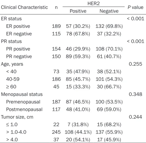
Clinical Characteristic n HER2 P value
Positive Negative
ER status < 0.001
ER positive 189 57 (30.2%) 132 (69.8%) ER negative 115 78 (67.8%) 37 (32.2%)
PR status < 0.001
PR positive 154 46 (29.9%) 108 (70.1%) PR negative 150 89 (59.3%) 61 (40.7%)
Age, years 0.255
< 40 73 35 (47.9%) 38 (52.1%) 40-59 186 85 (45.7%) 101 (54.3%)
≥ 60 45 15 (33.3%) 30 (66.7%)
Menopausal status 0.348
Premenopausal 187 87 (46.5%) 100 (53.5%) Postmenopausal 117 48 (41.0%) 69 (59.0%)
Tumor size, cm 0.244
≤ 1.0 22 7 (31.8%) 15 (68.2%)
> 1.0-4.0 245 108 (44.1%) 137 (55.9%) > 4.0 37 20 (54.1%) 17 (45.9%)
HER2: human epidermal growth factor receptor; ER: estrogen receptor); PR: progesterone receptor.
archived breast cancer tissue speci-mens and determine any relation-ships that these techniques have with clinical pathological character-istics in the patients.
Materials and methods
Patients and samples
Paraffin-embedded blocks samples
of tumors were retrieved from 304 patients who had breast carcinoma that was surgically resected between
2011 and 2014 at the Affiliated
staining in any proportion of tumor cells), 2+ (complete membrane staining that is either not
uniform or weak in intensity but with obvious
circumferential distribution in at least 10% of cells or intense, complete membrane staining
of ≤ 30% of invasive tumor cells), or 3+ (uniform
intense membrane staining of > 30% of inva-sive tumor cells).
HER2 FISH
FISH was performed with the Path Vysion HER2 DNA Probe Kit (Abbott Laboratories Ltd., Hong Kong) according to the instructions of
manufac-turer. This kit contains two fluorescently labeled
DNA probes, HER2 spectrum orange and CEP17 (chromosome 17 enumeration control probes direct-labeled with spectrum green), allowing simultaneous detection of the copy numbers of both HER2 and CEP17. The signal analysis was carried out using an Olympus BX 51 fluorescent microscope system. At least 30
tumor cell nuclei in at least three different areas of invasive carcinoma were counted for the numbers of HER2 and CEP17 signals. The HER2/CEP17 ratios were consistently interpret-ed according to the ASCO/CAP
recommenda-tions. HER2 gene expression was classified as
positive (HER2/CEP17 ratio > 2.2 or HER2 gene copy > 6.0), negative (HER2/CEP17 ratio < 1.8 or HER2 gene copy < 4.0), or equivocal (HER2/ CEP17 ratio 1.8-2.2 or HER2 gene copy
4.0-6.0). If the HER2/CEP17 ratio was classified as
equivocal, another 30 tumor cells were
count-ed and the final HER2/CEP17 ratio was calcu -lated from the 60 cells.
In all cases, IHC was performed as a primary test, followed by FISH. FISH assays were per-formed on all 3+ and 2+ cases, and on a small number of the 0/1+ cases. When there was a discrepancy between IHC and FISH, the result
of FISH was considered as definitive.
Statistical analysis
The Χ2 test was used to evaluate the correlation among clinical pathological features and HER2
gene amplification status as determined by
Results
Correlation of HER2 with clinic pathological characteristics
Among the 304 evaluable patients, 189 cases of them were ER positive by IHC. Data in Table 1 show that ER status has a significant nega -tive correlation with HER2 posi-tive rates (P < 0.001), and that the higher the expression level of ER, the lower the HER2 positive rates. Similarly, there is a negative correlation bet- ween PR status and HER2 positive rates (P < 0.001). HER2 positive rates tended to decrease with increasing age of the patients. The HER2 expression levels of premenopausal patients were higher than those of postmenopausal patients, but the results were not statistically
significant (P > 0.05). Furthermore, no signifi -cant relationship was found between HER2 positivity and tumor size (P > 0.05).
HER2 gene amplification and protein expres -sion
The concordance between HER2 FISH and IHC results are shown in Table 2. HER2 gene
ampli-fication was detected in 304 tumor samples
with HER2 to CEP17 ratios from 2.34 to 12.18. Almost half of the FISH positive samples (67 of 135, 49.6%) were IHC 3+, with an 84.8% con-cordance between FISH and IHC 3+, and 34.2%
of IHC 2+ were positive for HER2 amplification
by FISH. None of the IHC 0 cases proved FISH positive above an HER2/CEP17 ratio of 2.2, and only 2 tumors in the IHC 1+ tumors (2 of 22, 9.1%) were demonstrated HER2 expression positive, with ratios of 5.078 and 4.7, respec-tively. The rate of FISH-negativity within each group varied from 15.2% to 100%. There was a high level of consistency between IHC 0 and FISH; 100% of specimens were negative for
HER2 amplification. In short, 2 cases (IHC 1+) were amplified and 12 cases (IHC 3+) were non-amplified in a total of 111 tumors (after
[image:3.612.91.353.94.151.2]exclusion of IHC 2+), resulting in a concordance rate of 87.4% between IHC and FISH. The IHC Table 2. Immunochemistry and FISH Concordance expressed
as percentages of total specimens evaluated
FISH IHC 0 IHC 1+ IHC 2+ IHC 3+ Total
Positive 0 2 (9.1%) 66 (34.2%) 67 (84.8%) 135 Negative 10 (100%) 20 (90.9%) 127 (65.8%) 12 (15.2%) 169
Total 10 22 193 79 304
FISH. All P values were derived from the two-sided Fisher’s exact test and P < 0.05 was considered
and FISH results are shown in Figures 1 and 2, respectively.
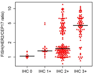
The distribution of FISH HER2/CEP17 ratios compared with IHC results is shown in Figure 3. The HER2/CEP17 ratio was from 0.73 to 12.18 in 304 tumor samples, and the maximum and minimum values of FISH ratio are all in the IHC 2+ group.
Discussion
[image:4.612.92.524.70.394.2]Since the FDA approval of the use of the Herceptin for the treatment of HER2-positive breast cancer, HER2-testing has become rou-tine in processing breast cancer specimens [14]. Furthermore, the ASCO/CAP Committee recommends that all patients with invasive breast cancer should be tested for either HER2
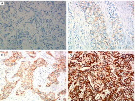
Figure 1. Immunohistochemical staining of HER2 protein expression in breast cancer tissue sections (× 100). A: IHC 0; B: IHC 1+; C: IHC 2+; D: IHC 3+.
[image:4.612.94.522.445.553.2]protein expression (IHC assay) or HER2 gene expression (ISH assay) using a validated HER2 test [7]. Although IHC and FISH are the most frequently used methods for HER2-testing in breast cancer [11], there are always discrepan-cies. According to the ASCO/CAP guidelines approximately 20% of the HER2 assays (partic-ularly those performed using IHC and FISH techniques) may be inaccurate. Correlation studies involving 2279 tumor samples with breast cancer showed a concordance of HER2 status between dual-color FISH and IHC was 87% [15]. The disparities in FISH and IHC assay
results have since been confirmed by some
other studies [12, 16]. The relationship between
HER2 protein expression and gene amplifica -tion in gastric cancer samples is also controver-sial [17]. In general, excellent concordance was shown in the IHC 0, IHC 1+ and IHC 3+ groups, while the most discrepancy was in the IHC 2+
group. The causes of the incongruence are like -ly to be multifactorial, such as issues with
spec-imen fixation, different sensitivity and specifici -ty of the antibodies used, and observer bias due to interpretation by different pathologists. The guidelines established by ASCO/CAP were very conducive to standardization of the testing algorithms used and introduced a revised scor-ing system aimed at improvscor-ing the concordance between IHC and ISH (in situ hybridization) results. This led to an improvement in its
accu-racy and utility to act as a predictive marker in
breast cancer [7].
In the present study, the HER2 protein
overex-pression (group IHC 3+) without gene amplifica
-tion was found in 15.2% of breast cancers,
while gene amplification without HER2 protein
overexpression (groups IHC 0 and IHC 1+) was detected in 6.2% of cases. After exclusion of group IHC 2+, the concordance rate between FISH and IHC was 87.4%. Similar results were seen in prior studies where the concordance rates (excluding 2+ samples) were closer to 80-90% [18, 19]. However, the IHC-FISH con-cordance rates are expected to be 95% [14].
We should take steps to ensure the highest
quality of testing. For instance, a high level of training, experience and attention to detail is required for interpretation of the assays for both FISH and IHC. Previous studies have indi-cated that improvement in concordance rate was ascribed to standardization of technical issues (such as pre-analytical processing and
specimen fixation), scoring and interpretation
[16], and it is recommended that concordance
testing should be annually confirmed [14]. The
HER2 testing is performed according to stan-dardized analytically validated protocols [7].
The significant aim for the detection of HER2 overexpression or amplification is to predict the trastuzumab response. The specific mecha -nism through which trastuzumab exerts its effects in vivo remains to be elucidated, but is
likely to include antibody-dependent cellular
[image:5.612.90.290.70.227.2]cytotoxicity, inhibition of the P13K-AKT path-way, inhibition of cell cycle progression, attenu-ation of cell signaling, inhibition of HER2 shed-ding and antiangiogenic effects [20, 21]. During the clinical trials with trastuzumab, it was observed that the maximal response rates were approximately 35% with response rates varying from 12-68% [20, 21]. The previous study showed that the response rate in tumor samples with 3+ staining by IHC was 35%, with no response in the IHC 2+ group. The response rates in patients with FISH-positive tumors were 34% compared with 7% in those with FISH-negative tumors [22]. Researchers have compared the clinical outcomes of IHC and FISH assays for prediction of response to trastuzumab therapy. The results indicate that compared with IHC, FISH is the preferred meth-od to select patients for trastuzumab therapy [23]. Other researchers have also suggested the use of FISH as a superior method [21]. The HER2-positive breast cancers with FISH ratios between 2.0 and 4.0 have similar responses to trastuzumab as patients whose tumors with
FISH ratios of 4.0 to 6.0 and 6.0 to 8.0, which is mentioned in a previous study [12]. ASCO/CAP recommends the patients with HER2 protein expression IHC 3+ or FISH-positive for HER2
gene amplification was eligible for treatment in
the adjuvant trastuzumab trials. The patients with IHC 0 or 1+ or FISH-negative were exclud-ed from therapy with trastuzumab and retest-ing was performed for specimens that tested IHC 2+ [14].
In our study, there was a significant inverse
association between expression of hormone
receptors (ER and PR) and HER2 amplification
(P < 0.001). However, no relationship was obser-
ved between HER2 amplification and age,
menopausal status and tumor size (P > 0.05). Estrogen is a crucial mitogen exerting its activ-ity by binding to the corresponding receptors and these have been shown to be present in 50-80% of breast carcinoma. The progesterone receptor is functionally similar to the estrogen receptor and is as valuable in predicting the behavior of invasive breast cancer [24]. A previ-ous study examined 3655 specimens of inva-sive breast carcinomas and showed that
expression of ER and PR were significantly
reduced in HER2 positive specimens compared with HER2 negative specimens [25]. Similar results were found in other studies where the
amplification of HER2 is correlated with a
reduction in the positivity of ER and PR [24, 26, 27]. Furthermore, the negative correlation between hormone receptor and HER2 was shown to be related to the fact that estrogens suppress HER2 through the ER [28]. Correlation of HER2 over-expression and tumor size was also studied by Shafaq Mujtaba et al. [26], who showed that HER2 over-expression increased with increasing tumor size, a result which is
reflected in our study. However, other research -ers did not show any association of HER2 with tumor size or age [27]. A correlation between HER2 and hormone receptors expression would be expected with age and menopausal status
since circulating estrogens are known to vary
before and after the menopause [28]. However, the results presented here are in agreement
with a prior study which showed no significant
association of age and HER2 status [29]. In summary, the data generated in this study demonstrate a relatively high rate of concor-dance for HER2 testing between FISH and IHC,
and there was a significant inverse association
between expression of hormone receptors (ER
and PR) and HER2 amplification. This study
therefore emphasizes the importance of pre-cisely assessing the HER2 status in breast can-cer patients prior to selection of patients for trastuzumab therapy. Improvement in the accu-racy of HER2 testing can only occur when stan-dardized analytically validated protocols are used. The results presented here suggest that further studies should be performed in a trastu-zumab-treated population.
Acknowledgements
The authors would like to thank Dr. Dev
Sooranna, Imperial College London, for helping to edit the manuscript. This study was
support-ed by the Guangxi Scientific Research and
Technical Development Program (Gui Ke Gong No.1298003-2-8).
Disclosure of conflict of interest
None.
Address correspondence to: Dr. Litu Zhang, De- partment of Medical Research, Affiliated Tumor Hospital of Guangxi Medical University, 71 He Di Road, Nanning 530021, P. R. China. Tel: +86-7715310593; E-mail: [email protected]
References
[1] Wilken JA, Badri T, Cross S, Raji R, Santin AD, Schwartz P, Branscum AJ, Baron AT, Sakhitab AI and Maihle NJ. EGFR/HER-targeted thera-peutics in ovarian cancer. Future Med Chem 2012; 4: 447-469.
[2] Sukawa Y, Yamamoto H, Nosho K, Ito M, Iga -rashi H, Naito T, Mitsuhashi K, Matsunaga Y, Takahashi T, Mikami M, Adachi Y, Suzuki H and Shinomura Y. HER2 expression and PI3K-Akt pathway alterations in gastric cancer. Diges-tion 2014; 89: 12-17.
[3] Rosa FE, Santos RM, Rogatto SR and Domingues MA. Chromogenic in situ hybridiza-tion compared with other approaches to evalu-ate HER2/neu status in breast carcinomas. Braz J Med Biol Res 2013; 46: 207-216. [4] Garrison LP Jr, Lalla D, Brammer M,
Babigumi-ra JB, Wang B and Perez EA. Assessing the po-tential cost-effectiveness of retesting IHC0, IHC1+, or FISH-negative early stage breast cancer patients for HER2 status. Cancer 2013; 119: 3113-3122.
combina-tion with capecitabine for previously treated metastatic breast cancer that overexpresses HER-2. Oncologist 2008; 13: 1114-1119. [6] Figueroa-Magalhaes MC, Jelovac D, Connolly
RM and Wolff AC. Treatment of HER2-positive breast cancer. Breast 2014; 23: 128-136. [7] Wolff AC, Hammond ME, Hicks DG, Dowsett M,
McShane LM, Allison KH, Allred DC, Bartlett JM, Bilous M, Fitzgibbons P, Hanna W, Jenkins RB, Mangu PB, Paik S, Perez EA, Press MF, Spears PA, Vance GH, Viale G and Hayes DF. Recommendations for human epidermal growth factor receptor 2 testing in breast can-cer: American Society of Clinical Oncology/Col-lege of American Pathologists clinical practice guideline update. J Clin Oncol 2013; 31: 3997-4013.
[8] Viale G, Slaets L, Bogaerts J, Rutgers E, van’t Veer L, Piccart-Gebhart MJ, de Snoo FA, Stork-Sloots L, Russo L, Dell’Orto P, van den Akker J, Glas A and Cardoso F. High concordance of protein (by IHC), gene (by FISH; HER2 only), and microarray readout (by TargetPrint) of ER, PgR, and HER2: results from the EORTC 10041/BIG 03-04 MINDACT trial. Ann Oncol 2014; 25: 816-823.
[9] Jacquemier J, Spyratos F, Esterni B, Mozzi-conacci MJ, Antoine M, Arnould L, Lizard S, Bertheau P, Lehmann-Che J, Fournier CB, Krieger S, Bibeau F, Lamy PJ, Chenard MP, Legrain M, Guinebretiere JM, Loussouarn D, Macgrogan G, Hostein I, Mathieu MC, Lacroix L, Valent A, Robin YM, Revillion F, Triki ML, Seaume A, Salomon AV, de Cremoux P, Porte-faix G, Xerri L, Vacher S, Bieche I and Penault-Llorca F. SISH/CISH or qPCR as alternative techniques to FISH for determination of HER2 amplification status on breast tumors core needle biopsies: a multicenter experience based on 840 cases. BMC Cancer 2013; 13: 351.
[10] Garcia-Murillas I, Lambros M and Turner NC. Determination of HER2 amplification status on tumour DNA by digital PCR. PLoS One 2013; 8: e83409.
[11] Krishnamurti U and Silverman JF. HER2 in breast cancer: a review and update. Adv Anat Pathol 2014; 21: 100-107.
[12] Sauter G, Lee J, Bartlett JM, Slamon DJ and Press MF. Guidelines for human epidermal growth factor receptor 2 testing: biologic and methodologic considerations. J Clin Oncol 2009; 27: 1323-1333.
[13] Fasching PA, Weihbrecht S, Haeberle L, Gaspa-ryan A, Villalobos IE, Ma Y, Ekici AB, Wachter DL, Hartmann A, Beckmann MW, Slamon DJ and Press MF. HER2 and TOP2A amplification in a hospital-based cohort of breast cancer pa-tients: associations with patient and tumor characteristics. Breast Cancer Res Treat 2014; 145: 193-203.
[14] Wolff AC, Hammond ME, Schwartz JN, Hagerty KL, Allred DC, Cote RJ, Dowsett M, Fitzgibbons PL, Hanna WM, Langer A, McShane LM, Paik S, Pegram MD, Perez EA, Press MF, Rhodes A, Sturgeon C, Taube SE, Tubbs R, Vance GH, van de Vijver M, Wheeler TM and Hayes DF. Ameri-can Society of Clinical Oncology/College of American Pathologists guideline recommenda-tions for human epidermal growth factor re-ceptor 2 testing in breast cancer. J Clin Oncol 2007; 25: 118-145.
[15] Lal P, Salazar PA, Hudis CA, Ladanyi M and Chen B. HER-2 testing in breast cancer using immunohistochemical analysis and fluores -cence in situ hybridization: a single-institution experience of 2,279 cases and comparison of dual-color and single-color scoring. Am J Clin Pathol 2004; 121: 631-636.
[16] Varga Z, Noske A, Ramach C, Padberg B and Moch H. Assessment of HER2 status in breast cancer: overall positivity rate and accuracy by fluorescence in situ hybridization and immuno -histochemistry in a single institution over 12 years: a quality control study. BMC Cancer 2013; 13: 615.
[17] He C, Bian XY, Ni XZ, Shen DP, Shen YY, Liu H, Shen ZY and Liu Q. Correlation of human epi-dermal growth factor receptor 2 expression with clinicopathological characteristics and prognosis in gastric cancer. World J Gastroen-terol 2013; 19: 2171-2178.
[18] Dybdal N, Leiberman G, Anderson S, McCune B, Bajamonde A, Cohen RL, Mass RD, Sanders C and Press MF. Determination of HER2 gene amplification by fluorescence in situ hybridiza -tion and concordance with the clinical trials immunohistochemical assay in women with metastatic breast cancer evaluated for treat-ment with trastuzumab. Breast Cancer Res Treat 2005; 93: 3-11.
[19] Reddy JC, Reimann JD, Anderson SM and Klein PM. Concordance between central and local laboratory HER2 testing from a community-based clinical study. Clin Breast Cancer 2006; 7: 153-157.
[20] Murphy CG and Modi S. HER2 breast cancer therapies: a review. Biologics 2009; 3: 289-301.
[21] Shah S and Chen B. Testing for HER2 in Breast Cancer: A Continuing Evolution. Patholog Res Int 2011; 2011: 903202.
[22] Vogel CL, Cobleigh MA, Tripathy D, Gutheil JC, Harris LN, Fehrenbacher L, Slamon DJ, Murphy M, Novotny WF, Burchmore M, Shak S, Stewart SJ and Press M. Efficacy and safety of trastu -zumab as a single agent in first-line treatment of HER2-overexpressing metastatic breast cancer. J Clin Oncol 2002; 20: 719-726. [23] Mass RD, Press MF, Anderson S, Cobleigh MA,
to HER2 detection by fluorescence in situ hy -bridization in women with metastatic breast cancer treated with trastuzumab. Clin Breast Cancer 2005; 6: 240-246.
[24] Azizun N, Bhurgri Y, Raza F and Kayani N. Com-parison of ER, PR and HER-2/neu (C-erb B 2) reactivity pattern with histologic grade, tumor size and lymph node status in breast cancer. Asian Pac J Cancer Prev 2008; 9: 553-556. [25] Lal P, Tan LK and Chen B. Correlation of HER-2
status with estrogen and progesterone recep-tors and histologic features in 3,655 invasive breast carcinomas. Am J Clin Pathol 2005; 123: 541-546.
[26] Mujtaba S, Haroon S, Faridi N and Lodhi FR. Correlation of human epidermal growth factor receptor 2 (HER-2/neu) receptor status with hormone receptors Oestrogen Receptor, Pro-gesterone Receptor status and other prognos-tic markers in breast cancer: an experience at tertiary care hospital in Karachi. J Pak Med As -soc 2013; 63: 854-858.
[27] Huang HJ, Neven P, Drijkoningen M, Paridaens R, Wildiers H, Van Limbergen E, Berteloot P, Amant F, Christiaens MR and Vergote I. Asso-ciation between HER-2/neu and the progester-one receptor in oestrogen-dependent breast cancer is age-related. Breast Cancer Res Treat 2005; 91: 81-87.
[28] Huang HJ, Neven P, Drijkoningen M, Paridaens R, Wildiers H, Van Limbergen E, Berteloot P, Amant F, Vergote I and Christiaens MR. Hor-mone receptors do not predict the HER2/neu status in all age groups of women with an oper-able breast cancer. Ann Oncol 2005; 16: 1755-1761.