ORIGINAL RESEARCH
The Artery of Bernasconi and Cassinari: A
Morphometric Study for Superselective
Catheterization
A.D. Banerjee H. Ezer A. Nanda
BACKGROUND AND PURPOSE: The artery of Bernasconi and Cassinari is an important infraclinoid branch of the internal cerebral artery. It is of neuroendovascular relevance in view of its supply to complex lesions such as meningiomas and arteriovenous malformations in the tentorial and falcoten-torial regions. The present microanatomic study attempts a morphometric elucidation of this slender but important branch of the meningohypophyseal trunk.
MATERIALS AND METHODS: The origin, course, dimensions, and related variations of the tentorial artery were studied in 10 formalin-fixed human cadaveric sides.
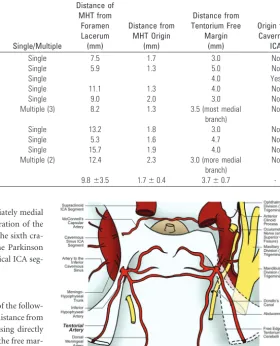
RESULTS: The tentorial artery originated from the meningohypophyseal trunk in all but 1 specimen, in which it arose directly from the intracavernous internal carotid artery. In 80% of specimens, it took origin as a single branch; as a bifurcation and trifurcation in 1 each. It was usually the terminal branch of the meningohypophyseal trunk (in 90%). In all, 5 distinct microvascular patterns were noted. The mean diameter of this artery was 0.7 mm (range, 0.3– 0.8 mm; SD,⫾0.1 mm). The mean length was 15.4 mm (range, 9 –23 mm; SD,⫾4.4 mm). Its mean distance from the origin of the meningohypophy-seal trunk was 1.7 mm (range, 1.3–2.3 mm; SD,⫾0.4 mm). The mean distance from the free edge of the tentorium was 3.7 mm (range, 3–5 mm; SD,⫾0.7 mm).
CONCLUSIONS:The artery of Bernasconi and Cassinari is an important vascular conduit to myriad neoplasms and vascular malformations in the vicinity of the tentorium cerebelli. In this era of advanced microneurosurgical techniques and superselective endovascular interventions, morphometric knowl-edge of this artery is important for precise and safe management of these lesions.
ABBREVIATIONS:DSA ⫽ digital substraction angiography; GSPN ⫽ greater superficial petrosal nerve; ICA⫽internal carotid artery; MHT⫽meningohypophyseal trunk; V1⫽ophthalmic division of trigeminal nerve, V2 ⫽ maxillary division of trigeminal nerve, V3⫽ mandibular division of trigeminal nerve
T
he tentorial artery is the most constant branch of the MHT that, in turn, arises from the proximal curvature of the intracavernous ICA.1It is important in neuroendovascular in-terventions for an entire spectrum of neoplasms and vascular lesions in the region of the tentorium cerebelli. Bernasconi and Cassinari2first reported the angiographic visualization of a ten-torial artery supplying tenten-torial meningiomas. Since then, similar angiographic3-5and anatomic1,6 observations have been pub-lished, but no morphometric study specific to the tentorial artery has been conducted. An anatomic study of this type is of utmost importance for imparting greater “precision” to the management of these lesions as well as in preventing inadvertent vascular com-promise to normal structures, especially in this era of advanced superselective endovascular interventions. The present micro-anatomic study is a step to address this issue.Materials and Methods
Five fresh human cadaveric heads were prepared for bilateral dis-section, yielding a total of 10 datasets. The internal carotid arteries,
the vertebral arteries, and the internal jugular veins of the speci-mens were cannulated and injected with colored silicon dye, as described previously.7 The specimens were kept submerged in
10% formalin for the next 48 hours. The prepared specimens were then dissected under variable magnification by using a neurosur-gical operating microscope (Carl Zeiss OPMI, Aalen, Germany), a high-speed drill (Midas Rex, Ft. Worth, Texas) and microinstru-ments, and a suction-irrigation system (Germed USA, New York, New York).
The heads were maintained in a head-holder clamp (Mayfield Skull Clamp, Cincinnati, Ohio) and screws and turned 45° to the opposite side, with the malar prominence being the most supe-rior point. A standard frontotemporal craniotomy was performed along with a zygomatic osteotomy. The temporal lobe was elevated extradurally to expose the floor of the middle fossa. This was performed in an anterior-to-posterior8 direction as the GSPN
was separated from the dura. The arcuate eminence initially was iden-tified along the petrous ridge. Extradural elevation was then contin-ued anteromedially to expose the region of the geniculate ganglion and the GSPN, which lay in the major petrosal groove and were cov-ered by a layer of connective tissue. The middle meningeal artery at the foramen spinosum was identified and divided to allow further release of the temporal dura from the middle fossa cranial base. The dura propria could then be elevated off the lateral wall of the cavernous sinus to expose the mandibular division of the trigeminal nerve (V3) as it exited the foramen ovale. Further extradural elevation exposed the posterior cavernous sinus region. A dural
inci-Received December 21, 2010; accepted after revisions January 8, 2011.
From the Department of Neurosurgery, Louisiana State University Health Sciences Center, Shreveport, Louisiana.
Please address correspondence to Anil Nanda, MD, FACS, Department of Neurosurgery, Louisiana State University Health Sciences Center in Shreveport, 1501 Kings Hwy, PO Box 33932, Shreveport, LA 71130-33932; e-mail: [email protected]
Indicates article with supplemental on-line video.
http://dx.doi.org/10.3174/ajnr.A2552
INTERVENTIONAL
ORIGINAL
sion was then made in the oculomotor triangle immediately medial and parallel to the oculomotor nerve with gentle separation of the cranial nerves from the reticular membrane, exposing the sixth cra-nial nerve and the horizontal segment of the ICA. The Parkinson triangle was then opened with identification of the vertical ICA seg-ment, proximal curvature, MHT, and its branches.
Measurement Parameters
Focusing on the tentorial artery, morphometric details of the follow-ing parameters were studied: 1) diameter and length, 2) distance from the MHT origin, 3) single or multiple, 4) whether arising directly from the cavernous segment ICA, and 5) distance from the free mar-gin of the tentorium cerebelli.
Measurements
The dimensions were measured by using calipers, and the values were compared with a template calibrated to the nearest 0.04 cm (accuracy).
Results
Origin, Course, and Relationship of the Tentorial Artery to Adjacent Structures
A meticulous dissection of the specimens through the Parkin-son triangle disclosed the slender tentorial artery, which arose from the MHT in 9 specimens and directly from the supero-medial wall of the supero-medial loop of the cavernous segment of the ICA in 1 specimen (Table). The third and fourth cranial nerves entered the dural roof of the cavernous sinus slightly behind the origin of the tentorial artery. Its origin from the MHT, near the posterior part of the roof of the cavernous sinus, was at a mean distance of 9.7⫾3.1 mm proximal to the entry point of the fourth cranial nerve into the cavernous sinus. In 80% of specimens, it took origin as a single branch; 7 of these from the MHT and in 1 specimen, directly from the cavernous ICA. In 1 specimen each, it took origin as a bifurcation and a trifurca-tion, respectively. It passed slightly forward to the roof of the cavernous sinus and then caudally and posterolaterally along the free edge of the tentorium. The artery sent tiny branches to the third and fourth cranial nerves and gasserian ganglion and blended within the 2 leaves of the tentorium caudally,
some-what medial to the free edge. In the 2 specimens where the artery was bifurcated or trifurcated, each branch blended sep-arately with the tentorium. The finer anastomoses with branches from the opposite counterpart were observed in most of our specimens.
The mean distance of the MHT origin from the foramen lacerum was 9.8⫾3.5 mm (range, 5.3–15.7 mm; Table). The MHT was “complete”9in 7 specimens (divided into 3 branch-es: the inferior hypophyseal, the dorsal meningeal, and the tentorial arteries, in that order); in 2 specimens, it showed incomplete branching patterns (the tentorial artery and the inferior hypophyseal artery in 1 specimen, and the tentorial artery as well as the dorsal meningeal artery in another speci-men). In 1 specimen, it was absent.
Thus, 5 distinct anatomic patterns of the tentorial artery were noted in our study: 1) single origin from a complete MHT; 2) single origin from an incomplete MHT (see On-line Video, demonstrating a specimen of a single trunk tentorial artery, originating from an incomplete variant of the MHT), 3) bifurcated origin from a complete MHT, 4) trifurcated or-igin from a complete MHT, and 5) direct oror-igin from cavern-ous ICA segment.
Figures 1–5 provide illustrations of different tentorial ar-tery variants in our study.
Fig 1.Illustration of cadaver specimen 1 depicts single origin of the tentorial artery from
a complete MHT on both sides.
Summary of the morphometric analysis of the tentorial artery
Specimen No.
Diameter
(mm) Length (mm) Single/Multiple
Distance of MHT from
Foramen Lacerum (mm)
Distance from MHT Origin
(mm)
Distance from Tentorium Free
Margin (mm)
Origin from Cavernous
ICA
1. Cadaver 1 (L) 0.6 12.5 Single 7.5 1.7 3.0 No
2. Cadaver 1 (R) 0.5 17.6 Single 5.9 1.3 5.0 No
3. Cadaver 2 (L) 0.8 16.1 Single 4.0 Yes
4. Cadaver 2 (R) 0.6 16.7 Single 11.1 1.3 4.0 No
5. Cadaver 3 (L) 0.3 23.0 Single 9.0 2.0 3.0 No
6. Cadaver 3 (R) 0.4
(largest branch)
21.0 (longest branch)
Multiple (3) 8.2 1.3 3.5 (most medial
branch)
No
7. Cadaver 4 (L) 0.5 13.0 Single 13.2 1.8 3.0 No
8. Cadaver 4 (R) 0.7 14.5 Single 5.3 1.6 4.7 No
9. Cadaver 5 (L) 0.6 9.0 Single 15.7 1.9 4.0 No
10. Cadaver 5 (R) 0.5
(larger branch)
10.3 (longer branch)
Multiple (2) 12.4 2.3 3.0 (more medial
branch)
No
[image:2.594.246.526.61.407.2]-Dimensions
The mean diameter of the tentorial artery was 0.7 mm (range, 0.3– 0.8 mm; SD,⫾0.1 mm). In the specimen with bifurcated origin, the diameter of the larger (superior) branch was 0.5 mm, and the diameter of the smaller branch was 0.2 mm. In the specimen with trifurcated origin, the diameter of the larg-est branch (middle) was 0.4 mm, of the inferior branch was 0.1 mm, and of the superior branch was 0.2 mm. The mean length was 15.4 mm (range, 9 –23 mm; SD,⫾4.4 mm). Its mean distance from the origin of the MHT was 1.7 mm (range, 1.3–
2.3 mm; SD,⫾0.4 mm). At the point where the tentorial artery blended with the tentorium, its mean distance from the free edge of the tentorium was 3.7 mm (range, 3–5 mm; SD,⫾0.7 mm). A summary of these morphometric observations is in the Table.
Discussion
Anatomy
The tentorial artery originates usually as the terminal branch of the meningohypophyseal trunk, the most proximal intra-cavernous ICA branch.1The MHT arises lateral to the dorsum sellae at or just before the apex of the medial loop of the intra-cavernous carotid where it turns forward after leaving the fo-ramen lacerum.10It divides near the roof of the cavernous sinus and typically gives rise to 3 branches: 1) the inferior hypophyseal artery that travels medially to supply the poste-rior pituitary capsule; 2) the dorsal meningeal artery that en-ters the dura of the posterior sinus wall and supplies the clival dura and sixth cranial nerve; and 3) the tentorial artery, also called the artery of Bernasconi and Cassinari. In our study, we found this complete pattern in 70% of specimens, similar to the study by Inoue et al.9Commenting on the origin of the tentorial artery, Reisch et al6noted a nonsingular branching pattern in 36% of specimens; we found the same in 20% of our specimens (bifurcated and trifurcated origin in 1 specimen each). In 1 of the specimens, there was a direct origin of this artery from the cavernous ICA. We also measured the average point of origin of the tentorial artery to be 9.7⫾3.1 mm prox-imal to the entry point of the fourth cranial nerve into the cavernous sinus and 1.7⫾0.4 mm from the origin of the MHT. Describing a similar artery in her cadaveric study, Iz-mailova11measured the diameter of that artery to be in the range of 0.6 – 0.8 mm; we found the average diameter to be 0.7⫾0.1 mm, with a mean length of 15.4⫾4.4 mm.
The tentorial artery courses posterolateral to the tento-rium, slightly lateral to the free edge. It sends branches to the proximal portion of the third and fourth cranial nerve as well as medial portion of the gasserian ganglion,1,12and anastomo-ses with the branches of the tentorial artery from the opposite side. Similar findings were observed in our study. Lang and Scha¨fer13noted that the tentorial artery runs 5 mm lateral to
Fig 2.Illustration of cadaver specimen 2 depicts single origin of the tentorial artery from
a complete MHT on the right side and from an incomplete MHT on the left side.
Fig 3.Illustration of cadaver specimen 3 depicts trifurcated origin of the tentorial artery
from a complete MHT on the right side and single origin of the tentorial artery from a complete MHT on the left side.
Fig 4.Illustration of cadaver specimen 4 depicts bifurcated origin of the tentorial artery
from a complete MHT on the right side and single origin of the tentorial artery from a complete MHT on the left side.
Fig 5.Illustration of cadaver specimen 5 depicts single origin of the tentorial artery from
[image:3.594.300.530.43.190.2] [image:3.594.55.284.43.192.2] [image:3.594.55.284.228.376.2] [image:3.594.55.282.421.568.2]the free edge of the tentorium cerebelli. In our dissections, we found this distance to be 3.7⫾0.7 mm.
In the presence of pathologic lesions supplied by the MHT and its branches, it is possible to visualize the different types of MHT based on conventional DSA, though the branching pat-terns could be better appreciated in 3D-DSA. As regards the tentorial artery branch of MHT, Wallace et al14have noted that if it is visualized to be longer than 40 mm, a related patho-logic lesion is considered probable. However, the tentorial ar-tery anatomy is usually not seen distinctly in normal angiograms.3
Historical Perspective.In summer 1956, Bernasconi and Cassinari made their observation of a “peculiar” artery supply-ing 5 of the 7 tentorial mensupply-ingiomas. This was a seminal find-ing in an era devoid of microcatheter and superselective cath-eterization techniques.2 They proposed this finding to be pathognomonic of tentorial meningiomas and thought it to be probably arising from the external carotid artery. Similar an-giographic observations were made by Wickbom and Stattin4 in 1958 and by Frugoni et al5in 1960 (including cases of fal-cotentorial and parasagittal meningiomas). However, Frugoni et al, in 1 of their specimens, found that this vessel did not fill when the external carotid artery was selectively punctured; this made them doubt the external carotid origin of this artery. In her landmark anatomic study in 1953, Izmailova11had already demonstrated an arterial branch (the third and lon-gest) arising from the “posterior trunk” of the intracavernous ICA, which “runs posteriorly to enter the 2 leaves of the ten-torium at its free margin.” A similar description was provided by Schnurer and Stattin15; in their specimens, this branch, which they called the marginal tentorial branch, was arising from the “anterior trunk” instead. Stattin16 further angio-graphically described this vessel to be arising from the intra-cavernous carotid siphon in 80% of tentorial meningiomas, by selective internal carotid arteriography. In 1964, Frugoni et al17finally proved the ICA origin of the tentorial artery in their seminal article.
Clinical Relevance
In addition to tentorial, posterior falcine and falcotentorial meningiomas, the tentorial artery has been implicated in myr-iad other pathologies, such as arteriovenous malformation and tentorial dural arteriovenous fistula,5,3,18 temporo-occip-ital glioblastoma,18,19falcotentorial angioma,20and temporo-occipital metastatic carcinoma.19
Selective catheterization and embolization of the MHT can be achieved by stabilizing the microcatheter within the MHT (and its branches) with a temporary balloon occlusion of the ICA at the site of origin of the MHT.
The embolizing material can be glue or a liquid agent (eg, ethanol). It has been considered prudent to use small aliquots of the embolizing agent along with frequent intraprocedural checking of vessel flow so as to avoid inadvertent damage to the inferior hypophyseal branch of the MHT and consequent damage to the pituitary gland.20
When a microcatheter cannot be positioned into the MHT, an indirect technique, described by Halbach et al,21can be used. According to their method, a temporary balloon occlu-sion of the ICA at the site of origin of the MHT is performed so as to reduce its blood supply to the lesion. Then, if the
embo-lizing agent is injected into a collateral artery supplying the lesion, the agent is found to be preferably perfusing into the lesion.
Because there exist a plethora of vascular and neoplastic pathologies that derive a major contribution of their blood supply from the tentorial artery, objective anatomic knowl-edge of this small artery and its variations is important for efficient endovascular management of these lesions.3,6
Following the seminal study by Parkinson,22the MHT has been widely held as an invariable single trunk dividing into 3 branches. Although it was considered to be controversial by some, there remained a distinct paucity of objective evidence to the contrary. We believe that the knowledge of 5 distinct anatomic patterns (including origin and branching patterns) involving the tentorial artery and its parent vessel of origin (the MHT), as depicted in our study, will help in enhancing the precision of selective injections of these important blood vessels as well as their interpretations.
The morphometric observations and measurements of in-dividual arteries and their variants are expected to facilitate the selection of superselective catheters of proper dimensions (in-cluding catheter diameters and tip angulations), suitable for each of these variations.
Our study also attempts to elucidate, in objective (mathe-matical) terms, the relationships of the MHT and its tentorial artery branch to the adjacent structures. With the advent of 3D fusion DSA,23we hope that these spatial elaborations will fur-ther facilitate and guide precise superselective catheterizations of these salient blood vessels in various pathologies.
Inadvertent damage to this vascular setup could lead to vascular compromise of salient anatomic structures, such as the third and fourth cranial nerves as well as the medial por-tion of the gasserian ganglion.12During superselective endo-vascular interventions involving the tentorial artery, inadver-tent puncture and embolization of the inferior hypophyseal branch or the MHT itself might put the normal functioning of the pituitary-hypothalamic axis in jeopardy.24Thus, an in-depth morphometric elucidation of the tentorial artery is of the essence in imparting greater safety to the neuroendovas-cular treatment of various complex lesions.
Conclusions
The artery of Bernasconi and Cassinari is an important arterial conduit for myriad vascular and tumorous pathologies in the region of the tentorium cerebelli. In this rapidly advancing era of superselective endovascular interventions, a thorough un-derstanding of its morphometric anatomy is of importance in efficient and safe management of these lesions.
Acknowledgments
We thank Ms. Lorry Tubbs for help and contribution toward the illustrations in this article.
References
1. Isolan G, De Oliveira E, Mattos JP.Microsurgical anatomy of the arterial com-partment of the cavernous sinus: analysis of 24 cavernous sinus.Arq Neurop-siquiatr2005;63:259 – 64
2. Bernasconi V, Cassinari V.Angiographical characteristics of meningiomas of
tentorium.Radiol Med1957;43:1015–26
4. Wickbom I, Stattin S.Roentgen examination of intracranial meningiomas.
Acta Radiol1958;50:175– 86
5. Frugoni P, Nori A, Galligioni F, et al.A particular angiographic sign in menin-giomas of the tentorium: the artery of Bernasconi and Cassinari. Neurochiru-rgia (Stuttg)1960;2:142–52
6. Reisch R, Vutskits L, Patonay L, et al.The meningohypophyseal trunk and its blood supply to different intracranial structures. An anatomical study.Minim Invasive Neurosurg1996;39:78 – 81
7. Sanan A, Abdel Aziz KM, Janjua RM, et al.Colored silicone injection for use in neurosurgical dissections: anatomic technical note.Neurosurgery1999;45: 1267–71; discussion 1271–74
8. Jittapiromsak P, Sabuncuoglu H, Deshmukh P, et al.Greater superficial petro-sal nerve dissection: back to front or front to back?Neurosurgery2009;64:253– 58, discussion 258 –59
9. Inoue T, Rhoton Al, Theele D, et al.Surgical approaches to the cavernous sinus: a microsurgical study.Neurosurgery1990;26:903–32
10. Jinkins JR.Atlas Of Neuroradiologic Embryology, Anatomy, and Variants.
Philadelphia: Lippincott Williams & Wilkins; 2000:299 –329
11. Izmailova IV.Arterii tverdoi oboloehki golovnogo mozga chelovecka.Arkh Anat Mozkva1953;30:41– 47
12. Krisht A, Barnett DW, Barrow DL, et al.The blood supply of the intracavern-ous cranial nerves: an anatomic study.Neurosurgery1994;34:275–79; discus-sion 279
13. Lang J, Scha¨fer K.The origin and ramifications of the intracavernous section of the internal carotid artery.Gegenbaurs Morphol Jahrb1976;122:182–202 14. Wallace S, Goldberg HI, Leeds NF, et al.The cavernous branches of the internal
carotid artery.Am J Roentgenol Radium Ther Nucl Med1967;101:34 – 46 15. Schnurer LB, Stattin S.Vascular supply of intracranial dura from internal
carotid artery with special reference to its angiographic significance.Acta Ra-diol (Diag)1963;1:441–50
16. Stattin S.Meningeal vessels of the internal carotid artery and their angio-graphic significance.Acta Radiol1961;55:329 –36
17. Frugoni P, Nori A, Galligioni F, et al.Further considerations on the Bernasconi and Cassinari’s artery and other meningeal rami of the internal carotid artery.
Neurochirurgia (Stuttg)1964;108:18 –23
18. Cortes O, Chase Ne, Leeds N.Visualization of tentorial branches of the inter-nal carotid artery in intracranial lesions other than meningiomas.Radiology
1964;82:1024 –28
19. Krayenbu¨hl H, Yasargil MG.Die Zerebrale Angiographie. Lehrbuch Fur Praxis.
Stuttgart, Germany: Georg Thieme Verlag; 1965
20. Phatouros CC, Higashida RT, Malek AM, et al.Embolization of the meningo-hypophyseal trunk as a cause of diabetes insipidus.AJNR Am J Neuroradiol
1999;20:1115–18
21. Halbach VV, Higashida RT, Hieshima GB, et al.Embolization of branches arising from the cavernous portion of the internal carotid artery.AJNR Am J Neuroradiol1989;10:143–50
22. Parkinson D.Collateral circulation of the cavernous carotid artery: anatomy.
Can J Surg1964;7:251– 68
23. Gailloud P, Oishi S, Murphy K.Three-dimensional fusion digital subtraction angiography: new reconstruction algorithm for simultaneous three-dimen-sional rendering of osseous and vascular information obtained during rota-tional angiography.AJNR Am J Neuroradiol2005 Apr;26:908 –11.
24. Bernasconi V, Cassinari V, Gori G.Diagnostic value of the tentorial arteries of the carotid siphon. (Angiographic study of a case of falcotentorial angioma).